ABSTRACT
Objective: We aimed to determine whether bone marrow-derived mesenchymal stem cells (BDMSCs) effectively attenuate the degeneration of human nucleus pulposus cells (NPCs).
Methods: Four NPC lines were obtained from 3 subjects who underwent spinal surgery for cervical disc herniation (n=1) or lumbar disc herniation (n=2). For co-culture wells without contact, BDMSCs and adipose-derived mesenchymal stem cells (ADMSCs) were seeded on tissue culture plates and maintained for 3 days. Senescence-associated β-gal (SA-β-gal) staining was represented as a percentage of the total number of stained cells (%). The cells with intracellular lipid droplets (LDs) were represented as the percentage of the number of cells with LDs. Glycosaminoglycan (GAG) secretion was measured at 450 nm, using a commercial kit, to analyze optical density.
Results: The ratio of cells stained with SA-β-gal to the total number of cells reduced significantly when co-cultured with BDMSCs and ADMSCs (p<0.001 vs. p<0.001). The proportion of NPCs containing LDs was lower when co-cultured with BDMSCs than with ADMSCs (p<0.001). The optical density related to GAG secretion was lower in BDMSCs and ADMSCs co-cultured with NPCs than in the controls (p<0.001 vs. p<0.001).
Conclusion: SA-β-gal staining showed significant attenuation of degenerative changes in NPCs co-cultured with BDMSCs. Moreover, the unexpected increase in LDs was significantly higher in NPCs co-cultured with ADMSCs than in those co-cultured with BDMSCs. However, GAG secretion was significantly decreased in NPCs co-cultured with MSCs.
Keywords: Bone marrow mesenchymal stem cells; Adipose-derived mesenchymal stem cells;
Nucleus pulposus cell; Co-culture
INTRODUCTION
Low-back pain (LBP) is one of the most common conditions in clinical practice and in chronic cases that may cause significant morbidity, disability, and lost productivity.10) LBP can be caused by various factors such as sprains, degenerative spondylosis, spinal instability, intervertebral prolapse, and osteoporosis. However, the exact mechanism of LBP is not clear.
Other than trauma, degenerative changes in the spine are the main causes of back pain.
Laboratory Research
Received: Aug 9, 2020 Revised: Sep 9, 2020 Accepted: Sep 15, 2020 Address for correspondence:
Ki-Jeong Kim
Department of Neurosurgery & Spine Care, Seoul National University Bundang Hospital, 82 Gumi-ro 173beon-gil, Bundang-gu, Seongnam 13620, Korea.
E-mail: [email protected]
Copyright © 2020 Korean Neurotraumatology Society
This is an Open Access article distributed under the terms of the Creative Commons Attribution Non-Commercial License (https://
creativecommons.org/licenses/by-nc/4.0/) which permits unrestricted non-commercial use, distribution, and reproduction in any medium, provided the original work is properly cited.
ORCID iDs Sang Hoon Yoon
https://orcid.org/0000-0003-0212-4819 Dae Hee Kim
https://orcid.org/0000-0002-8312-4847 Sam Cho
https://orcid.org/0000-0002-5745-9563 Ki-Jeong Kim
https://orcid.org/0000-0001-8547-8545 Conflict of Interest
The authors have no financial conflicts of interest.
Sang Hoon Yoon 1,2, Dae Hee Kim 1,2,3,4, Sam Cho 1,2,4, and Ki-Jeong Kim 2
1Department of Neurosurgery, Armed Forces Capital Hospital, Seongnam, Korea
2Department of Neurosurgery & Spine Care, Seoul National University Bundang Hospital, Seongnam, Korea
3Neurosurgical Laboratory, Seoul National University Bundang Hospital, Seongnam, Korea
4Research Institute, Sociotech Co. Ltd., Seongnam, Korea
Evaluation of Bone Marrow-derived
Stem Cells and Adipose-derived Stem
Cells Co-cultured on Human Nucleus
Pulposus Cells: A Pilot Study
Degenerative changes in the intervertebral discs (IVDs) is a natural aging phenomenon, and it has been evidenced to begin in the late teens. Whether the deterioration of natural IVDs due to aging is pathological remains controversial. Since magnetic resonance imaging (MRI) is used as a universal method for the diagnosis of spinal diseases, it has become clear that degenerative changes in the IVDs are changes associated with aging.1,19) However, current medical, physical, and surgical treatment options have often had limited improvement, most of which are unpredictable and frequently have unsatisfactory results.
IVDs have 3 distinct tissue structures: the central gelatinous nucleus pulposus (NP), which is rich in proteoglycans and collagen type II; the surrounding fibrocartilaginous annulus fibrosus (AF), which is rich in collagen type I; and the superior and inferior cartilaginous endplates.16) The role of weight-bearing from the IVD decreases with the degenerative changes in the disc, which in turn causes pain23) and neurological abnormalities in the patient.5) Therefore, attenuation of degeneration or promotion of regeneration of the disc is important for load sharing and pain reduction.8)
In vitro studies suggest that the regenerative potential of mesenchymal stem cells (MSCs) may result from interactions between MSCs and nucleus pulposus cells (NPCs), which upregulate extracellular matrix (ECM) protein synthesis15,20); however, the mechanism by which this is achieved has not been established yet. Nonetheless, it is widely known that MSCs contribute to tissue repair and regeneration by differentiating into the phenotypes of the host cells.7,11,14) Adipose-derived mesenchymal stem cells (ADMSCs) and bone marrow-derived mesenchymal stem cells (BDMSCs) exhibit regenerative abilities on NPCs. In order to confirm the
regenerative effect, we conducted an exploratory pilot study to confirm the degree of involvement of ADMSCs or BDMSCs in NPC degeneration or regeneration by co-culturing them with NPCs. It is known that the positive rate of senescence-associated β-gal (SA-β-gal) staining and the rate at which lipid droplets (LDs) are observed in all NPCs are related to the degree of NPC degeneration.2-4,6) Changes in the amount of glycosaminoglycan (GAG) secretion in the ECM and lipid content in the cells, which is considered a degenerative marker, such as morphology and lipofuscin, were determined along with SA-β-gal staining to confirm the effect on NPCs. In this study, we aimed to determine the MSC type exhibiting a high regenerative effect with low adverse effects on NPCs in co-culture for a short period.
MATERIALS AND METHODS
Isolation and characterization of NPCs and preparation of ADMSCs and BDMSCs After obtaining informed consent from patients and approval from the Armed Forces Capital Hospital Institutional Review Board (AFCH-19-IRB-011), NPCs were separated from discarded NP tissue from 3 subjects who underwent spinal surgery for either cervical disc herniation (n=1) or lumbar disc herniation (n=2). In total, 4 NP cell lines were used and compared. The NPCs from 2 subjects who had undergone lumbar discectomy and anterior cervical discectomy and fusion, respectively, were obtained at the third passage and then co- cultured with MSCs (n=2). Further, the NPCs from the other individual who had undergone lumbar discectomy were obtained at the second passage (n=1) and the third passage (n=1) co-cultured with MSCs.
By analyzing the patient's preoperative condition and MRI, the disc condition was determined by the Pfirrmann grading system,13) and the characteristics of each patient's condition and NPC were classified. The preoperative diagnosis revealed 2 cases of lumbar disc herniation and one case of cervical disc herniation. Demographic data are shown in TABLE 1.
MRI
The MRIs of the lumbar spine were obtained on a 1.5-T scanner (Siemens Impact Expert;
Siemens Medical Systems, Erlangen, Germany) using a dedicated receive-only spine coil. The imaging protocol included T2-weighted FRFSE (repetition time [TR] 3,498 ms/echo time [TE] 92.3 ms) images with the following parameters: matrix, 416×256; field of view, 300×300 mm; slice thickness, 4 mm; echo train length (ETL), 19 (the first echo of this sequence was discarded), and axial T2-weighted axial FSE scans (TR 3,498 ms/TE 92.3 ms; matrix, 384×256;
field of view, 180×180 mm; ETL, 18). All the sequences were acquired without fat saturation.
Primary cell culture and preparation of MSC
NPCs were isolated as described above; NP tissues were enzymatically digested with 0.2%
pronase (Roche, Indianapolis, IN, USA) in Dulbecco's Modified Eagle's Medium (DMEM);
Nutrient Mixture (DMEM/F12, Gibco-BRL, Grand Island, NY, USA) containing 5% fetal bovine serum (FBS; HyClone, Logan, UT, USA) for 2 hours at 37°C. The tissues were washed with Hank's Balanced Salt Solution (HBSS) and then digested with 0.2% collagenase type II (Sigma-Aldrich, St. Louis, MO, USA). After digestion, the suspended cells were filtered through a sterilized 0.45-μm cell filter to minimize cell aggregates. All cell culture was performed under a humidified atmosphere containing 5% CO2 at 37°C and used at passage 3 for all experiments.
Human BDMSCs (ATCC® PCS-500-012) and human ADMSCs (ATCC® PCS-500-011; ATCC) were purchased from the American Type Culture Collection (ATCC, Manassas, VA, USA).
BDMSC complete culture medium (ATCC® PCS-500-030) plus one MSC growth kit (ATCC® PCS-500-041) and ADMSC complete culture medium (ATCC® PCS-500-030) plus one MSC growth kit (ATCC® PCS-500-040) were used to cultivate both MSCs; this medium consists of 485 mL Human MSC Basal Medium, 35 mL Human MSC-Qualified FBS, 0.5 mL penicillin/
streptomycin-amphotericin and 6 mL L-alanyl-L-glutamine. The cells were incubated in a humidified atmosphere containing 5% CO2 at 37°C, with media changed 2–3 times a week.
The tissue samples were washed 3 times using HBSS (WELGENE, Gyeongsan, Korea) containing 1% (v/v) antibiotics–antimycotics (Gibco-BRL) for 15 minutes. Then, the tissues were digested with 0.2% (w/v) pronase (Roche) for 2 hours. The tissues were washed 3 times with HBSS and then digested with 0.2% collagenase type II (Sigma-Aldrich). The digested mixture was strained with a cell strainer (40 mm pore size; Becton Dickinson, Franklin Lakes, NJ, USA), centrifuged for 5 minutes at 2,000 rpm, and washed with HBSS twice to TABLE 1. Demographic data of patients
Demographic data Subject number
#1 #2 #3 #4
Age/sex 43/M 48/M 20/M
Disc level of NPCs obtained L4/L5 C5/C6 L4/L5
Cell passage 2 3 3 3
Pfirrmann grade 3 3 4 3
Smoking Y Y Y N
DM N N N N
NPC: nucleus pulposus cells, DM: diabetes mellitus.
remove any remaining collagenase. The washed NP cells were seeded in culture plates and maintained in DMEM; Nutrient Mixture (DMEM/F12; Gibco-BRL) containing 10% (v/v) FBS (HyClone) and 1% (v/v) penicillin/streptomycin (Gibco-BRL) until the cells were confluent.
A total of 300,000 NP cells at passage 0 were dissolved in TRIzol (Gibco-BRL) and frozen at
−70°C until further evaluation for β-galactosidase staining and GAG secretion.
Co-culture of MSCs on NPCs
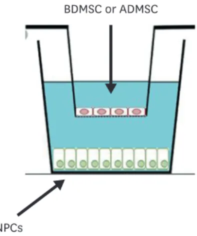
All co-cultures were conducted in triplicate in 6-well plates (Corning Incorporate, Corning, NY, USA) and using 0.4-μm pore size, high pore density, polyester membrane tissue culture treated polystyrene. Disc NPC control wells were prepared with 7.5×10⁵ cells per cm2. For co-culture wells without contact, BDMSCs and ADMSCs were seeded on tissue culture inserter plates, whereas disc NPCs were seeded in the base of the appropriate wells of a 6-well plate. Cells were seeded at ratios of 75:25 disc NP cells/MSCs (2.5×10⁵ cells per cm2). Co- cultured cells were maintained for 3 days (FIGURE 1).
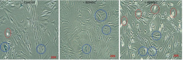
Senescence-associated β-galactosidase (SA-β-gal) staining and LD count When NPCs had been cultured to 80%–90% confluence, SA-β-gal staining (Cell Signaling Technology, Danvers, MA, USA) was performed according to the manufacturers protocol to analyze the rate of degeneration of cells in the ADMSCs and BDMSC co-culture groups.
Briefly, cells were washed with 1× phosphate buffered saline (PBS) 3 times and then fixed with fixative solution for 10–15 minutes at 20–25°C, washed twice with PBS, and stained with X-Gal containing SA-β-gal working solution overnight at 37°C without CO2. Quantification analysis was performed using microscopy by determining the average percentage of control cells in 4 randomly selected fields for each well. The number of cells stained with SA-β-gal was determined as a percentage of the total number of cells (%).
The total number of cells on a single slide stained with SA-β-gal was determined, and the number of cells for which intracellular LDs were observed were presented as a percentage of the total number of cells with droplets (%) (FIGURE 2).
BDMSC or ADMSC
NPCs
FIGURE 1. Co-culture system. BDMSCs and ADMSCs (pink, elliptical nucleus) co-culturing on NPCs (green, spherical nucleus). For co-culture wells without contact, BDMSCs or ADMSCs were seeded on tissue culture plates, whereas NPCs were seeded at the bottom of the appropriate wells of a 6-well plate.
BDMSC: bone marrow-derived mesenchymal stem cell, ADMSC: adipose-derived mesenchymal stem cell, NPC:
nucleus pulposus cell.
GAG secretion analysis
At >90% confluence of the human NPCs, they were cultured for 48 hours in DMEM/F12 (1:1) containing 1% FBS. Cultured samples were collected and washed twice with Dulbecco's Phosphate-Buffered Saline (Gibco, Thermo Fisher Scientific, Bohemia, NY, USA), and then centrifuged for 15 minutes at 3,000 rpm (2,095 g). The supernatant was cleared of any debris and the remaining supernatant was transferred to a clean tube. The prepared samples were added to the test sample well; HRP-conjugate reagent was added to each well, followed by incubation for 60 minutes at 37°C. Following 5 washes for 5 minutes, chromogen solution mixture was added to each well, followed by incubation at 37°C for 15 minutes. Finally, GAG secretion was measured as absorbance at 450 nm (MyBiosource, San Diego, CA, USA).
Statistical analysis
All the quantitative experiments were conducted in triplicate (SA-β-gal staining, LD count) or quadruplicate(GAG secretion assay) but the number of cell lines was small (n=4), so nonparametric statistics were applied to perform statistical analysis. Mann-Whitney U test was used to determine statistical significance between control and experimental groups. Kruskal- Wallis one-way analysis of variance was performed to determine significance among control and all experimental groups. A p-value less than 0.05 was considered statistically significant.
RESULTS
The older patient showed a higher rate of SA-β-gal staining of NPCs and more degenerative changes in NP according to the Pfirrmann grading system.
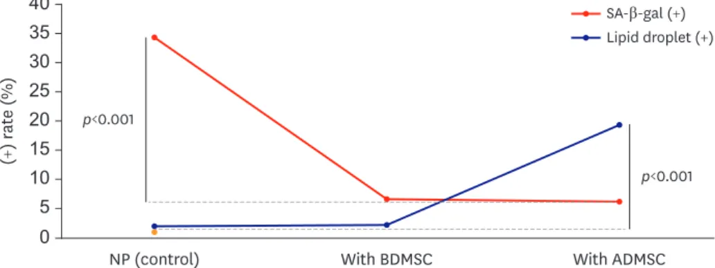
The proportion of cells stained with SA-β-gal was significantly lower when NPCs were co- cultured with either BDMSCs (43.27% vs. 6.95%, p<0.001) or ADMSCs (43.27% vs. 7.41%, p<0.001) (FIGURE 3). But there was no statistical significance between NPCs groups co- cultured with BDMSCs and ADMSCs (p=0.744).
The LDs observed in the NP control cell lines that had undergone primary cell culture were measured and represented less than 8.54% of all NPCs, regardless of the grade of
Control + BDMSC + ADMSC
FIGURE 2. SA-β-gal staining. The blue circle shows NPCs stained with SA-β-gal, and the red circle shows NPCs containing lipid droplets.
SA-β-gal: senescence associated-β-gal, NPC: nucleus pulposus cell, BDMSC: bone marrow-derived mesenchymal stem cell, ADMSC: adipose-derived mesenchymal stem cell.
degeneration, the patient's age or the MRI of the cell. However, for co-culture with BDMSCs, up to 10.23% of all NPCs had LDs. In the co-culture with ADMSCs, up to 33.3% of NPCs had LDs; therefore, LDs were increased, while SA-β-gal decreased, within the NPCs (FIGURE 3).
For co-culture with BDMSCs, the number of cells containing LDs was found to be statistically significantly lower than for co-culture with ADMSCs (5.24% vs. 13.41%, p<0.001). Cells tended to have a longer nucleus and cytoplasm in NPCs co-cultured with ADMSCs (FIGURE 2).
Optical density related to GAG secretion tended to be significantly lower for co-culture of BDMSCs and ADMSCs with NPCs than that of NPCs control group (p<0.001, p<0.001) (FIGURE 4).
DISCUSSION
MSCs of various origins show therapeutic effects in the attenuation of degenerative changes in NPCs.1,9,11,21) With the progress of regenerative medicine and tissue engineering, cell-based
40 35 30 25 20 15 10 5
0 NP (control) With BDMSC With ADMSC
p<0.001
p<0.001
(+) rate (%)
SA-β-gal (+) Lipid droplet (+)
FIGURE 3. Ratio of NPCs positive for SA-β-gal staining and the proportion of NPCs having lipid droplets. The ratio of SA-β-gal-stained cells was significantly more reduced in co-culture with MSCs than in the control group (p<0.001 vs. p<0.001). Moreover, when co-cultured with ADMSCs, the observed proportion of lipid droplets was significantly increased (p<0.001).
SA-β-gal: senescence associated-β-gal, NP: nucleus pulposus, NPC: nucleus pulposus cell, MSC: mesenchymal stem cell, ADMSC: adipose-derived mesenchymal stem cell, BDMSC: bone marrow-derived mesenchymal stem cell.
120 110 100 90 80 70 60 ECM change (GAG secretion) Optical density (OD/control) 50
Control BDMSC ADMSC
p<0.001
p<0.001 p=0.589
115.485 100.0
92.675 83.463
73.567
91.242
75.953
58.125 96.951
FIGURE 4. GAG secretion test. In both, BDMSCs and ADMSCs co-culture groups, the optical density decreased significantly when compared to the control group (p<0.001, p<0.001).
GAG: glycosaminoglycan, BDMSC: bone marrow-derived mesenchymal stem cell, ADMSC: adipose-derived mesenchymal stem cell, ECM: extracellular matrix, OD, optical density.
therapies are being explored with the aim of repairing degenerated IVDs and restoring IVD homeostasis.18) Despite preliminary results showing positive effects of MSC-based strategies in IVD regeneration, the exact mechanisms by which this regeneration occurs have not been completely determined yet. In IVD regeneration, MSCs serve as feeders or nursing cells to stimulate NP cells, both in vitro and in vivo.17,22) However, it has not been reported yet whether transplanted MSCs have any biophysical effects on endogenous NPCs. MSCs have the ability to modulate their microenvironment, including local endogenous cells, by secreting cytokines, growth factors, and chemokines such as stromal cell-derived factor-1, to facilitate regeneration.12) Vadalà et al.21) suggested that human MSCs obtain a more chondrogenic gene expression profile after being co-cultured with NPCs and influence the mRNA levels of human NPCs.
Although various studies have been conducted on MSCs, the effects of co-culturing ADMSCs and BDMSCs with NPCs have not been sufficiently investigated. While it is assumed that all MSCs will show similar characteristics, in this study, we had a clear understanding of the differences that could be observed in MSCs if they have different origins and proceeded with experiments accordingly. Data acquisition for this study was based on results and assumptions discussed in previous studies,2,3,6) There are a number of other biochemical differences between healthy discs and those altered by age and degeneration, such as an increase in the proportion of non-collagenous protein in both the NP and the AF,2) and the presence of LDs and the derived pigment, lipofuscin, in aged or degenerate human discs.2,3,6) Lipofuscin tends to accumulate more in the NP than in the AF,2) which results in the disc becoming progressively more pigmented with age, ranging in color from white in the immature form to yellow/brown in old age.2,3,6) Based on this preliminary study, it was assumed that when co-culturing MSCs with NPCs, it was possible to determine the SA-β-gal staining rate and the change in the lipid content of NPCs due to the MSC co-culture effects.
Based on the results of this study, it was confirmed that when NPCs were co-cultured with BDMSCs, the phenotype of markers related to degenerative changes was significantly improved, and there was a significant change in the GAG content in the ECM. However, our study could not confirm the gene expression levels of GAG; therefore, it was not possible to confirm the difference in the internal chemical fraction of GAG and to specifically determine which substance is changed significantly. Nonetheless, we believe that co-culturing BDMSCs with NPCs has been sufficiently shown to attenuate degenerative changes in NPCs. Further co-culturing studies and more diverse studies need to be conducted to verify these results.
CONCLUSION
When co-culturing NPCs with BDMSCs, SA-β-gal staining was used to show a statistically significant attenuation of degenerative changes. Moreover, the unexpected adverse effects of increased LD were significantly higher in NPCs co-cultured with ADMSCs. However, when NPCs were co-cultured with MSCs, GAG secretion was significantly decreased, and further studies need to be conducted to confirm these results. This is a pilot study on the effects of co-culturing BDMSCs and ADMSCs with NPCs collected from only 3 patients, and further studies need be performed to investigate the effects of BDMSCs.
ACKNOWLEDGMENTS
We would like to thank Editage (www.editage.jp) for English language editing. This work was supported by grant No. 02-2012-016 from the Seoul National University Bundang Hospital (SNUBH) Research Fund.
REFERENCES
1. Allon AA, Schneider RA, Lotz JC. Co-culture of adult mesenchymal stem cells and nucleus pulposus cells in bilaminar pellets for intervertebral disc regeneration. SAS J 3:41-49, 2009
PUBMED | CROSSREF
2. Banga I. Investigations of fluorescent peptides and lipofuscins of human intervertebral disc relating to atherosclerosis. Atherosclerosis 22:533-541, 1975
PUBMED | CROSSREF
3. Dickson IR, Happey F, Pearson CH, Naylor A, Turner RL. Variations in the protein components of human intervertebral disk with age. Nature 215:52-53, 1967
PUBMED | CROSSREF
4. Dimri GP, Lee X, Basile G, Acosta M, Scott G, Roskelley C, et al. A biomarker that identifies senescent human cells in culture and in aging skin in vivo. Proc Natl Acad Sci U S A 92:9363-9367, 1995 PUBMED | CROSSREF
5. Erwin WM, Hood KE. The cellular and molecular biology of the intervertebral disc: a clinician's primer. J Can Chiropr Assoc 58:246-257, 2014
PUBMED
6. Eyre DR. Biochemistry of the intervertebral disc. Int Rev Connect Tissue Res 8:227-291, 1979 PUBMED | CROSSREF
7. Ferrari G, Cusella-De Angelis G, Coletta M, Paolucci E, Stornaiuolo A, Cossu G, et al. Muscle regeneration by bone marrow-derived myogenic progenitors. Science 279:1528-1530, 1998
PUBMED | CROSSREF
8. Galbusera F, van Rijsbergen M, Ito K, Huyghe JM, Brayda-Bruno M, Wilke HJ. Ageing and degenerative changes of the intervertebral disc and their impact on spinal flexibility. Eur Spine J 23 Suppl 3:S324-S332, 2014 PUBMED | CROSSREF
9. Hildner F, Concaro S, Peterbauer A, Wolbank S, Danzer M, Lindahl A, et al. Human adipose-derived stem cells contribute to chondrogenesis in coculture with human articular chondrocytes. Tissue Eng Part A 15:3961-3969, 2009
PUBMED | CROSSREF
10. Kivitz AJ, Gimbel JS, Bramson C, Nemeth MA, Keller DS, Brown MT, et al. Efficacy and safety of tanezumab versus naproxen in the treatment of chronic low back pain. Pain 154:1009-1021, 2013 PUBMED | CROSSREF
11. Kotton DN, Ma BY, Cardoso WV, Sanderson EA, Summer RS, Williams MC, et al. Bone marrow-derived cells as progenitors of lung alveolar epithelium. Development 128:5181-5188, 2001
PUBMED
12. Liu X, Duan B, Cheng Z, Jia X, Mao L, Fu H, et al. SDF-1/CXCR4 axis modulates bone marrow mesenchymal stem cell apoptosis, migration and cytokine secretion. Protein Cell 2:845-854, 2011 PUBMED | CROSSREF
13. Pfirrmann CW, Metzdorf A, Zanetti M, Hodler J, Boos N. Magnetic resonance classification of lumbar intervertebral disc degeneration. Spine 26:1873-1878, 2001
PUBMED | CROSSREF
14. Pittenger MF, Mackay AM, Beck SC, Jaiswal RK, Douglas R, Mosca JD, et al. Multilineage potential of adult human mesenchymal stem cells. Science 284:143-147, 1999
PUBMED | CROSSREF
15. Richardson SM, Walker RV, Parker S, Rhodes NP, Hunt JA, Freemont AJ, et al. Intervertebral disc cell- mediated mesenchymal stem cell differentiation. Stem Cells 24:707-716, 2006
PUBMED | CROSSREF
16. Risbud MV, Schoepflin ZR, Mwale F, Kandel RA, Grad S, Iatridis JC, et al. Defining the phenotype of young healthy nucleus pulposus cells: recommendations of the Spine Research Interest Group at the 2014 annual ORS meeting. J Orthop Res 33:283-293, 2015
PUBMED | CROSSREF
17. Sakai D, Mochida J, Iwashina T, Watanabe T, Nakai T, Ando K, et al. Differentiation of mesenchymal stem cells transplanted to a rabbit degenerative disc model: potential and limitations for stem cell therapy in disc regeneration. Spine 30:2379-2387, 2005
PUBMED | CROSSREF
18. Sakai D, Andersson GB. Stem cell therapy for intervertebral disc regeneration: obstacles and solutions.
Nat Rev Rheumatol 11:243-256, 2015 PUBMED | CROSSREF
19. Singh K, Masuda K, Thonar EJ, An HS, Cs-Szabo G. Age-related changes in the extracellular matrix of nucleus pulposus and anulus fibrosus of human intervertebral disc. Spine 34:10-16, 2009
PUBMED | CROSSREF
20. Sobajima S, Vadala G, Shimer A, Kim JS, Gilbertson LG, Kang JD. Feasibility of a stem cell therapy for intervertebral disc degeneration. Spine J 8:888-896, 2008
PUBMED | CROSSREF
21. Vadalà G, Studer RK, Sowa G, Spiezia F, Iucu C, Denaro V, et al. Coculture of bone marrow mesenchymal stem cells and nucleus pulposus cells modulate gene expression profile without cell fusion. Spine 33:870- 876, 2008
PUBMED | CROSSREF
22. Yamamoto Y, Mochida J, Sakai D, Nakai T, Nishimura K, Kawada H, et al. Upregulation of the viability of nucleus pulposus cells by bone marrow-derived stromal cells: significance of direct cell-to-cell contact in coculture system. Spine 29:1508-1514, 2004
PUBMED | CROSSREF
23. Zheng CJ, Chen J. Disc degeneration implies low back pain. Theor Biol Med Model 12:24, 2015 PUBMED | CROSSREF