http://dx.doi.org/10.12671/jkfs.2014.27.3.206
206
Copyright ⓒ 2014 The Korean Fracture Society. All rights reserved.
This is an Open Access article distributed under the terms of the Creative Commons Attribution Non-Commercial License (http://creativecommons.org/licenses/
by-nc/3.0) which permits unrestricted non-commercial use, distribution, and reproduction in any medium, provided the original work is properly cited.
Received November 25, 2013 Revised May 12, 2014 Accepted May 12, 2014
Address reprint requests to: Kook Jin Chung, M.D.
Department of Orthopaedic Surgery, Hallym University Kangnam Sacred Heart Hospital, Hallym University College of Medicine, 1 Singil-ro, Yeongdeungpo-gu, Seoul 150-950, Korea
Tel: 82-2-829-5435ㆍFax: 82-2-834-1728 E-mail: [email protected]
Financial support: None. Conflict of interest: None.
슬개골 분쇄 골절 치료에서 긴장대 강선 고정 및 부가적인 환형 강선 고정술의 결과
이영민⋅정국진 ⋅황지효⋅김홍균⋅윤용현
한림대학교 의과대학 강남성심병원 정형외과학교실
Results of Tension Band Wiring and Additional Circumferential Wiring in Treatment of Comminuted Patella Fracture
Young Min Lee, M.D., Kook Jin Chung, M.D. , Ji Hyo Hwang, M.D., Ph.D., Hong Kyun Kim, M.D., Yong Hyun Yoon, M.D.
Department of Orthopaedic Surgery, Kangnam Sacred Heart Hospital, Hallym University College of Medicine, Seoul, Korea
Purpose: The purpose of this study is to evaluate the results of tension band wiring and additional circumferential wiring in treatment of comminuted patella fractures.
Materials and Methods: A retrospective study of 67 patients with follow-up period longer than six months who underwent tension band wiring and additional circumferential wiring for comminuted patellar fracture from January 2004 to December 2012 was conducted. Analysis was based on radiological evaluation of bony union and articular surface displacement, and clinically by evaluating the postoperative function of the knee joint using the Levack scoring system.
Results: Only one case out of 67 (1.5%) showed nonunion without metal breakage while good bone union was achieved in all other cases. Excluding the nonunion case, range of motion was 90 degrees minimum, 135 maximum, 129 on average.
Average displacement was less than 2 mm, and 64 out of 67 cases showed satisfactory outcome with excellent functional score according to the Levack scoring system.
Conclusion: Tension band wiring and additional circumferential wiring technique for treatment of comminuted patella frac- tures can be considered as an effective treatment for achievement of good bone union and restoration of normal knee function.
Key Words: Patella, Comminuted fractures, Tension band wiring, Cerclage wiring
서 론
슬개골은 대퇴사두근, 건과 슬개건을 연결하는 해부학적 골조직으로 대퇴사두근의 수축에 의한 장력이 슬개골을 통 해 슬개건으로 전달되어 슬관절이 신전되는 신전 기전의 중요한 역할을 담당한다.1,2) 슬개골의 골절이 발생할 경우 슬관절 신전 기능의 소실이 유발되므로 정확한 치료가 예
Fig. 1. (A) Preoperative radiographs of a 37-year-old male patient show comminuted and displaced fracture of patella. (B) Postoperative radiographs show well reduced bony fragments by tension band wiring and additional circumferential wiring.
후에 중요한 영향을 미친다. 불완전 골절 및 비전위 골절 에는 장하지 석고 고정을 통한 보존적 치료를 할 수 있으 며 전위와 분쇄의 정도가 심하지 않은 경우에는 관혈적 정 복 및 내고정술이 일반적인 치료 방법으로 잘 알려져 있고 최근에는 최소 침습적 수술법 등을 통한 치료가 가능하다 고 보고되었다.3) 전위가 심한 분쇄 골절에서는 정확한 관 절면의 정복 및 견고한 내고정을 통한 조기 관절 운동을 위해 관혈적 정복 및 견고한 내고정이 필수적이다.4,5) 슬개 골의 전위가 심한 분쇄 골절의 경우 합병증으로 무혈성 괴 사,6) 불유합,7) 슬개 대퇴 관절염 및 이로 인한 슬관절 전 방부 통증7) 등이 발생할 수 있으며 슬개골의 부분적 절제 술, 슬개건의 이전술 및 슬개골 전절제술이 필요하게 되어 하지 기능에 많은 장애를 초래하게 된다.8-11) 저자들은 슬 개골의 심한 전위가 동반된 분쇄 골절에서 긴장대 강선 고 정(tension band wiring) 및 부가적인 환형 강선 고정 (circumferential wiring)을 통한 내고정술을 통한 임상적 결과 및 문헌 고찰을 보고하고자 한다.
대상 및 방법
1. 연구 대상
저자들은 2004년 1월부터 2012년 12월까지 슬개골 골절 에 대해 수술적 치료를 받은 환자 중 슬개골 분쇄 골절로 진단되어 수술을 시행받았던 환자에서 최소 6개월 이상 추 시가 가능하였던 67예 환자를 대상으로, 방사선 사진, 입원
및 외래 기록지, 수술기록지를 이용하여 후향적 분석을 시 행하였다. 슬개골 골절의 분류는 골절의 형태에 따라 분류 하였으며12) 분쇄 골절은 단순 골절과는 달리 전위와 무관 하게 골절 주위로의 골절편(bony fragment)이 관찰되거나 슬개골 골절 위치와 상관없이 골절선이 두 개 이상인 경우 로 정의하였다. 평균 추시 기간은 19개월(6-62개월)이었으 며, 남자가 45명, 여자가 22명이었고, 평균 나이는 남자 54 세, 여자 55.2세였다. 손상 원인은 실족이 50예로 가장 많 았고, 교통사고가 13예, 추락 사고가 4예였다. 개방성 골절 은 5예였다.
2. 수술 술기
수술은 지혈 압박대를 착용 후 골절부위를 중심으로 한 횡절개를 하였으며 개방창이 존재하는 경우 그 형태에 따 라 절개 형태를 달리하였다. Towel clamp를 이용하여 골 절편의 정복을 시행 후 K-강선 2개를 골절면에 가급적 수 직으로 고정한 후 슬관절 굴곡 0도-30도에서 대퇴 사두건 및 슬개 인대의 긴장을 최소화하고 긴장대 강선 방법으로 고정하였고 골편 수에 따라서 K-강선을 추가로 고정한 이 후 부가적으로 환형 강선 고정술을 시행하였다(Fig. 1). 고 정력을 확인하고자 부드럽게 슬관절을 90도까지 굴곡하여 안정성을 확인하였다.
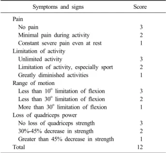
Table 1. Scoring System (According to Levack) Symptoms and signs Score
Pain
No pain 3
Minimal pain during activity 2
Constant severe pain even at rest 1 Limitation of activity
Unlimited activity 3
Limitation of activity, especially sport 2
Greatly diminished activities 1
Range of motion
Less than 10o limitation of flexion 3 Less than 30o limitation of flexion 2 More than 30o limitation of flexion 1 Loss of quadriceps power
No loss of quadriceps strength 3
30%-45% decrease in strength 2
Greater than 45% decrease in strength 1
Total 12
3. 재활
수술 후 다음날부터 장하지 부목 고정하여 대퇴사두근 등척 운동을 시행하였고 2주부터 굴곡운동을 시작하였으며 수술 후 3주부터 신전 상태로 전 체중부하를 허용하였다.
수술 후 4주부터는 90도 이상 가능한 한 굴곡을 시행하였 고, 수술 후 6주 후에는 제한 없이 굴곡을 허용하였다.
4. 평가 방법
측면 방사선 사진상 관절면의 전위 정도를 수술 전과 수술 후에 측정하였다. 전위 정도는 두 골편의 관절면 사 이의 전후 거리를 전위(displacement)라 하였으며13) 1 mm 미만의 부드러운 관절면을 보일 때를 우수(excellent), 1-2 mm의 전위를 보일 때는 양호(good), 2-3 mm의 전위는 보통(fair), 그 이상을 초과할 때는 불량(poor)으로 정의하 였다. 그 외 수술 후 결과 판정은 객관성을 부여하기 위해 Levack scoring system14)을 이용하여 슬관절 기능을 평가 하였다(Table 1). 9점 초과 시 양호(good), 6-9점 시 보통 (fair), 6점 미만일 때는 불량(poor)으로 정의하였다. 슬관 절 운동 범위는 절대치와 건측에 비교하여 각도를 측정하 여 어느 정도 제한이 있는지 확인하였다.
결 과
총 67예 중 수술 후 1예에서 강선 파열과 동반한 불유 합이 있었으며 이는 같은 수술 방법을 통한 재수술로 골유
합을 얻었다(Fig. 2). 그 외 모든 경우에서 수술 후 내 고 정의 실패로 인한 정복 소실은 관찰되지 않았으며, 골유합 을 얻었던 방사선 사진의 측면상에서 평균 전위 정도는 수 술 전 6.3 mm (2.8-14.7 mm)에서 수술 후 0.3 mm (0.1- 1.2 mm)로 감소하여 우수(excellent) 64예, 양호(good) 2예 로 평가되었으며, 불유합되었던 1예는 수술 전 8.5 mm에 서 수술 후 0.7 mm로 우수하였으나 3개월 때 12.4 mm로 전위되어 불량으로 포함시켰다. 관절 운동 범위는 90도에 서 135도까지 평균 129도였다.
임상 평가에서 Levack scoring system을 사용하였으며 통증 정도는 평균 2.39 (표준 편차, ±0.53), 활동 제한 정 도는 평균 2.35 (표준 편차, ±0.48) 관절 운동 범위는 평 균 2.52 (표준 편차, ±0.54) 대퇴 사두근 근력 소실 정도 는 평균 2.52 (표준 편차, ±0.54)였다. 양호(good)가 47예 (12점 19예, 11점 23예, 10점 5예), 보통(fair) 19예(9점 15 예, 8점 3예, 7점 1예), 불량(poor)이 1예(5점 1예)로 만족 할 만한 결과를 얻었다. 정복 소실의 1예(1.5%)로 인해 재 수술 이외에는 재수술을 요할만한 수술 절개 부위 감염이 나 골수염, 내 고정물의 실패, 골 괴사 및 관절 강직의 소 견은 없었으며 임상적으로 70% 이상에서 양호 이상의 결 과를 얻었다. 고정물의 피부 자극을 호소하는 경우는 13예 였으나 피부 괴사는 없었으며 경미한 통증을 호소하여 조 기 제거술은 시행하지 않았다. 정복 소실 1예의 경우 수술 후 관절 운동의 강직으로 인한 무리한 수동적 관절 운동으 로 인해 발생하였다.
고 찰
다른 장관골에 비해 슬개골은 우리 몸에서 가장 큰 종 자골로서 다른 종자골에 비해 그 역할이 크며 대퇴 사두근 지렛대(lever arm)의 지렛목(fulcrum) 역할을 하여 슬관절 의 신전력을 증가시키는 역할을 한다. 따라서 슬개골에 문 제가 발생하면 신전 기능에 문제가 생기며 이는 무릎에 상 당한 문제를 초래할 수 있다. 따라서 골절의 치료 목적은 신전 기전의 연속성을 확보하고 슬개골 기능을 보존시키고 슬개골 골절로 인한 대퇴 슬개골 관절염을 줄이고자 하는 것이다.
슬개골 골절의 치료 방법은 1-2 mm 이하의 비전위 골 절의 경우에는 슬관절 혈종의 제거 후 석고 고정을 이용한 장하지 석고 고정으로 치료가 가능하지만 2 mm 이상의 전위가 있는 골절의 경우 소실된 슬관절의 신전 기능의 회 복을 위해서는 반드시 수술적 치료가 필요하게 된다. 슬개 골 골절의 수술 후 정상적인 슬관절 기능 회복에는 최소 6-12개월이 걸리는 것으로 알려져 있으며15) 슬개골 골절 후 골유합과 기능의 회복에 대한 예후는 일반적으로 좋다
Fig. 2. (A) Preoperative radiographs of a 43-year-old man show a comminuted patella fracture by a fall from a standing height. (B) Immediate postoperative radiographs show that the fragments are well stabilized by tension band wiring and additional circumferential wiring. (C) Loss of reduction was observed at four months after surgery. (D) Reoperation was performed using the same technique and good reduction of the fragments was achieved.
고 알려져 있다.
수술적 치료의 골절 고정 방법은 골절편의 형태에 따라 다양한데 환형 강선 고정술, 종 강선 고정술(longitudinal wiring), 나사 고정술, 장력대 고정술 등이 있으며 이러한 술식을 약간씩 변형시켜서 수술하는 경우도 있다. 과거의 경우는 대부분 강선을 통한 고정이 대부분의 치료였으나 최근 국내에서 보고된 논문들은 이러한 고식적인 치료 방 법보다는 단순 골절일 때 Kim 등13)은 유관 나사 단독 사 용으로 좋은 결과를 보고하였고 Ha와 Sun,16) Choi 등17) 및 Kang 등18)은 유관 나사못과 강선을 사용하여 슬개골 정복 후 양호한 결과를 얻었다고 보고하였다. 또한 골절의 비침습적 치료(minimal invasive ostesynthesis)가 대두되면 서 관절경을 이용한 정복술에 대한 논문도 소개되었다.19) 그러나 이러한 논문에서 심한 분쇄 골절인 경우는 많은 제
한이 있으며 분쇄 골절의 경우에는 골절 편의 연부조직의 부착을 유지하면서 골절 편을 정복하기 위해 간접적 정복 의 방법을 시도할 수 있다. 하지만 이 경우 슬개골 관절 연골의 부정확한 정복으로 인한 슬개골 관절 연골의 불일 치를 초래하게 되고 관절 내 골절의 특성으로 관절 연골의 손상으로 인한 외상성 관절염으로 발전할 수 있음은 잘 알 려져 있다.20) 이러한 외상성 관절염은 비전위 골절 또는 단순 골절보다 분쇄 골절에서 흔히 발생하며 이 경우 슬관 절 신전 기전의 소실이 20%-49%에서 발생하고, 슬개골 전 절제술을 시행하게 되는 경우 불량한 예후를 보이며21) 정 상적인 슬관절 기능을 보이는 경우는 단 5%에 불과한 것 으로 알려져 있다.6) 따라서 좀 더 덜 침습적인 치료일수록 치료 결과가 나빠질 수 있다. 과거 강선 고정술에 대한 연 구에서는 Ha 등22)은 12예의 골절에서 11예가 양호하다고
보고한 바가 있으며, Chung 등23)은 29예의 슬개골 골절 환자에서 8예(27%)의 합병증이 발생한다고 보고한 바 있 다. 그러나 저자들은 타 연구에 비해 환자 대상이 충분함 에도 불구하고 훨씬 더 만족스러운 결과를 얻었다. 저자들 이 수술에 이용한 술식은 비록 수술 절개창이 크고 수술 시간과 출혈량이 많을 수 있는 고식적인 방법이나 최근 대 두되고 있는 비침습적 술식에 비해 슬개골 관절면의 해부 학적 정복 및 견고한 고정을 얻을 수 있으며 또한 이를 통 한 조기 관절 운동을 통해 만족스러운 슬관절의 운동 범위 의 제한 및 관절염과 같은 합병증을 최소화하였기에 슬개 골의 분쇄 골절 치료의 만족스러운 술식으로 판단되어 보 고하는 바이다.
저자들의 경우 타 연구에 비해 환자 수도 충분하며 우 수한 치료 결과를 보였으나 다른 슬개골 분쇄 골절의 다른 술식의 대조군을 설정하여 전향적으로 비교하였다면 이러 한 치료 방법의 상대적인 우수성을 좀 더 정확히 평가할 수 있을 것으로 생각되며 또한 평균 추시 기간이 19개월로 슬개골 골절 이후 발생한 슬개 대퇴 관절염을 정확히 평가 하기에 충분한 추시 기간이 되지 않아 이를 평가하기에 제 한이 있었다는 점이 한계점으로 생각된다.
결 론
슬개골 분쇄 골절에서 관혈적 정복을 통한 관절면의 해 부학적 정복 및 긴장대 강선 고정과 부가적인 환형 강선 고정술을 시행하고 임상적, 방사선학적으로 양호한 결과를 얻어 슬개골 분쇄 골절에서 긴장대 강선고정 및 부가적인 환형 강선 고정술은 좋은 치료법의 하나로 생각된다.
References
1) Carson WG Jr, James SL, Larson RL, Singer KM, Winternitz WW: Patellofemoral disorders: physical and radiographic evaluation. Part II: radiographic examination.
Clin Orthop Relat Res, (185): 178-186, 1984.
2) Insall J, Goldberg V, Salvati E: Recurrent dislocation and the high-riding patella. Clin Orthop Relat Res, 88:
67-69, 1972.
3) Pritchett JW: Nonoperative treatment of widely displaced patella fractures. Am J Knee Surg, 10: 145-147, 1997.
4) Harris RM: Fractures of the patella. In: Bucholz RW, Heckman JD ed. Rockwood and Green’s fractures in adults. vol. 2. 5th ed. Philadelphia, Lippincott Williams &
Wilkins: P1775e99, 2001.
5) Muller ME, Allgower M, Schneider R, et al: Manual of
internal fixation. Techniques recommended by the AO group. Berlin, Springer-Verlag: 248-253, 1979.
6) Scott JC: Fractures of the patella. J Bone Joint Surg Br, 31: 76-81, 1949.
7) Nummi J: Fracture of the patella. A clinical study of 707 patellar fractures. Ann Chir Gynaecol Fenn Suppl, 179:
1-85, 1971.
8) Depalma AF, Flynn JJ: Joint changes following ex- perimental partial and total patellectomy. J Bone Joint Surg Am, 40: 395-413, 1958.
9) Dobbie RP, Ryerson S: The treatment of fractured pa- tella by excision. Am J Surg, 55: 339-373, 1942.
10) Einola S, Aho AJ, Kallio P: Patellectomy after fracture.
Long-term follow-up results with special reference to functional disability. Acta Orthop Scand, 47: 441-447, 1976.
11) Shim SS, Leung G: Blood supply of the knee joint. A microangiographic study in children and adults. Clin Orthop Relat Res, (208): 119-125, 1986.
12) Canale ST, Beaty JH: Campbell's operative orthopedics.
12th ed. Philadelphia, Elsevier Mosby: 2681, 2013.
13) Kim JM, Yoo JS, Kwon YJ, Cheon JO: Treatment of transverse patellar fracture with cannulated screws. J Korean Fract Soc, 20: 149-153, 2007.
14) Levack B, Flannagan JP, Hobbs S: Results of surgical treatment of patellar fractures. J Bone Joint Surg Br, 67:
416-419, 1985.
15) Duthie HL, Hutchinson JR: The results of partial and total excision of the patella. J Bone Joint Surg Br, 40:
75-81, 1958.
16) Ha CW, Sun JI: The treatment of patellar fracture with modified tension band wiring, using cannulated screws. J Korean Fract Soc, 17: 117-121, 2004.
17) Choi SK, Ham DK, Yim MS, Kim KY, Shin HS:
Treatment of displaced transverse patellar fractures with cannulated screws and figure-eight wiring. J Korean Fract Soc, 18: 149-154, 2005.
18) Kang S, Chung PH, Hwang CS, Kim JP, Kim YS, Parke CS: Transverse fracture through screw site after cannulated screw fixation in vertical patella fracture: a case report. J Korean Fract Soc, 19: 96-99, 2006.
19) Ko SH, Cho SD, Na HY, et al: Arthroscopic-assisted re- duction and internal fixation of patella fractures. J Korean Soc Fract, 16: 482-489, 2003.
20) Gekeler EO, Quaranra AV: Patellectomy for degener-
ative arthritis of the knee: late results. J Bone Joint Surg Am, 44: 1109-1114, 1962.
21) Jakobsen J, Christensen KS, Rasmussen OS: Patellecto- my--a 20-year follow-up. Acta Orthop Scand, 56: 430-432, 1985.
22) Ha KI, Hahn SH, Chung MY, Park HS: Treatment of
patella fracture using wiring method. J Korean Soc Fract, 2: 229-233, 1989.
23) Chung YK, Yoo JH, Park YW, Roh GC: Operative treatment of comminuted fracture of the patella. J Korean Soc Fract, 9: 970-976, 1996.
Copyright ⓒ 2014 The Korean Fracture Society. All rights reserved.
This is an Open Access article distributed under the terms of the Creative Commons Attribution Non-Commercial License (http://creativecommons.org/licenses/
by-nc/3.0) which permits unrestricted non-commercial use, distribution, and reproduction in any medium, provided the original work is properly cited.
http://dx.doi.org/10.12671/jkfs.2014.27.3.206
슬개골 분쇄 골절 치료에서 긴장대 강선 고정 및 부가적인 환형 강선 고정술의 결과
이영민⋅정국진 ⋅황지효⋅김홍균⋅윤용현
한림대학교 의과대학 강남성심병원 정형외과학교실
목 적: 슬개골 분쇄 골절에 있어서 긴장대 강선 고정(tension band wiring) 및 부가적인 환형 강선 고정(circumferential wiring)을 통한 치료 결과를 보고하고자 한다.
대상 및 방법: 2004년 1월부터 2012년 12월까지 슬개골 분쇄 골절로 긴장대 강선 고정과 부가적인 환형 강선 고정술을 시행받 았던 환자 중 6개월 이상 추시가 가능하였던 67명의 환자를 대상으로 후향적 분석을 통해 방사선학적 골유합, 관절면 전위 정도, 임상적으로 Levack scoring system을 이용하여 수술적 치료 후 슬관절의 기능을 평가하였다.
결 과: 총 67예 중 수술 후 내고정의 정복 소실로 인한 불유합 1예(1.5%)를 제외한 모든 경우에서 골유합을 얻었으며, 평균 관절 운동 범위는 90도에서 135도까지 평균 129도였다. 평균 전위 정도는 불유합인 경우를 제외하고는 2 mm 이하였으며 Levack scoring system을 이용하여 슬관절 기능을 평가한 결과는 총 67예 중 64예가 우수로 만족할 만한 결과를 얻었다.
결 론: 슬개골 분쇄 골절에서 긴장대 강선 고정과 부가적인 환형 강선 고정술은 만족스러운 골유합을 얻고 정상적인 슬관절 기능을 회복할 수 있는 좋은 치료법의 하나로 생각된다.
색인 단어: 슬개골, 분쇄 골절, 긴장대 강선 고정, 환형 강선 고정
접수일 2013. 11. 25 수정일 2014. 5. 12 게재확정 2014. 5. 12 교신저자 정 국 진
서울시 영등포구 신길로 1, 한림대학교 의과대학 강남성심병원 정형외과학교실 Tel 02-829-5435, Fax 02-834-1728, E-mail [email protected]
212