Brief Report
804 Ann Dermatol
Received September 9, 2016, Revised October 22, 2016, Accepted for publication October 31, 2016
Corresponding author: Won Park, Division of Rheumatology, Department of Internal Medicine, Inha University, 366 Seohae-daero, Jung-gu, Incheon 22332, Korea. Tel: 82-32-890-3483, Fax: 82-32-890- 2237, E-mail: [email protected]
This is an Open Access article distributed under the terms of the Creative Commons Attribution Non-Commercial License (http://creativecommons.org/
licenses/by-nc/4.0) which permits unrestricted non-commercial use, distribution, and reproduction in any medium, provided the original work is properly cited.
Copyright © The Korean Dermatological Association and The Korean Society for Investigative Dermatology
https://doi.org/10.5021/ad.2017.29.6.804
Palisaded Neutrophilic Granulomatous Dermatitis in a Patient with Systemic Sclerosis-Rheumatoid Arthritis Overlap Syndrome
Kyong-Hee Jung, Sangho Jeong, Seong-Ryul Kwon, Mie Jin Lim, Jiyeon Gwon, Jeonghyun Shin
1, Won Park
Division of Rheumatology, Department of Internal Medicine, 1Department of Dermatology, Inha University, Incheon, Korea
Dear Editor:
Palisaded neutrophilic granulomatous dermatitis (PNGD) is a rare pathohistologic diagnosis that has been associated with various autoimmune diseases1. However, to our know- ledge, the occurrence of PNGD in patients with systemic sclerosis (SSc) /rheumatoid arthritis (RA) overlap syndrome has not been reported so far. There are many reports about skin manifestations of RA and SSc. However, re- ports about skin manifestations of their overlap syndrome are few. SSc-RA overlap syndrome is a rare autoimmune disease and has a distinct genetic, immunological, and clinical entity2. Herein, we report a case of PNGD in a pa- tient with SSc-RA overlap syndrome.
A 63-year-old woman presented with complaints of severe tenderness of both soles for 2 months. Symptoms devel- oped after repeated heel walking as an exercise to im- prove muscle strength. About 6 years prior, she was diag- nosed with interstitial lung disease. Approximately 1 year prior, she presented with hand and facial swelling and multiple joints pain. She complained of Raynaud’s phe- nomenon and swollen hands. Sclerodactyly, telangiectases, and arthritis were also observed. The laboratory evalua- tion showed positive results for anti-Scl 70 antibody (Ab).
The level of anti-cyclic citrullinated peptide Ab was 205.6 IU/ml and that of rheumatoid factor was 390.0 IU/ml.
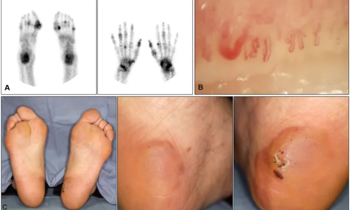
99mTc bone scintigraphy showed abnormal increased joints uptake (Fig. 1A). Nailfold capillary microscopy showed a dilatated and tortuous capillary loops and giant capillary (Fig. 1B). The patient was diagnosed with SSc-RA overlap
syndrome. When the patient was admitted with foot pain, 5-cm erythematous, annular tender edematous nodules were observed on both soles (Fig. 1C). The histopatho- logic findings revealed diffuse histiocytes infiltrations in- terstitially and palisading with neutrophils, nuclear dusts, and degenerated collagen bundles in the entire dermis (Fig. 2). Immunohistochemical staining for CD68 showed positive results for the infiltrated histiocytes. These find- ings were consistent with PNGD. We continued treatment for her underlying SSc-RA overlap syndrome, added dap- sone 25 mg bid for PNGD. She was also advised to avoid weight-bearing activity and cold exposure. Soon after, the lesions seemed to have improved, but worsened 3 months later, prompting us to prescribe additional methylpredni- solone and increase dosage of dapsone. About 5 months later, the lesions improved, but in the next 6 months she developed new lesions on both legs. A retrial of dapsone improved her skin within 2 weeks.
PNGD is a type of reactive granulomatous dermatitis asso- ciated with connective tissue diseases, lymphoproliferative diseases, and medications3. The lesion sometimes occurs af- ter repeated trauma, and trauma may be involved in the deposition of immune complexes3. The differential diag- nosis of PNGD includes small vessel vasculitis, neu- trophilic dermatoses, granuloma annulare, and interstitial granulomatous dermatitis3,4. PNGD is known as a self-lim- iting benign disease, and management of the underlying disease is most important. Unlike other PNGD cases, this patient showed a waxing and waning clinical course. The
Brief Report
Vol. 29, No. 6, 2017 805 Fig. 1. (A) Whole-body bone scan shows increased uptake in both wrists, hands, ankles, and feet. (B) Nailfold capillary microscopy shows dilatated and tortuous capillary loops and giant capillary. (C) Erythematous annular nodules on both soles.
Fig. 2. (A) The low power view shows diffuse palisading granulo- matous infiltrations in the entire dermis (H&E, ×40). (B) Interstitial and palisading histiocytes infiltration with degenerated collagen bundles surrounded by neutrophils (inset,
×400) in the deep dermis (H&E,
×200).
reason for a different course may be that SSc-RA overlap syndrome has a greater disease burden than that in patients with limited SSc5. Although PNGD with SSc-RA overlap syndrome is very rare, clinical awareness of this combina- tion would allow an early diagnostic and therapeutic approach.
ACKNOWLEDGMENT
This work was supported by Inha University Research Grant.
CONFLICTS OF INTEREST
The authors have nothing to disclose.
REFERENCES
1. Bremner R, Simpson E, White CR, Morrison L, Deodhar A.
Palisaded neutrophilic and granulomatous dermatitis: an unusual cutaneous manifestation of immune-mediated disorders. Semin Arthritis Rheum 2004;34:610-616.
Brief Report
806 Ann Dermatol
Received March 16, 2016, Revised November 3, 2016, Accepted for publication November 7, 2016
Corresponding author: Hei Sung Kim, Department of Dermatology, Incheon St. Mary’s Hospital, College of Medicine, The Catholic University of Korea, 56 Dongsu-ro, Bupyeong-gu, Incheon 21431, Korea. Tel: 82-32-280-5700, Fax: 82-32-506-9514, E-mail: [email protected]
This is an Open Access article distributed under the terms of the Creative Commons Attribution Non-Commercial License (http://creativecommons.org/li- censes/by-nc/4.0) which permits unrestricted non-commercial use, distribution, and reproduction in any medium, provided the original work is properly cited.
Copyright © The Korean Dermatological Association and The Korean Society for Investigative Dermatology 2. Szücs G, Szekanecz Z, Zilahi E, Kapitány A, Baráth S,
Szamosi S, et al. Systemic sclerosis-rheumatoid arthritis overlap syndrome: a unique combination of features suggests a distinct genetic, serological and clinical entity. Rheumatology (Oxford) 2007;46:989-993.
3. Rosenbach M, English JC 3rd. Reactive granulomatous dermatitis: a review of palisaded neutrophilic and granu- lomatous dermatitis, interstitial granulomatous dermatitis, interstitial granulomatous drug reaction, and a proposed
reclassification. Dermatol Clin 2015;33:373-387.
4. Kim YS, Lee JH, Lee JY, Park YM. Interstitial granulomatous dermatitis associated with rheumatoid arthritis. Ann Dermatol 2016;28:395-397.
5. Moinzadeh P, Aberer E, Ahmadi-Simab K, Blank N, Distler JH, Fierlbeck G, et al. Disease progression in systemic sclerosis-overlap syndrome is significantly different from limited and diffuse cutaneous systemic sclerosis. Ann Rheum Dis 2015;74:730-737.
https://doi.org/10.5021/ad.2017.29.6.806
A Case of Segmental (Zosteriform) Juvenile Xanthogranuloma
Seok Hoon Moon, Sang Hyun Cho, Jeong Deuk Lee, Hei Sung Kim
Department of Dermatology, Incheon St. Mary’s Hospital, College of Medicine, The Catholic University of Korea, Incheon, Korea
Dear Editor:
A 14-year-old boy presented with asymptomatic skin nodules. Clinical examination revealed multiple, 0.3∼0.5 cm-sized, brown to skin-colored nodules in a band-like fashion along the left side of the waist (Fig. 1). The lesions were said to have appeared 6 months ago and have been increasing in size and number. He denied the history of trauma or other cutaneous inflammation. There were no systemic symptoms such as fever and he had no family history of any skin diseases. There were no evidence of systemic organ involvement, including eyes and bones.
The clinical differential diagnoses included prurigo nod- ularis, steatocystoma multiplex, segmental leiomyoma and juvenile xanthogranuloma (JXG). A 4-mm punch biopsy was taken from lesions on the back and left flank. Histo- pathologic examination showed dense lymphohistiocytic infiltration in the dermis. Touton-type giant cells with
foamy cytoplasm were present. The overlying epidermis was normal. Histiocytic cells were stained with CD68 (Fig. 2). S-100 stain was negative. Laboratory tests were normal including lipid profile. Based on these findings, a final diagnosis of segmental distribution of JXG was made.
Patient was lost for follow-up after the initial visit.
JXG is the most common form of the non-langerhans cell histiocytosis. It is a self-limiting disorder which typically occurs during infancy or childhood. It is known to dis- appear within months to years without any treatment. JXG typically presents as a solitary, yellow-brown papule or nodule commonly affecting the head, neck, and trunk.
Though the lesions disappear spontaneously without any treatment, it is at times associated with systemic disorders, such as neurofibromatosis and myeloproliferative dis- orders1. When the lesions are confined to the skin, com- plete removal is suggested only for cosmetic purpose.