INTRODUCTION
Among the important functions of B cells are antigen reco- gnition, antigen presentation, antibody production, and im- mune regulation. The surface markers of B cells include im- munoglobulin (Ig) M and IgD, which bind antigen, as well as CD19, CD20 and CD21 (1). Recent results suggest that the strength of the signal generated through the antigen bind- ing receptor determines the fate of B cells, that is, whether they exist as immunoglobulin-secreting B-1 cells or as con- ventional B-2 cells (2, 3).
The neck has complicated anatomic structures, with lymph node influx from the neck itself, the face, the oral cavity, the upper extremities, the chest, and the abdomen via the thoracic duct. The neck contains about 30% of the lymphoid tissue and lymph nodes in the entire body. Consequently, these lym- phoid structures have a major role as a primary defense bar- rier against bacteria and other foreign pathogens and against the spread of malignant tumors. Although the pathology of lymph nodes has been extensively investigated, there have been few studies of normal lymph nodes (4, 5). In addition, little is known about B cells of cervical lymph nodes, perhaps because these cells are considered identical to all other lym- phoid B cells and because of the ethical issues involved in obtaining sufficient cervical lymph nodes of normal individ- uals. Cervical lymph nodes, however, are exposed to contin- ual antigenic stimulation from the naso- and/or oro-pharynx.
In the present study, we therefore compared the characteris-
tics of normal murine cervical lymph node B cells with those of B cells of spleen and peritoneal fluid, which consist of B- 2 and B-1 cells (6-8).
MATERIALS AND METHODS Animals
Male BALB/cByJ mice 8-14 week of age were obtained and housed at least 1 week before experimentation. Mice were cared for and handled at all times in accordance with institu- tional animal care committee.
B cell purification and cell count
B cells were obtained from cervical lymph nodes, spleen and peritoneal fluid. Cell suspensions were depleted of T cells by treatment with anti-Thy-1.2 antibody plus rabbit com- plement. They were then plated on plastic dishes to deplete macrophage. Red blood cells and nonviable cells were remo- ved by sedimentation over Lympholyte-M (Cedarlane, Ontario, Canada). The resulting B cells were cultured in Roswell Park Memorial Institute medium 1640 (BioWhittaker, Walker- sville, MD, U.S.A.) supplemented with 5% heat-inactivated fetal bovine serum (Sigma-Aldrich, St. Louis, MO, U.S.A.), 10 mM Hepes (pH7.2) (Calbiochem-Novabiochem, San Di- ego, CA, U.S.A.), 50 M 2-ME (Sigma-Aldrich), 2 mM L-
Seung Geun Yeo*, Joong Saeng Cho*, Dong Choon Park
Department of Obstetric and Gynecology, College of Medicine, The Catholic University of Korea, Suwon;
Department of Otolaryngology*, College of Medicine, Kyung Hee University, Seoul , Korea
Address for correspondence Dong Choon Park, M.D.
Department of Obstetrics and Gynecology, St. Vincent’s Hospital, The Catholic University of Korea, 93-6 Gi-dong, Paldal-gu, Suwon 442-723, Korea
Tel : +82.31-244-9054 , Fax : +82.31-254-7481 E-mail : [email protected]
391
B Cells in Murine Cervical Lymph Nodes are Conventional B-2 Cells
We investigated the characteristic features of cervical lymph node B cells to deter- mine whether their behavior differs from that of B cells located elsewhere, because cervical lymph nodes may be exposed to continual antigenic stimulation from the naso- and/or oropharynx. B cells were isolated from cervical lymph nodes, spleen and peritoneal fluid of mice, cultured in medium, and exposed to various stimuli. The expression of various surface molecules characteristic of lymphoid B cells was assay- ed by flow cytometry, and immunoglobulin secreted into the culture supernatants was evaluated by enzyme-linked immunosorbent assay. B220+ cells were cultured in medium alone or with lipopolysaccharide, and their entrance into S phase in res- ponse to stimuli was measured by proliferative assays. Phenotypic characteristics of cervical lymph node B cells included CD5low, CD23high, CD43low, B7.1low, B7.2low, and Syndecan-1low. Unstimulated lymphoid B cells did not secrete immunoglobulin, but, upon stimulation, secretion of IgM was increased more than secretion of IgA and IgG. B cells actively entered S phase after 48 hr stimulation. These results show that B cells in cervical lymph nodes are conventional B2 cells, like splenic B cells.
Key Words : B-Lymphocytes; Lymph Nodes; Flow Cytometry; Enzyme-linked Immunosorbent Assay; Cell Proliferation
Received : 26 September 2005 Accepted : 7 December 2005
glutamine (Invitrogen Co., Japan), 100 U/mL penicillin, and 100 g/mL streptomycin (Invitrogen Co., Japan).
B cells then were divided into four groups: First, 0.5×10⁶ cells for their expression of B cell surface glycoproteins. Sec- ond, 2.0×10⁶cells for cell survival rate. Third, 2.0×10⁶cells for the in vitro ELISA assay to measure the amounts of immu- noglobulin and B cell differentiation. Fourth, 1.0×10⁶cells for cell proliferation assay to evaluate the entering of S phase.
Flow cytometry: cell surface staining
Purified B cells or lymphocytes from the cervical lymph node, spleen and peritoneal fluid were assessed by immunoflu- oresent staining and flow cytometric analysis (Becton Dickin- son Corp., Sunnyvale, CA, U.S.A.) to determined the char- acteristic features of cells. For staining of B cells, B cells were incubated with fluorescein isothiocyanate (FITC)-conjugat- ed monoclonal anti-CD220, phycoerythrin (PE)-conjugated monoclonal antibodies which anti-CD5, anti-CD23, anti- CD43, anti-CD80 (B7.1), anti-CD86 (B7.2), anti-IgM, or anti-CD 138 (Syndecan-1) (BD PharMinogen, San Diego, CA, U.S.A.).
Survival rate
Cell viability was determined by trypan blue exclusion (Sigma-Aldrich) and counting cells with a hemocytometer on days 2 and 5.
Enzyme-linked immunosorbent assay (ELISA)
Total secreted IgA, IgG and IgM were measured by ELISA.
B cells divided into 4 groups; B cells with medium, B cells with lipopolysaccharide (LPS), B cells with IL-4 and B cells with soluble CD40L/ CD8 which is a fusion protein consist- ing of the extracellular domains of CD40L and CD8 (6), and collected supernatant at day 1, 2, 3, and 5. The 96 well flat- bottom trays were coated with each goat anti-mouse Ig (H+L) (Southern Biotechnology Associates, Inc., Birmingham, AL, U.S.A.) in coating buffer (Na2CO3plus NaHCO3plus NaN3) and incubated overnight at 4℃. Plates were washed six times.
Applied samples make serial dilutions for standard curve in PBS/Tween/BSA and followed by 3 hr incubation at room temperature. After washing, we added horseradish peroxidase- labeled affinity-purified goat anti-mouse IgM, anti-mouse IgG, and anti-mouse IgA in PBS/Tween/BSA. After the wash- ing, a substrate solution, 2,2′-AZINO-Bis (Sigma-Aldrich) was added to the wells and plates were read on microplate reader (Emax, Vienna, Virginia, U.S.A.) at 414 nm.
Proliferative assay
3×10⁴of B cells were cultured in 0.2 mL in well flat bot- tomed microtiter plates (Costar, Cambridge, MA, U.S.A.) for
24 hr and 48 hr, at 37℃CO2incubator, respectively. Tritium incorporation was assessed after exposure to 0.5 Ci of [3H]
thymidine (Dupont Co, NEN Research Products, Boston, MA, U.S.A.) during the last 6 hr of culture. All experimen- tal and control conditions were carried out in quadruplicate.
Tritium incorporation was measured by MicroBeta Windows Workstation (1450 Microbeta; Liquid scintillation & Lumi- nescence counter). Results are reported as mean counts/min values for quadruplicate cultures; SEM cpm values were generally less than 10% of mean values.
RESULTS Surface phenotype
Whole B cells have been classified as B-1 and B-2 cells.
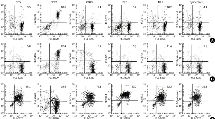
Flow cytometry showed that almost all lymph node B cells had the B-2 phenotype, which included CD5low, CD23high, CD43low, CD80 (B7.1)low, CD86 (B7.2)low, and Syndecan-1low, similar to the phenotype in normal spleen (Fig. 1).
Lymph node B cells do not live longer
Upon stimulation, lymph node B cells showed 60±5.1%
survival on day 2 and 25±3.2% survival on day 5, whereas unstimulated lymph node B cells showed 39±4.1% and 10±2.3% survival on days 2 and 5, respectively. These sur- vival rates were no higher than those of splenic and peritoneal B cells (Fig. 2).
B cells of cervical lymph nodes and spleen secrete immunoglobulin in response to stimulation
We found that unstimulated B cells from cervical lymph nodes and spleen did not spontaneously secrete immunoglob- ulin. These cells secreted IgM upon stimulation with LPS (3,300 ng/mL on day 3 and 4,286 ng/mL on day 5), CD40L plus anti-CD8 antibody (1,147 ng/mL on day 3 and 1,392 ng/mL on day 5) or IL-4 (1,017 ng/mL on day 5) (Fig. 3). In contrast, unstimulated peritoneal B cells secreted IgM, and secreted higher concentrations of IgM than either splenic or lymph node B cells (Fig. 4).
Stimulated cervical lymph node and splenic B cells enter S phase on day 2
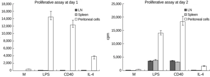
Unstimulated B cells of cervical lymph nodes and spleen did not enter S-phase, as shown by incorporation of [3H] thy- midine, after 24 hr in culture, but actively entered S phase after 48 hr of stimulation. These cells incorporated [3H] thy- midine upon stimulation with soluble CD40L/CD8 (229
×103±23×103cpm at 24 hr and 3,526×103±307×103 cpm at 48 hr), LPS (256×103±25×103cpm at 24 hr and
2,790×103±254×103cpm at 48 hr), or IL-4 (171×103
±18×103cpm at 24 hr and 256×103±28×103cpm at 48 hr) (Fig. 5). Although recombinant IL-4 was originally identified as a growth and differentiation factor for conven- tional B cells, it did not stimulate cervical lymph node and splenic B cells to enter S phase. Thus, it seems unlikely that cervical lymph node B cells are especially primed by antigenic stimulation to enter the cell cycle.
DISCUSSION
B cells are responsible for antibody-mediated immunity, which is also known as humoral immunity. These cells are not evenly distributed in the blood, bone marrow, spleen,
thymus, and peripheral lymphoid tissues. Rather, the ratio of B cells to T cells varies by the tissue or organ. For example, human B cells are seldom found in the thymus and, in the blood, T cells are outnumbered by B cells by a ratio of 8:1.
This ratio changes to 1:1 in the spleen, whereas B cells out- number T cells by a ratio of 1:3 in bone marrow. We found that the ratio of B cells to T cells in murine cervical lymph nodes was 1:3.1 by negative selection methods and 1:2.5 by flow cytometry (1).
B cells have been classified as B-1 and B-2 cells. B-1 cells are distinguished from the more abundant conventional B (B-2) cells by expression of the pan-T cell surface glycopro- tein, CD5. Additional identifying phenotypic characteristics of B-1 cells include surface Ig (sIg) Mhigh, sIgDlow, B220low, CD 23low, and CD43high. In contrast, B-2 cells express Ig (sIg) Mlow,
A
C B
FL1-B220 100 101 102 103 104
FL2-CD5
104 103 102 101 100
FL1-B220 100 101 102 103 104
FL2-CD5
104 103 102 101
100
FL1-B220 100 101 102 103 104
FL2-CD23
104 103 102 101
100
FL1-B220 100 101 102 103 104
FL2-CD43
104 103 102 101
100
FL1-B220 100 101 102 103 104
FL2-B7.1
104 103 102 101
100
FL1-B220 100 101 102 103 104
FL2-B7.2
104 103 102 101
100
FL1-B220 100 101 102 103 104
FL2-Syndecan-1
104 103 102 101 100 FL1-B220
100 101 102 103 104
FL2-CD23
104 103 102 101
100
FL1-B220 100 101 102 103 104
FL2-CD43
104 103 102 101
100
FL1-B220 100 101 102 103 104
FL2-B7.1
104 103 102 101
100
FL1-B220 100 101 102 103 104
FL2-B7.2
104 103 102 101
100
FL1-B220 100 101 102 103 104
FL2-Syndecan-1
104 103 102 101 100
CD5 CD23 CD43 B7.1 B7.2 Syndecan-1
Fig. 1.Antigen surface expression on (A) cervical lymph node (B) spleen (C) peritoneal fluid B cells. B cells of BALB/cByJ mice were harvested and stained with Abs specific for B220, CD5, CD23, CD43, B7.1, B7.2, IgM, and Syndacan-1. Expression of functional molecules on B cells was analyzed by two-color flow cytometry.
Fig. 2.Survival rates of stimulated and unstimulated B cells.
%
70 60 50 40 30 20 10
0 2 days 5 days
%
70 60 50 40 30 20 10
0 2 days 5 days
%
80 70 60 50 40 30 20 10
0 2 days 5 days
Survival rate of LN B cells Survival rate of splenic B cells Survival rate of peritoneal B cells Medium Stimulation Medium
Stimulation Medium
Stimulation
3.5
86.1 5.0
19.8 82.4
72.1 5.7
80.2 5.3
52.2 11.4
16.8 5.1
80.6 2.1 3.2 10.2 4.9
sIgDhigh, B220high, CD23high, and CD43low(3, 9). We found that cervical lymph node B cells expressed CD5low, CD23high, CD 43low, CD80 (B7.1)low, CD86 (B7.2)lowand Syndecan-1low, sug- gesting that these cells are comparable to B-2 cells.
We previously reported that splenic B cells are exclusively B-2 cells and peritoneal B cells are mainly B-1 cells (7). B-1 cells represent 10-25% of the B cells found in adult human peripheral blood and lymphoid organs. They are the princi- pal lymphocyte population in the peritoneal cavity and rep- resent a small proportion of splenic B cells, but they are absent from the peripheral blood in adult mice (2). Although B-1 (B220+CD5+) cells have been reported to be absent from murine lymph nodes (10), we found that 3.5% of the B cells in murine cervical lymph nodes were B220+CD5+cells (1).
Our phenotypic data, however, represent only a single point
in time, and we cannot rule out the possibility that B-1 cells are generated in murine cervical lymph nodes and then depart.
When B cells are activated by T-dependent, T cell produc- ing cytokines or T-independent mitogens, they proliferate or differentiate into antibody producing plasma cells or mem- ory cells (11-13). After stimulation with antigen, B cells first produce IgM and IgD and thereafter switch to secrete IgG, IgA or IgE, a switching that follows a DNA recombination event (14). The class of Ig produced by B cells differs by organ sites, stimuli and/or pathophysiologic conditions. Thus, most of human B cells in the middle ear and nasal mucosa primari- ly secrete IgA, both under normal and pathologic conditions, whereas other B cells in these organs secrete IgG rather than IgM. In contrast, nasal polyps contain B cells capable of spon- taneous, high-rate IgA secretion, followed by secretion of IgG and IgM (15, 16). These findings suggest that the mid- dle ear mucosa and nasal mucosa contain distinct immune
cpm
18,000 16,000 14,000 12,000 10,000 8,000 6,000 4,000 2,000 0
M LPS CD40 IL-4
Fig. 5.Stimulation of B cell entry into S phase. Primary murine B cells were cultured for 24 hr and 48 hr with medium alone, LPS (25 g/mL), CD40L (1:10) plus anti-CD8 antibody (1:40), or interleukin (IL)-4 (50 U/mL). [3H] thymidine was added during the last 6 hr in culture, and incorporation of label was assessed. Mean values of quadruplicate cultures are shown along with lines indicating the standard errors of the means.
Proliferative assay at day 1 LN Spleen Peritoneal cells
cpm
25,000
20,000
15,000
10,000
5,000
0
M LPS CD40 IL-4
Proliferative assay at day 2 LN Spleen Peritoneal cells
ng/m
4,500 4,000 3,500 3,000 2,500 2,000 1,500 1,000 500 0
Medium LPS CD40L IL-4
Fig. 3.IgM production by murine cervical lymph node B cells. Cells (0.5×10⁶per group) were cultured in fetal bovine serum (FBS)- containing culture medium with or without stimulation for 5 days.
Supernatants were collected and assayed for IgM secretion using an enzyme-linked immunosorbent assay (ELISA).
Ig secretion of LN at day 5 lgM lgG lgA
ng/mL
12,000
10,000
8,000
6,000
4,000
2,000
0
Medium LPS CD40 IL-4
Fig. 4.IgM production by peritoneal B cells. Assays were perfor- med as shown in the legend to Fig. 5.
IgM secretion at day 5 LN Spleen Peritoneal cells
systems, which exhibit features similar to those at other mu- cosal sites. For example, IgG producing adenoid CD5+and CD5-B cells were most abundant in adenoids, followed by IgA-producing and IgM-producing cells (17).
We found that B cells in murine cervical lymph nodes se- creted mainly IgM, followed by IgG and IgA, when stimu- lated with LPS, a T-independent mitogen; soluble CD40L/
CD8 , a stimulus provided by T cells; or IL-4, a cytokine produced by T cells. These finding suggests that B cells in cervical lymph nodes are stimulated or respond differently than B cells at the nasal and middle ear mucosal surfaces. In contrast, our finding, that murine cervical lymph node B cells do not secrete any Ig spontaneously, was unexpected. This result suggests that cervical lymph node B cells may not be subjected to continual antigenic stimulation from the naso- or oro-pharynx. Rather, it is likely that these cells have the same characteristic features as splenic B cells.
There are several differences between B-1 and B-2 cells concerned with Ig secretion (2, 7). In normal individuals B-1 cells are responsible for the production of most nonimmune serum IgM, as well as producing substantial amounts of rest- ing IgA. B-1 cell-derived Ig often recognizes discrete micro- bial cell wall determinants, suggesting that these cells pro- duce natural antibodies, representing a set of specificities en- coded in the germline and evolutionarily retained. These nat- ural antibodies provide, at low affinity, a degree of serologi- cal protection against a range of microorganisms prior to the immunization that accompanies microbial pathogenesis (18).
Primary B cells are normally arrested in the G0phase of the cell cycle (6, 7, 19, 20). Following the crosslinking of antigen specific surface Ig receptors on B cells, a number of early me- tabolic changes occur, which eventuate in cell cycle progres- sion and entry into S phase.
B-1 and B-2 cells also show different proliferation respons- es. We found that murine cervical lymph node B cells respon- ded in vitro to LPS or CD40L/CD8 in terms of cell cycle entry. While they did not proliferate actively after 1 day, they proliferated at 2 days. We had conjectured that lymph node B cell would be longer lived than splenic B cells, due to con- tinuous stimulation from the naso- or oropharynx; however, their survival rates, with or without stimulation, were not high, suggesting that murine cervical lymph node B cells have the characteristics of B-2 cells.
The phenotypic characteristics of nodal B cells include CD 5low, CD23high, CD43low, B220high, sIg Mlow, sIgDhigh, Mac-1low, CD80 (B7.1)low, CD86 (B7.2)low, and CD138 (Syndecan-1)low. Our finding, that 3.5% of B cells were B220+CD5+suggests that a small population of B-1 cells is present in cervical lymph nodes. Unlike our findings with peritoneal B cells, which spontaneously produce Ig and enter S phase after 1 day of stimulation, we did not observe these characteristics in cells from murine cervical lymph nodes. Despite our expectation that these lymph node B cells would be continually stimu- lated by antigens from the naso- or oro-pharyngeal mucosa,
we found no evidence for unusual surface phenotype, differ- entiation, survival rate or proliferation. These results indicate that cervical lymph node B cells correspond to conventional B-2 cells and that they have similar characteristics to splenic B cells (B-2), but not to peritoneal B cells (B-1).
ACKNOWLEDGEMENT
We thank Dr. Thomas L. Rothstein for teaching and criti- cal comments.
REFERENCES
1. Kim JB, Yeo SG, Kim SW, Cho JS, Cha CI. Characteristic features of immune B cells in murine cervical lymph node. Korean J Oto- laryngol 2005; 48: 241-6.
2. Rothstein TL. Cutting Edge: Commentary: Two B-1 or Not To Be One. J Immunol 2002; 168: 4257-61.
3. Hayakawa K, Hardy RR. Development and function of B-1 cells. Curr Opin Immunol 2000; 12: 346-53.
4. Beckstead JH. The evaluation of human lymph nodes, using plastic sections and enzyme histochemistry. Am J Clin Pathol 1983; 80: 131-9.
5. Hsu SM, Cossman J, Jaffe ES. Lymphocyte subsets in normal human lymphoid tissues. Am J Clin Pathol 1983; 80: 21-30.
6. Gavin MF, Laura AS, William DH, Kejian Y, Rachel MG, Babara SN. Splenic and peritoneal B-1 cells differ in terms of transcription- al and proliferative features that separate peritoneal B-1 from splenic B-2 cells. Cellular Immunol 2001; 213: 62-71.
7. Yeo SG, Cho JS, Park DC, Rothstein TL. B-1 cells differ from con- ventional B (B-2) cells. Immune Network 2004; 4: 155-60.
8. Martin F, Kearney JF. B-1 cells: similarities and differences with other B cell subsets. Curr Opin Immunol 2001; 195-201.
9. Robert B, Henry HW. Origins and functions of B-1 cells with notes on the role of CD5. Annu Rev Immunol 2002; 20: 253-300.
10. Casola S, Otipoby KL, Alimzhanov M. B cell receptor signal strength determines B cell fate. Nat Immunol 2004; 5: 317-27.
11. Barr TA, Carlring J, Heath AW. CD40 antibody provides the CD40 signal to B cells, but does not substitute for T cell help in responses to TD antigens. Vaccine 2005; 23: 3477-82.
12. Davey EJ, Thyberg J, Conrad D, Severinson E. Expression and pro- duction of interleukin 4 in B-cell chronic lymphocytic leukaemia. Leuk Lymphoma 2001; 42: 689-98.
13. Kerr WG, Heller M, Herzenberg LA. Analysis of lipopolysaccharide- response genes in B-lineage cells demonstrates that they can have differentiation stage-restricted expression and contain SH2 domains.
Proc Natl Acad Sci USA 1996; 93: 3947-52.
14. Beisner DR, Chen IL, Kolla RV, Hoffmann A. Cutting edge: innate immunity conferred by B cells is regulated by caspase-8. J Immunol 2005; 175: 3469-73.
15. Segura AS, Brieva JA, Rodriguez C. Regualtion of immunoglobulin secretion of plasma cells infiltrating nasal polyps. Laryngoscope 2000; 110: 1183-8.
16. Suenaga S, Kodama S, Ueyama S, Suzuki M, Mogi G. Mucosal im- munity of the middle ear: Analysis at the single cell level. Laryngo- scope 2001; 111: 290-6.
17. Arita M, Kodama S, Suzuki M, Mogi G. Single cell analysis of ade- noid CD5+B cells and their protective contributions to nasopharyn- geal immunity. Laryngoscope 2003; 113: 484-91.
18. Marianne B, Andrey PP, Tara S, Michael CC, Jianzhu C. A critical role of natural immunoglobulin M in immediate defense against sys-
temic bacterial infection. J Exp Med 1998; 12: 2381-6.
19. Piatelli MJ, Tanguay D, Rothstein TL, Chiles TC. Cell cycle control mechanism in B-1 and B-2 lymphoid subsets. Immunol Res 2003; 27:
31-52.
20. Tanguay DA, Colarusso TP, Doughty C, Pavlovic-Ewer S, Rothstein TL, Chiles TC. Cutting edge: Differential signaling requirements for activation of assembled cyclinD3-cdk4 complexes in B-1 and B-2 lymphocyte subsets. J Immunol 2001; 166: 4273-7.