INTRODUCTION
Peripheral neuropathy is one of the rare complications of lymphoma. The most common causes of lymphoma-related neuropathy are local infiltration of lymphoma and proliferation of the lymphoma inside of peripheral nerve tissue (1). Other causes of lymphoma-related neuropathy are loss of dorsal root ganglion cells, (2) the neurotoxic effects of drugs such as vin- cristine, (3) and viral infections such as HIV (4). However, para- neoplastic syndrome is a rare cause of lymphoma-related neuro- pathy (3). We report a case of peripheral sensory loss, which may be due to a lymphoma-induced paraneoplastic syndrome.
CASE REPORT
The thirty-four year old male was presented to the hospi- tal with tingling or burning sensations in both hands and feet that began 20 months ago. The symptom first started from his feet. Recently, the burning had progressed to his hands.
There was no past history of alcoholism or any long-term me- dication use.
On neurological examination, all the cranial nerve functions, including pupillary light reflexes, were normal. Motor strength of the upper and lower limbs was grade V by the NMC clas- sification. Both hands and feet had severe loss of vibration and position sensation compared to the chin and slight loss of pain and thermal sensation compared to the face.
Deep tendon reflexes of bilateral upper and lower limbs were reduced and there was no Babinski sign. Tinnel’s sign was
negative in bilateral wrists and ankles. There was no abnormal sweating in the face and trunk, and changing body position did not affect blood pressure significantly.
On admission, there were two palpable masses found in the left inguinal area, but the patient could not clarify when they first appeared. The masses were measured 2 cm and 4 cm in diameter, and were partially movable and non-tender. Just after the admission, his blood laboratory values were: WBC 22,500/ L, RBC 5.27×10⁶/ L, hemoglobin 15.8 g/dL and platelets 329,000/ L. His WBC differential counts were: seg- mented neutrophils 84% and lymphocytes 12%. Electrolytes and liver function tests were normal. Hemoglobin A1c was 5.7%, and CRP was increased to 2.13 mg/dL. In his cerebro- spinal fluid (CSF), WBC count was 6/mL, and 92% of them were lymphocytes. CSF protein was 42.3 g/dL and glucose was 53 mg/mL. CSF cytology was negative for malignant cells.
Other tests, specifically cryoglobulins, antinuclear antibody, and anti- Sm antibody, anti-SS-A, anti-SS-B antibody, and ANCA were all negative. In addition, antibodies to HIV were negative by ELISA. Several tests to rule out other acquired polyneuropathies were performed and all results were normal:
TSH 4.7 U/mL, free T40.87 ng/dL, vitamin B12311 pg/mL, folate 1.1 ng/mL.
Bone marrow biopsy was negative for neoplastic lymphoid cell infiltration. No monoclonal immunoglobulins were found on immunofixation electrophoresis of serum and urine. Indi- rect immunofluorescent stain of triple tissue slides (stomach, cerebellum, kidney) showed no known paraneoplastic antibod- ies, including anti-Hu, anti-Ri, anti-Yo, and antiamphiphysin antibodies.
Byeong Cheol Oh, Young-Min Lim, Young Mee Kwon*, Shin Kwang Khang*, Kwang Kuk Kim
Departments of Neurology and Diagnostic Pathology*, University of Ulsan College of Medicine, Asan Medical Center, Seoul, Korea
Received : 2 January 2003 Accepted : 24 February 2003
Address for correspondence Kwang Kuk Kim, M.D.
Department of Neurology, University of Ulsan, College of Medicine, Asan Medical Center, 388-1 Pungnap-dong, Songpa-gu, Seoul 138-736, Korea Tel : +82.2-3010-3444, Fax : +82.2-474-4691 E-mail : [email protected]
130 J Korean Med Sci 2004; 19: 130-3
ISSN 1011-8934
Copyright � The Korean Academy of Medical Sciences
A Case of Hodgkin's Lymphoma Associated with Sensory Neuropathy
Peripheral neuropathies occur in lymphoma patients. Causes of neuropathy include chemotherapy, opportunistic infections, and the lymphoma itself. We report a patient with lymphoma whose chief complaint was a sensory loss in the hands and feet.
Electrophysiologic studies and sural nerve biopsy showed sensory polyneuropathies.
We hypothesize that this neuropathy is associated with lymphoma-related ganglio- nopathy, and among the possible causes, we suspect that a systemic cause such as a paraneoplastic syndrome is the most likely pathogenic etiology. However, further follow-up will be necessary to see whether sensory symptoms change with lymphoma treatment.
Key Words :Lymphoma; Polyneuropathies; Paraneoplastic Syndromes
Lymphoma Associated with Sensory Neuropathy 131
Electrophysiological studies showed that the action poten- tials of sensory nerve in all limbs were not detectable but the parameters for motor nerves, including F-waves, were with- in normal limits (Table 1). On plain chest radiograph and chest CT scan, there were no pathological findings suggesting metastases. However, on abdominal CT scan, multiple lym- phadenopathy was found in the left inguinal canal, para-aor- tic space, and pelvic cavity.
After the admission, biopsies of the left inguinal mass and the left sural nerve were performed. Grossly, the inguinal mass measured 8.5 cm in its greatest dimension, and it was well- encapsulated, grayish tan, and solid with fresh fish appearance.
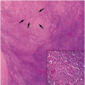
Microscopically, the mass exhibited diffuse effacement of lymph node architecture and multiple granulomas (Fig. 1). Many large atypical mononuclear or binuclear cells, including Reed-Stern- berg cells, were scattered among many mature lymphocytes,
DL (R/L), msec Amp (R/L), mV NCV (R/L), m/s F-wave (R/L), msec Motor
Median N. W 3.5/3.5 16.13/12.21 27.9/28.2
E 14.51/11.23 55/52
A 15.21/11.46 54/79
Ulnar N. W 2.5/2.3 11.39/14.95 27.3/26.3
BE 11.55/14.90 54/52
AE 11.23/15.00 53/63
A 11.63/13.95 75/56
Peroneal N. Ank 5.7/4.7 1.556/8.178 54.1/48.6
F 1.788/6.706 41/44
Tibial N. Ank 4.0/4.3 22.58/18.07 51.7/49.5
P 13.23/12.66 43/41
Sensory
Median N. F-W NP
Ulnar N. F-W NP
Sup. peroneal N. NP
Sural N. NP
N, nerve; sup, superficial; DL, distal latency; Amp, amplitude; NCV, nerve conduction velocity; R, right; L, left; W, wrist; E, elbow; A, axillary; BE, below elbow; AE, above elbow; Ank, ankle; F, fibular; P, popliteal; F-W, finger to wrist; NP, no potential.
Table 1.Nerve conduction study. There are no producible potentials of the sensory nerves. However, the parameters of the motor nerves are nearly normal
Fig. 2.Lymph node biopsy (H&E stain, ×200). Reed-Sternberg cells (solid arrow) are scattered among a background of many mature lympocytes. Arrow inside inset shows mitosis.
Fig. 1.Lymph node biopsy (H&E stain, ×20). Microscopically, the mass exhibits diffuse effacement of lymph node architecture and granulomas. Inset is the enlarged picture (×200) of the area indi- cated by arrow.
132 B.C. Oh, Y.-M. Lim, Y.M. Kwon, et al.
rare eosinophils, and plasma cells (Fig. 2). By immunohisto- chemical staining, the large atypical cells were tested positive for CD15 and CD30, and the diagnosis of Hodgkin’s disease lymphocyte rich (classical) type, was made from the inguinal mass (5).
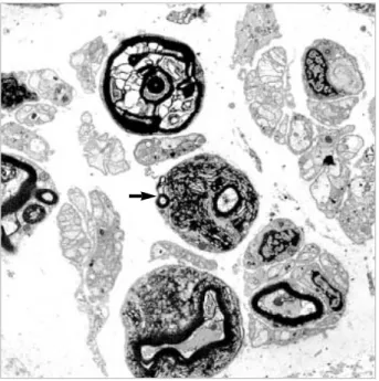
The sural nerve biopsy showed several myelin digestion chambers, suggesting axonal degeneration on modified Gomori stain (Fig. 3). There was no evidence of vasculitis or inflam- matory neuropathy. There was no neoplastic lymphoid cell infiltration of the sural nerve. On electron microscopy, the sural nerve showed the loss of small and large myelinated fibers with axonal sprouting and myelin ovoids, suggesting an axonal neuropathy (Fig. 4).
Patient was diagnosed with stage IIB Hodgkin’s lymphoma and discharged after one cycle of chemotherapy with adriamycin and bleomycin.
DISCUSSION
This patient had clinical symptoms suggestive of a sensory polyneuropathy, and electrophysiological and pathological studies confirming this diagnosis. We sought for the mech- anisms that could link lymphoma to peripheral neuropathy in this case because we could find no other obvious cause for the sensory loss of the patient.
The incidence of peripheral neuropathy is approximately 1- 2% in patients with lymphoma (6). However, Walsh et al. re- ported that 35% of lymphoma patients have electrophysiolog- ical evidence of neuropathy regardless of their symptoms (7).
Previous reports in the Korean literatures have described the lymphoma patients with the associated peripheral neuro- pathies (8, 9). However, the mechanism in this previous report was direct infiltration of the nerves by the lymphoma (9). In contrast, in the current case there was no evidence of direct tu- mor infiltration.
There are several reports that tried to relate lymphoma and neuropathy. Schold et al. observed neuronal degeneration in anterior horn cells and posterior spinal columns and segmen- tal demyelination of spinal roots and brachial and lumbar plexi from the autopsy of the patients suffered with lymphoma (10).
They suggested that these autopsy findings are due to radia- tion therapy and opportunistic viral infection. Thomas et al.
reported the infiltration of lymphocytes in the epineurium of sural nerve from a lymphoproliferative disorder patient (11).
They suggested the possibility that this finding is due to a systemic effect of circulating monoclonal immunoglobulins.
Plante-Bordeneuve et al. proposed sensory neuropathies asso- ciated with lymphoma correspond to a variant of Hodgkin’s disease-associated inflammatory demyelinating polyneuropa- thy rather than to a true paraneoplastic sensory ganglionitis (12). However, Lee et al. reported the case of a patient with CNS lymphoma, who had severe peripheral polyneuropathy (13). This patient had an IgM kappa monoclonal paraproteine- mia, and his symptoms improved with the treatment of the lymphoma and the decreased paraprotein levels, suggesting a monoclonal immunoglobulin as a possible cause of his neuro- pathy.
In this case, we suspected systemic causes rather than the local infiltration of lymphoma cells or compression by tumor
Fig. 3.Sural nerve biopsy (modified Gomori trichrome stain, ×100).
The sural nerve biopsy shows several myelin digestion chambers (arrow), suggesting axonal degeneration.
Fig. 4.Sural nerve biopsy (EM findings, ×7,000). The sural nerve shows loss of small and large myelinated fibers and degenerating axons with myelin ovoids (arrow), suggesting axonal neuropathy.
Lymphoma Associated with Sensory Neuropathy 133
mass as an explanation for the sensory loss because the patient had symmetric pure sensory polyneuropathy. Cryoglobulin- emia and paraneoplastic antibody-mediated nerve damage are likely systemic pathogenic mechanisms of polyneuropathy (14). Despite negative findings on paraneoplastic antibody and paraprotein tests in this patient, we could not rule out the possibility of a systemic cause because there may be other paraproteins that cannot be measured by standard serum elec- trophoresis, and there may be other paraneoplastic antibodies that we could not measure.
In this case, we suspect that an antibody against some anti- gen expressed by primary sensory neurons may induce axon- al degeneration after passing through weak points in the blood brain barrier near spinal nerve roots or dorsal root ganglia. In this case, evoked sensory nerve action potentials are diffusely absent. These findings support the possibility of ganglion- opathies. And the reason this patient had sensory deficits rather than motor deficits is that this antibody chiefly affect- ed dorsal root ganglion neurons. However, the ganglionopa- thy of paraneoplastic syndrome may rarely occur after lym- phocyte infiltration of the dorsal root ganglia (15).
So, further evaluation will be necessary to clarify the patho- genic causes of this case by following up whether his symp- toms change as lymphoma treatment progress.
REFERENCES
1. Vital C, Vital A, Julien J, Rivel J, deMascarel A, Vergier B, Henry P, Barat M, Reiffer J, Broustet A. Peripheral neuropathies and lym- phoma without monoclonal gammopathy: A new classification. J Neu- rol 1990; 237: 177-85.
2. Sterman AB, Schaumburg HH, Asbury AK. The acute sensory neu- ronopathy syndrome: a distinct clinical entity. Ann Neurol 1980; 7:
345-8.
3. Dumitru D, Amato AA, Zawarts MJ. Electrodiagnostic medicine. 2nd ed. Philadelphia: Hanley & Belfus 2002; 992-3.
4. Gherardi RK, Chretien F, Delfau-Larue MH, Authier FJ, Moulignier
A, Roulland-Dussoix D, Belec L. Neuropathy in diffuse infiltrative lymphocytosis syndrome. An HIV neuropathy, not a lymphoma. Neu- rology 1998; 50: 1041-4.
5. Chen, Y-T, Godwin TA, Mouradian JA. Immunohistochemistry and gene rearrangement studies in the diagnosis of malignant lymphomas:
A comparison of 152 cases. Hum Pathol 1991; 22: 1249-57.
6. McLeod JG. Peripheral neuropathy associated with lymphomas, le- ukemias, and polycythemia vera. In Dyck PJ, Thomas PK, Griffin JW:
Peripheral neuropathy, 3rd ed. Philadelphia: W.B. Saunders 1993;
1591-8.
7. Walsh JC. Neuropathy associated with lymphoma. J Neurol Neurosurg Psychiatry 1971; 34: 42-50.
8. Kim HW, Park SY. A case of angiocentric T cell lymphoma accom- panied with multiple erythematous nodules, subcutaneous mass on the right face and peripheral polyneuropathy. Korean J Hematol 1997; 32: 140-5.
9. Kim CH, Paik KW. A case report of infiltrative polyneuropathy asso- ciated with lymphoma. J Korean Acad Rehabil Med 2001; 25: 724-8.
10. Schold SC, Cho ES, Somasundaram M, Posner JB. Subacute motor neuronopathy: A remote effect of lymphoma. Ann Neurol 1979; 5: 271- 87.
11. Thomas FP, Vallejos U, Foitl DR, Miller JR, Barrett R, Fetell MR, Knowles DM, Latov N, Hays AP. B cell small lymphocytic lymphoma and chronic lymphocytic leukemia with peripheral neuropathy: two cases with neuropathological findings and lymphocyte marker anal- ysis. Acta Neuropathol 1990; 80: 198-203.
12. Plante-Bordeneuve V, Baudrimont M, Gorin NC, Gherardi RK. Sub- acute sensory neuropathy associated with Hodgkin’s disease. J Neu- rol Sci 1994; 121: 155-8.
13. Lee SM, Harper P, Luthert P, Hughes RAC. Primary CNS lymphoma in association with IgM kappa paraproteinaemia and peripheral polyneu- ropathy: A case report. Eur Neurol 1995; 35: 237-9.
14. Gemignani F, Marchesi G, Di Giovanni G, Salih S, Quaini F, Nobile- Orazio E. Low-grade non-Hodgkin B-cell lymphoma presenting as sensory neuropathy. Eur Neurol 1996; 36: 138-41.
15. McLeod JG. Paraneoplastic neuropathies. In Dyck PJ, Thomas PK, Griffin JW. Peripheral neuropathy, 3rd ed. Philadelphia: W.B. Saun- ders 1993; 1583-90.