• 검색 결과가 없습니다.

Bile-Acid Induced Apoptosis in HL-60 Leukemia Cells

N/A
N/A
Protected

Academic year: 2021

Share "Bile-Acid Induced Apoptosis in HL-60 Leukemia Cells"

Copied!
8
0
0

로드 중.... (전체 텍스트 보기)

전체 글

(1)

of Cancer Prevention 1998; 3(2): 107-114

INTRODUCTION

Apotosis is a distinct mode of cell death that is responsible for deletion of cells in normal tissues: it also occurs in specific pathologic conditions. A characteristic biochemical feature of apoptosis is double-strand cleavage of nuclear DNA at the linker regions between nucleosomes leading to the production of oligonucleosomal fragments. In many, although not all of the circumstances where apoptosis occurs, it is suppressed by inhibitors of mes- senger RNA and protein synthesis.

1)

Separation and quantitation of bile acids [chenodeoxycholic acid (CDCA), dehydrocholic acid (DHCA), lithocholic acid (LCA) and ursodeoxycholic acid (UDCA)] from bile are accomplished using high-performance liquid chromatography.

2)

Bile acids have several functions; 1) bile acids are the principal endogenous ligands for male-specific and female-specific sterole-binding protein in rat liver cells,

3)

2) UDCA and LCA enhance receptor-dependent low-density lipoprotein uptake and degradation in isolated hamster hepatocytes.

4)

3) UDCA induces cellular chloramphenicol acetyltransferase (CAT) acti- vities in a glucocorticoid receptor-dependent fashion but weak rather than synthetic dexamethasone,

5)

4) LCA affects on the

Bile-Acid Induced Apoptosis in HL-60 Leukemia Cells

Kwang-Rok Kim, Chang-Mo Kang

1

, Jin Hyen Baek You Mie Lee and Kyu-Won Kim

Department of Molecular Biology,

1

Department of Biology, Pusan National University, Pusan 609-735, Korea

We investigated that lithocholic acid (LCA) or ursodeoxycholilc acid (UDCA), components of bile salt, induced apoptosis in HL-60 cells. The growth of HL-60 cells was reduced by these compounds in a time- and a concentration-dependent manners. Moreover, the degree of DNA fragmentation was increased depending on the treatment time and molar concentration of LCA or UDCA on agarose gel electrophoresis. Morphologically, we examined the formation of membrane- enclosed apoptotic bodies in LCA or UDCA-treated HL-60 cells by haemato- xylin/eosin staining. The expression of c-myc was down-regulated after 24 h treatment of LCA or UDCA. In case of bcl-2, there was no change of the expression after 24 h treatment of LCA or UDCA. The DNA fragmentation was reduced when HL-60 cells were treated with 0.5 μM cycloheximide (CHX) and LCA or UDCA.

Therefore, bile acids, LCA and UDCA, induced apoptosis in HL-60 cells, which required de novo protein synthesis.

ꠏꠏꠏꠏꠏꠏꠏꠏꠏꠏꠏꠏꠏꠏꠏꠏꠏꠏꠏꠏꠏꠏꠏꠏꠏꠏꠏꠏꠏꠏꠏꠏꠏꠏꠏꠏꠏꠏꠏꠏꠏꠏꠏꠏꠏꠏꠏꠏꠏꠏꠏꠏꠏꠏꠏꠏꠏꠏꠏꠏꠏꠏꠏꠏꠏꠏꠏꠏꠏꠏꠏꠏꠏ Key Words: HL-60 leukemia cells, Apoptosis, LCA, UDCA, c-myc

Corresponding author Kyu-Won Kim, Department of: Molecular Biology, Pusan National University, 30 Jangjun-dong Keumjung-ku, Pusan 609-735, Korea

(2)

releasing of intracellular Ca

2+

in liver cells. The effect is neither mediated by Ins (1,4,5) P

3

nor via activation of the Ins (1,4,5) P

3

receptor, but it is specific for the membrane of the internal pool.

6)

The structure of bile acids is very similar with that of glucocorticoid. Glucocorticoid hormones also stimulate thymocyte apoptosis

7)

by a mechanism that is dependent on a sustained increase in cytosolic Ca

2+

level. Glucocorticoid treatment of these cells causes a G1 arrest and eventual cell death.

8)

A few genes (bcl-2, myc, ras, jun and fos etc.) are known to engage apoptosis.

9 12)

Constitutive c-myc expression prevents growth arrest in serum-deprived Rat-1 fibroblast and induces cell death. This results insisted that deregulated c-myc expression is a potent inducer of programmed cell death (apoptosis) when combined with a block to cell proliferation.

13)

Bile acids induce apoptosis in Epstein-Barr virus-transformed lymphoid cells.

Since these cells contain high levels of bcl-2 protein, the product of a proto-oncogene that maintains cellular viability,

14)

the apoptosis- inducing action of bile salts appears to circumvent the protective role of bcl-2.

In this study, the bile acid components, LCA and UDCA were used to examine whether these components induce apoptosis in HL-60 cells biochemically and morphologically. Especially, the effects of UDCA or LCA on the apoptosis- related gene regulation were investigated.

Futhermore, the effect of cycloheximide (CHX), protein inhibitor, on LCA or UDCA-induced apoptosis was examined in HL-60 cells.

MATERIALS AND METHODS

1) Cell culture

The HL-60 cells were cultured in a similar manner as described by Collins et al.

15)

Stock cultures were maintained by growth as suspension cultures and passed by 2 days in RPMI-1640 medium, supplemented with heat inactivated 10% fetal bovine serum (FBS), 100

units/ml penicillin, and 100 μg/ml streptomycin at 37

o

C in humidified atmosphere containing 5%

CO

2

. Cells were suspended a 5×10

5

cells/ml in RPMI-1640 and reagents were added so that final concentration of ethanol did not exceed 0.015%. At the end of incubation, the cells were harvested by centrifugation at 200×g for 10 min.

2) Morphological characterization of cell death

The cells were harvested and suspended 2×10

6

cells/ml in PBS. The cells were cytocen- trifuged onto slide glass at 700 rpm for 10 min (Cytospin, SHANDON) and then fixed in 95%

ethanol for 30 min at room temperature. Sample was rinsed with distilled water. It was stained with haematoxylin and eosin, and examined under the light microscope (×1000).

3) DNA fragmentation assay

DNA isolation was done as described by Goels et al.

16)

Samples was treated for 3 h at 37

o

C with 300 μg/ml boiled bovine pancreatic RNase, and loaded onto 1.5% (w/v) agarose horizontal slab gels containing 10 μg/ml ethidium bromide. DNA from 2×10

6

cells was loaded into each lane.

Electrophoresis was performed in TAE buffer for 4 h at 3 V/cm. Gels were photographed under UV light with instant films.

17)

4) Quantification of DNA fragmentation

The extent of DNA fragmentation was

determined as follows.

17,18)

Briefly, 3×10

6

cells

in complete medium were centrifuged (200×g, 5

min, 4

o

C) and the pellet was lysed in 400 μl

hypotonic lysing buffer (10 mM Tris [pH 8.0], 20

mM EDTA, 0.5% Triton X-100) for 15 min. The

lysate was centrifuged at 13000×g for 20 min at

4

o

C and the chromatin was separated from

fragmented DNA. The supernatant was removed,

and the pellet resuspended in 400 μl of lysing

buffer. DNA from both pellet and supernatant

was precipitated by the addition of 100 μl of 2.5

N perchloric acid at 4

o

C during overnight. After

centrifugation at 13000×g for 20 min, the

(3)

supernatant was discarded, and 0.4 ml of 0.5 N perchloric acid was added. The DNA was hydrolyzed by incubation at 70

o

C for 20 min. The amount of DNA was quantified by the dipheny- lamin method.

19)

Percent fragmentation refers to the ratio of DNA in the supernatant to the total DNA recovered in the supernatant plus pellet.

Data are represented as the mean of 3 samples per data point.

5) Northern analysis

Total cellular RNAs were isolated by the acid- guanidinium thiocyanate-phenol-chloroform ex- traction method.

20)

The Northern blotted nylon membrane was hybridized with

32

P-labeled probe. The filter was dried and exposed to X-ray film for 2 to 5 days.

Fig. 1. Growth rate of LCA or UDCA-treated HL-60 cells. The cells were cultured with LCA at the various concentrations for 24 hr (A). The cells were cultured with UDCA at the various concentrations for 24 hr (B). The cells were cultured with 100 μM LCA and 500 μM UDCA for the various period (C). The cell number was counted by trypan blue staining. Data are represented as the mean of 3 samples per data point.

Fig. 2. LCA or UDCA-induced DNA fragmentation in HL-60 cells. A and B show DNA fragmentation on

1.5 % agarose gel in LCA-treated HL-60 cells at the various concentrations for 24 hr and for the various

periods, respectively. The LCA-treated cells was compared with the UDCA-treated cells (C). Lane 1 is

control, lane 2 is 100 μM LCA-treated cells, lane 3 is 200 μM UDCA-treated cells and lane 4 is 500 μM

UDCA-treated cells.

(4)

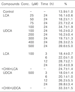
Table 1. DNA fragmentation rate in bile-acid treated HL- 60 cells

ꠧꠧꠧꠧꠧꠧꠧꠧꠧꠧꠧꠧꠧꠧꠧꠧꠧꠧꠧꠧꠧꠧꠧꠧꠧꠧꠧꠧꠧꠧꠧꠧꠧꠧꠧꠧꠧꠧꠧꠧꠧꠧꠧꠧꠧꠧꠧꠧ

Compounds Conc. ( M) μ Time (h) % ꠏꠏꠏꠏꠏꠏꠏꠏꠏꠏꠏꠏꠏꠏꠏꠏꠏꠏꠏꠏꠏꠏꠏꠏꠏꠏꠏꠏꠏꠏꠏꠏꠏꠏꠏꠏꠏꠏꠏꠏꠏꠏꠏꠏꠏꠏꠏꠏ

Control - - 13.9±1.0

LCA 25 24 16.1±0.6

50 24 18.2±1.1

75 24 23.7±2.4

100 24 34.7±1.9

UDCA 100 24 16.2±0.2

200 24 16.2±0.4

300 24 19.7±1.3

400 24 28.3±2.5

500 24 39.6±5.0

LCA 100 3 18.4±0.7

6 22.0±1.0

12 28.7±2.1

24 33.4±0.9

*CHX+LCA - - 24.7±1.4

UDCA 500 3 18.0±1.4

6 20.1±1.0

12 26.2±5.5

24 39.8±3.4

*CHX+UDCA - - 33.3±1.5

ꠏꠏꠏꠏꠏꠏꠏꠏꠏꠏꠏꠏꠏꠏꠏꠏꠏꠏꠏꠏꠏꠏꠏꠏꠏꠏꠏꠏꠏꠏꠏꠏꠏꠏꠏꠏꠏꠏꠏꠏꠏꠏꠏꠏꠏꠏꠏꠏ

*100 M LCA or 500 M UDCA-treated cells with 0.5 μ μ M CHX for 24 hr after 4 hr treatment of 0.5 M CHX.

μ μ

Data are represented as the mean of 3 samples per data point.

RESULTS

Treatment of LCA resulted in dose-dependent decrease of cell growth rate (Fig. 1A). When HL-60 cells were treated with 100 μM LCA for various periods, the cell growth rate decreased according to the time (Fig. 1C). Similarly, treatment of UDCA caused the growth arrest of HL-60 cells (Fig. 1B and C). All experiments were started with the same cell number and living cell number was counted by trypan blue staining method. All experiments were done with 0.015% ethanol-treated cell as a control. LCA or UDCA reduced the growth rate of HL-60 cells in a dose and a time-dependent manners.

DNA fragmentation is the biochemical property of apoptosis. HL-60 cells were treated with bile acids and then DNA was isolated and analyzed by gel electrophoresis. All components of bile acids induced DNA fragmentation and especially UDCA and LCA highly induced the DNA fragmentation (data not shown). However, further experiments were done with LCA and UDCA, because of the fact that LCA induced a differentiation in F9 teratocarcinoma stem cells in our laboratory and UDCA induced cellular CAT activities in a glucocorticoid receptor-dependent fashion.

5)

Fig. 3. Morphological change of HL-60 cells by LCA or UDCA treatment. Control cells (A), 100 μM

LCA-treated cells for 24 hr (B) and 500 μM UDCA-treated cells for 24 hr (C) were cytocentrifuged onto

slide glass at 700 rev/min for 10 min and then fixed in 95% ethanol for 30 min at room temperature. Sample

was rinsed with distilled water. It was stained with hae matoxylin and eosin, and examined under the light

microscope (×1000). Arrowheads indicate apoptotic cells.

(5)

When the cells cultured with various concen- tration of LCA for 24 h, DNA fragmentation was increased in proportion to molar concentration of LCA (Fig. 2A, Table 1). In case of treatments with 100 μM LCA for various time, DNA fragmentation was augmented time dependently (Fig. 2B, Table 1). Similarly, treatment of UDCA increased DNA fragmentation in HL-60 cells (Table 1). But LCA have a similar effect on DNA fragmentation at a lower concentration than that of UDCA (Fig. 2C). This result explained LCA or UDCA induced dose and time-dependent apoptosis in a biochemical point of view.

After treated with 100 μM LCA or 500 μM UDCA for 24 h, HL-60 cells were attached to the slide glass by cytospin and stained with haematoxylin and eosin, and then examined by light microscopy (×1000). LCA or UDCA induced a chromatin condensation, apoptotic body, and cytosol shrinkage (Fig. 3). Control cells did not show any change in morphology.

The expression of the gene related to apoptosis

was investigated by Northern analysis. When the cells cultured with 100 μM LCA for various periods, c-myc expression reduced after 1 h (Fig. 4A). Especially, expression of c-myc abruptly reduced at 24 h treatment with 100 μM LCA . But the expression of bcl-2 appeared no change (Fig. 4A). UDCA had the same results as those of LCA (Fig. 4B). These results explained that LCA or UDCA-induced apoptosis may have closely relation to c-myc expression but not have any direct concern with bcl-2 expression.

When the cells cultured with 100 μM LCA or 500 μM UDCA in condition of continuous expo- sure of 0.5 μM cycloheximide for 24 h after 4 h treatment of cycloheximide, cycloheximide reduced LCA or UDCA-induced DNA fragmen- tation (Fig. 5, Table 1). CHX alone did not change the expression of c-myc. CHX increased c-myc expression of the cells treated with 100 μM LCA or 500 μM UDCA for 24 h (Fig. 6). From this result, c-myc expression may have a very close relation to apoptosis and the apoptosis Fig. 4. Expression of c-myc and bcl-2 in the LCA or UDCA-treated cells. A and B showed the expression of c-myc and bcl-2 in the 100 μM LCA-treated cells and 500 μM UDCA-treated cells for various period, respectively.

Lane 1 is control. Lane 2, 3, 4, 5 and 6 are LCA or UDCA-treated cells for 1, 3, 6, 12 and 24 hr, respectively.

An ethidium bromide stain of the gel prior to transfer is shown. 18S rRNA and 28S rRNA were used as control.

(6)

induced by LCA or UDCA needs de novo protein synthesis.

DISCUSSION

From Fig. 1, LCA and UDCA reduced the growth rate of HL-60 cells. But, as shown in Fig.

2 and 3, LCA and UDCA can induce apoptosis in HL-60 cells. It is suggested that these compounds could be used as agents for cancer therapy because most of the cytotoxic anti- cancer drugs in current use have been shown to induce apoptosis in susceptible cells. The fact that disparate agents, which interact with different targets, induce cell death with some common features (endonucleolytic cleavage of DNA and chromatin condensation) suggests that cytotoxicity is determined by the ability of the cell to engage this so-called ‘programmed’ cell death. The mechanism of the coupling of a stimulus is not known, but modulation of this

coupling may affect the outcome of drug treatment.

As shown in Fig. 4, LCA or UDCA reduced c-myc expression. In recent report, the transient transfection of human leukemic cells with con- stitutively expressed c-myc-containing cons- tructs inhibited dexamethasone-induced cell death.

21)

Similarly, c-myc suppression seems to be involved in the apoptosis of HL-60 cells. The reduction of this gene probably triggers the pathway of apoptosis.

However, LCA or UDCA did not induce any change of bcl-2 expression in HL-60 cells (Fig.

4). LCA or UDCA induced apoptosis may not have direct relation with bcl-2 gene expression.

Overexpression of bcl-2 can prevent apoptosis depending on nerve growth factor but not depending upon ciliary neurotrophic factor in neurons.

22)

These results suggest the existence of multiple independent intracellular mechanisms Fig. 5. Suppression of DNA fragmentation in LCA

or UDCA-treated HL-60 cells by CHX. This figure shows the DNA fragmentation on the agarose gel (lane 1: control, lane 2: 0.5 μM CHX-treated cells for 28 hr, lane 3: 100 μM LCA-treated cells with 0.5 μM CHX for 24 hr after 4 hr treatment of 0.5 μM CHX, lane 4: 100 M LCA-treated cells without 0.5 μ μM CHX for 24 hr after 4hr treatment of 0.5 μM CHX, lane 5: 100 μM LCA-treated cells for 24 hr, lane 6: 500 μM UDCA- treated cells with 0.5 μM CHX for 24 hr after 4 hr treatment of 0.5 μM CHX, lane 7: 500 M UDCA- treated cells without 0.5 μ μM CHX for 24 hr after 4 hr treatment of 0.5 μM CHX, lane 8: 500 μM UDCA- treated cells for 24 hr).

Fig. 6. Change of c-myc expression by CHX. This

panel shows change of c-myc expression by CHX

(lane 1: 0.015% ethanol treated cell for 24 hr, lane

2: 0.5 μM CHX-treated cells for 28 hr, lane 3: 100

μM LCA-treated cells with 0.5 μM CHX for 24 hr

after 4 hr treatment of 0.5 μM CHX, lane 4: 100 μM

LCA-treated cells for 24 hr, lane 5: 500 μM

UDCA-treated cells with 0.5 μM CHX for 24 hr after

4 hr treatment of 0.5 μM CHX, lane 6: 500 μM

UDCA-treated cells for 24 hr). An ethidium bromide

stain of the gel prior to transfer is shown. 18S rRNA

and 28S rRNA were used as control.

(7)

of apoptosis, some of which can be prevented by bcl-2 and others of which are unaffected by this gene. Bcl-x, a bcl-2-related gene can function as a bcl-2-independent regulator of programed cell death.

23)

LCA or UDCA-induced apoptosis possibly have a mechanism unrelated to bcl-2.

HL-60 cells originally have an abnormal p53.

24)

So, these cells have no relations to the mechanism of p53-dependent apoptosis.

Recently, it was reported that p53 is not an activator of apoptosis- mediator genes, but rather represses genes necessary for cell survival or a component of the enzymatic machinery for apoptotic cleavage.

25)

Agents that initiate DNA-strand breakage kill thymocytes by the p53-dependent pathway. The p53-independent stimuli include several physiological cell-deletion signals, namely glucocorticoid, calcium associated activation and aging in vitro.

26)

So, apoptosis induced by LCA and UDCA may have a novel p53- independent pathway.

As indicated in Fig. 5 and 6, LCA or UDCA- induced apoptosis may need de novo protein synthesis. In these cases, new protein synthesis might be required in order to generate an appropriate triggers to activate an endogenous mechanism of cell death. It is supposed that there is a killer gene. Recently, from C. elegans to plants, various apoptosis-related genes have been found. During the development of the nematode Caenorhabditis elegans, 131 cells undergo programmed cell death. Fourteen genes have been identified that function in different steps of the genetic pathway of programmed cell death in C. elegans. Two of these genes, ced-3 and ced-4, play essential roles in either the initiation or execution of the cell death program.

CED-3 protein acts as a cystein protease in controlling the onset of programmed cell death in C. elegans and its non-serine-rich portions are similar to human interleukin-1 -converting β enzyme (ICE).

27)

Overexpression of ICE causes Rat-1 cells (rat fibroblasts) to undergo program- med cell death.

28)

In plants, the hypersensitive

response to pathogens involves rapid cell death, which is hypothesized to arise from the activation of a cell death program. Arabidopsis thaliana have an accelerated cell death (ACD) gene called ACD2, involved in a pathway that negatively regulates a genetically programmed hypersensitive response.

29)

The killer gene may take major role for apoptotic signal.

When differentiation in LCA or UDCA-induced HL-60 was investigated by NBT reduction and morphological observation, LCA or UDCA did not induce the differentiation to granulocyte or macrophages within 24 hr (data not shown).

From this result, LCA or UDCA-induced apop- tosis probably have the different mechanism from the differentiation of HL-60 cells.

As a result, LCA and UDCA can induce apoptosis in HL-60 cells and will supply the chance to take a step forward to solving the mechanism related with apoptosis.

ACKNOWLEDGMENTS

This study was supported by Pusan National University Research Grant, 1995, Korea.

REFERENCES

1) Martin DP, Schmidt RE, DiStefano PS, Lowry OH, Carter JG, Johnson DM Jr. Inhibitors of protein synthesis and RNA synthesis prevent neuronal death caused by nerve growth factor deprivation.

J Cell Biol 1988; 106: 829-844.

2) Nakayama F, Nakagaki M. Quantitative determination of bile acids in bile with reversed- phase high-performance liquid chromatography. J Chromatogr 1980; 183: 287-293.

3) Grob PM, David E, Warren TC, DeLeon RP, Farina PR, Homon CA. Characterization of a receptor for human monocyte-derived neutrophil chemotactic factor/interleukin-8. J Biol Chem 1990; 265:

8311-8316.

4) Bouscarel B, Fromm H, Ceryak S, Cassidy MM.

Ursodeoxycholic acid increases low-density lipoprotein binding, uptake and degradation in isolated hamster hepatocytes. J Biochem 1991;

280: 589-598.

(8)

5) Tanaka H, Makino I. Ursodeoxycholic acid- dependent activation of the glucocorticoid receptor. Biochem Biophys Res Commun 1992;

188: 942-948.

6) Combettes L, Berthon B, Doucet E, Erlinger S, Claret M. Characteristics of bile acid-mediated Ca

2+

release from permeabilized liver cells and liver microsomes. J Biol Chem 1989; 264:

157-167.

7) Wyllie AH. Glucocorticoid-induced thymocyte apoptosis is associated with endogenous endonuclease activation. Nature 1980; 284:

555-556.

8) Eastman-Reks SB, Vedeckis YW. Glucocorticoid inhibition of c-myc, c-myb, and c-Ki-ras expression in a mouse lymphoma cell line. Cancer Research 1986; 46: 2457-2462.

9) Wagner J, Small MB, Hay N. Myc-mediated apoptosis is blocked by ectopic expression of Bcl-2. Mol Cell Biol 1993; 13: 2432-2440.

10) Milner AE, Grand RJA, Waters CM, Gregory CD.

Differential effects of BCL-2 on survival and pro- liferation of human B-lymphoma cells following gamma-irradiation Oncogene 1993; 8: 3385-3391.

11) Selvakumaran M, Lin HK, Sjin RTT, Reed JC, Lie- bermann DA, Hoffman B. The novel primary response gene MyD118 and the proto-oncogenes myb, myc, and bcl-2 modulate transforming growth factor beta 1-induced apoptosis of myeloid leukemia cells. Mol Cell Biol 1994; 14:

2352-2360.

12) Colotta F, Polentarutti N, Sironi M, Mantovani A.

Expression and involvement of c-fos and c-jun protooncogenes in programmed cell death induced by growth factor deprivation in lymphoid cell lines. J Biol Chem 1992; 267: 18278-18283.

13) Evan GI, Wyllie AH, Gilbert CS, Littlewood TD, Land H, Brooks M, Waters CM, Penn LZ, Hancock DC. Induction of apoptosis in fibroblasts by c-myc protein. Cell 1992; 69: 119-128.

14) Allsopp TE, Wyatt S, Paterson HF, Davies AM. The proto-oncogene bcl-2 can selectively rescue neurotrophic factor-dependent neurons from apoptosis. Cell 1993; 73: 295-307.

15) Collins SJ, Gallo RC, Gallagher RE. Continuous growth and differentiation of human myeloid leukaemic cells in suspension culture. Nature 1977; 270: 347-349.

16) Goelz SE, Hamilton AR, Vogelstein B. Purification of DNA from formaldehyde fixed and paraffin embedded human tissue. Biochem Biophys Res Comm 1985; 130: 118-126.

17) Kaufmann SH. Induction of endonucleolytic DNA

cleavage in human acute myelogenous leukemia cells by etoposide, camptothecin, and other cytotoxic anticancer drugs: a cautionary note.

Cancer Res 1989; 49: 5870-5878.

18) Sentman CL, Shutter JR, Hockenbery D, Kanagawa O, Korsmeyer SJ. bcl-2 inhibits multiple forms of apoptosis but not negative selection in thymocytes. Cell 1991; 67: 879-888.

19) Andruski JE, Blumstein SE, Burton M. The effect of subphonetic differences on lexical access.

Cognition 1994; 52: 163-187.

20) Chomczynski P, Sacchi N. Single-step method of RNA isolation by acid guanidinium thiocyanate- phenol-chloroform extraction. Anal Biochem 1987; 58: 541- 548.

21) Thulasi R, Harbour DV, Thompson EB.

Suppression of c-myc is a critical step in gluco- corticoid-induced human leukemic cell lysis. J Biol Chem 1993; 268: 18306-18312.

22) Kane DJ, Sarafian TA, Bredesen DE. Bcl-2 inhibition of neural death: decreased generation of reactive oxygen species. Science 1993; 262:

1274-1277.

23) Boise LH, Gonzáles-Garcia M, Postema CE, Ding L, Lindstem T, Turka LA, Mao X, Nuñez G, Thompson CB. bcl-x, a bcl-2-related gene that functions as a dominant regulator of apoptotic cell death. Cell 1993; 74: 597-608.

24) Slingerland JM, Minden MD, Benchimol S.

Mutation of the p53 gene in human acute myelo- genous leukemia. Blood 1991; 77: 1500-1507.

25) Caelles C, Helmberg A, Karin M. p53-dependent apoptosis in the absence of transcriptional activation of p53-target genes. Nature 1994; 370:

220-223.

26) Clarke AR, Purdie CA, Harrison DJR. Morris G, Bird CC, Hooper ML, Wyllie AH. Thymocyte apoptosis induced by p53-dependent and independent pathways. Nature 1994; 362: 849-851.

27) Yuan J, Shaham S, Ledoux S, Ellis HM, Horvitz HR. The C. elegans cell death gene ced-3 encodes a protein similar to mammalian inter- leukin-1 beta-converting enzyme. Cell 1993; 75:

641-652.

28) Miura M, Zhu H, Rotello R, Hartwieg EA, Yuan J.

Induction of apoptosis in fibroblasts by IL-1 beta- converting enzyme, a mammalian homolog of the C. elegans cell death gene ced-3. Cell 1993; 75:

653-660.

29) Greenberg JT, Guo A, Klessig DF, Ausubel FM.

Programmed cell death in plants: a pathogen-

triggered response activated coordinately with

multiple defense functions. Cell 1994; 77: 551-563.

수치

Fig. 1. Growth rate of LCA or UDCA-treated HL-60 cells. The cells were cultured with LCA at the various concentrations for 24 hr (A)
Fig. 3. Morphological change of HL-60 cells by LCA or UDCA treatment. Control cells (A), 100 μM LCA-treated cells for 24 hr (B) and 500 μM UDCA-treated cells for 24 hr (C) were cytocentrifuged onto slide glass at 700 rev/min for 10 min and then fixed in 95
Fig. 6. Change of c-myc expression by CHX. This panel shows change of c-myc expression by CHX (lane 1: 0.015% ethanol treated cell for 24 hr, lane 2: 0.5 μM CHX-treated cells for 28 hr, lane 3: 100 μM LCA-treated cells with 0.5 μM CHX for 24 hr after 4 hr

참조

관련 문서

Effects of Anthriscus sylvestris Hoffmann aqueous layer (ASAL) on cell apoptosis and PARP activity in NIH/3T3 fibroblast and KB, FaDu oral cancer cells by

Simulation waveforms at startup of the suggested converter employing 1-SL cells and 2-stage

Number employed Month and

Glutamate excitotoxicity induced by excessive activation of NMDA receptor causes various damage to cells, which leads to cell death.. In previous studies, increased ROS

Activation of Autophagy by Mangiferin Protects Auditory Hair Cells from Oxidative stress Induced Ototoxicity.. Gyeongmin

Taken together, these results suggested that latex containing the ficin inhibited the cell growth and induced apoptosis by caspase and Bcl-2 family signaling pathway in

Activated Foxo3a can inhibit the activation of p53 by DNA damage [46] and Resulted in a significant down-regulation of both p21 and p53 expression in Foxo3a- siRNA U2OS cells

Excess charge induced by the transfer of electrons are compensated by the presence of ions of opposite sign..