1Department of Oral & Maxillofacial Surgery, College of Dentistry, Chosun University
2Department of Oral and Maxillofacial Surgery, Section of Dentistry, Seoul National University Bundang Hospital
3College of Technology, Yeungnam University
국
국문문초초록록
본 연구의 목적은 실패한 임프란트를 임상 및 주사전자현미경으로 분석함으로써 임프란트의 형태와 표면에 따른 실패 요인을 분석하는 데 있다.
2003년 6월부터 2005년 12월까지 분당서울대학교병원 치과와 조선대학교 치과병원 구강악안면외과에서 임프란트 시술 후 제거된 임프란트들과 타치과에서 시술 후 심미적 합병증 혹은 신경 손상으로 인해 의뢰되어 제거하였던 임프란트들을 대상으 로 의무기록지, 진료의뢰서 및 방사선 사진을 참고하여 다음의 사항들을 조사하였다. 타병원에서 시술되었던 증례들은 의료 분쟁과 담당의사의 자료 제공 미협조로 인해 일부 조사가 불가능한 항목들이 있었다. 표면처리 방법에 따른 골유착 정도의 분 석, 제조회사에 따른 골유착 정도의 분석, 성별과 나이에 따른 골유착 정도의 분석, 골대체물 종류와 사용 여부에 따른 골유착 정도의 분석을 시행하였다.
여러 가지 표면처리 방법에 따라 다양한 결과가 얻어졌는데, 얻어진 결과만 보고 판단한다면, 종류를 알 수 없는 샘플들에서 비교적 높은 골유착을 보였다. 그 이유는 대부분 타 병원에서 시술된 후 심미적 합병증, 신경손상, 보철적 합병증, 골수염 등 으로 인해 의뢰되어 골유착이 이루어진 것들을 제거하였기 때문에 사료된다. 그 다음으로 RBM 표면이 골유착에 있어서 none부터 excellent까지 비교적 고르게 분포되어 있으며, 다른 처리 방법에 비하여 우수한 것으로 나타났다. 성별에 따른 골 유착 정도를 비교하여 뚜렷한 경향을 볼 수 없지만 대체적으로 여성 환자에서 넓은 분포의 골유착 정도를 나타냈고, 남성 환 자에서는 골유착이 낮은 쪽으로 분포되어 있었다. 골이식술을 시행한 경우 대체적으로 임프란트의 골유착이 불량한 것으로 나와 있으나 원래 환자의 골질 상태가 나빠 골대체물을 사용하였기 때문에 이러한 결과가 나온 것으로 생각된다.
향후 실패한 임프란트에 대한 임상적인 요소와 함께 조직학적인 평가 등이 필요하리라 사료된다.
Analysis of failed implants
Young-Jong Kim1, Su-Gwan Kim1, Young-Kyun Kim2, Suk-Young Kim3
조선대학교 치과대학 구강악안면외과학교실, 1, 분당서울대학교병원 치과 구강악안면외과2, 영남대학교 공과대학3
Introduction
K nowledge of the concept of osseointegration has enhanced the success of dental implants owing to improved understanding of the concept of bone stress and bone response. Longitudinal clinical studies report 10-year success rates of 81~85%
for implants in the maxilla and 98~99% for implants in the anterior mandible1). In 1989, the main causes of implant failure were the selection of inappropriate patients and the accumulation of residue owing to the use of improper prosthetic restoration materials. Many investigators have reported individual points of view and clinical observations concerning implant failure.
Risk factors that may be involved in the early as well as mid-long term failure of implants are very numerous, and clinicians should be familiar with such risk factors well, make efforts to avoid them if possible, and be able to explain it sufficiently to patients if it failed or complications were developed.
Some implant failures occur for different reasons, including impaired healing, microbial contamination, or mechanical failures, such as fracture of the implant. In many long-term studies of implants, fractures have been reported2,3). Subsequent analysis4,5) of failed
contribute to the evolution of implant systems and the development of measures to prevent failures.
Analyses of retrieved implants provide a unique opportunity to evaluate osseointegration around implants that have been in function for long periods6).
Histological reports in the literature of retrieved dental implants from humans are rare and often presented as case reports7).
The purpose of this study was to analyze the causes of implant failure with respect to implant type and surface treatment by using scanning electron microscopy to examine the surfaces of failed implants.
Materials and Methods
From June 2003 to December 2005, of 32 implants removed after implanting at the Bundang Seoul National University Dental Clinics and Chosun University Dental Hospital referred to us for its esthetic complications or neurological injuries after implanting and removed at our hospital, by reviewing their medical record, diagnosis request, and radiographs, the following items were examined.
For the cases performed at other hospitals, some items were unable to examine because of medical disputes as well as the lack of cooperation in
Ⅱ I
charge of the patient.
The analysis of the level of osseointegration according to the surface treatment methods, the analysis of the level of osseointegration depending on gender and age, and the analysis of the level of osseointegration depending on the type of bone substitute material as well as with or without its use, etiology of implant removal were performed.
Results
1. The analysis of the level of osseointegration according the surface treatment methods
Various results were obtained depending on diverse surface treatment methods, and evaluating based on the obtained results only, relatively high osseointegration was obtained in others (unknown). Next, RBM (resorbable blast media) was distributed relatively evenly from none to excellent, and it is considered to be superior to other treatment methods. It is considered that perhaps, the surface roughness was substantial and the fixation force by mechanical interlocking was high, and thus good results were obtained.
All samples treated with other methods (SLA, ABE, DAE, A) showed poor osseointegration. The difference from the RBM samples is only the difference of acid etching of the blasting surface and anodizing. Generally, blasting samples
exhibit macro-roughness and the etching samples exhibit micro-roughness.
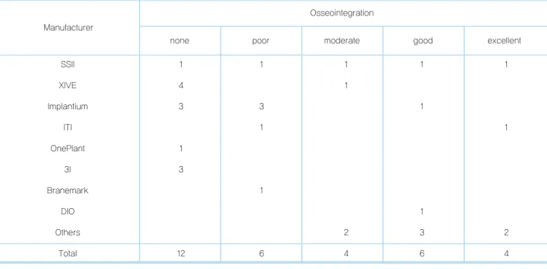
The SSII Osstem�(Ostem, co. Seoul Korea) samples underwent RBM treatment showed a relatively even level of osseointegration as mentioned above, on the other hand, the products of other companies were treated by the SLA (Xive, Implantium, ITI), ABE (Oneplant), DAE (3i Osseotite), and A (TiUnite) methods in most cases, and thus a low level of osseointegration was shown.
2. The analysis of the level of osseointegration according to gender and age
By comparing the level of osseointegration depending on gender, a distinct trend could not be detected, nonetheless, in female patients, generally, the level of osseointegration in a wide distribution was shown, and in male patients, their osseointegration was distributed in the lower side.
3. The analysis of the level of
osseointegration according to the type of bone substitute and with or without its use
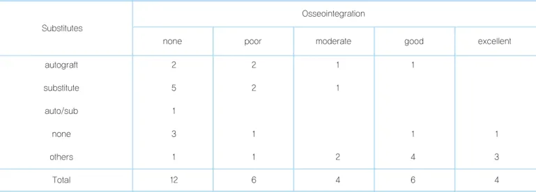
The use of autologous bone or artificial bone were shown to be in the poor side of the adhesion of implants to bone generally, however, the original bone condition of such patients was poor and thus bone substitutes were used, hence, it could not be concluded that osseointegration was poor because of the use of bone substitutes.
Ⅲ
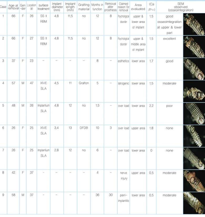
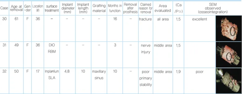
Table 1. Patient and retrieved implant data
1 66 F 26 4.8 11.5 no 12 8
Case Age atremovalGen -derLocation
(#)
surface treatment
Implant diameter
(mm)
Implant length
(mm)
Grafting material
Months in function
Removal after prosthesis
Claimed reason for
removal
Area evaluated
(Ca /P�)
SEM observed (osseointegration) Psychological
disorder
Psychological disorder
esthetics
upper &
lower area of implant
good osseoimtegration at upper & lower
part 1.5
2 66 F 27 4.8 11.5 no 12 8 upper &
middle area of implant
excellent 1.5
3 37 F 23 - - - - 8 - lower area 1.7 good
Iatrogenic
4 57 M 47 4.5 11 Grafton 5 - lower area 1.5 moderate
SS II RBM
SS II RBM
XIVE SLA
over load
5 48 M 26 Implantium 4.8 12 no 1.5 - lower area 2.2 poor
SLA
over load
6 26 F 25 XIVE 3.4 13 DFDB 10 3 upper area 1.8 none
SLA
over load
7 26 F 25 Implantium 2.8 12 no 6 - lower area 0 none
SLA
nerve injury
8 42 F 37 - - - - 4 - upper area 0.5 moderate
peri- implantitis
9 58 M 37 - - - - 36 30 lower area 0.5 moderate
10 40 M 16 4.8 11.5 5 - Case Age atremovalGen
-derLocation (#)
surface treatment
Implant diameter
(mm)
Implant length
(mm)
Grafting material
Months in function
Removal after prosthesis
Claimed reason for
removal
Area evaluated
(Ca /P�)
SEM observed (osseointegration) maxillary
sinus perforation,
poor primary stability fracture
peri- implantitis
lower area 1.5 moderate
11 39 M 15 96 - middle
area
excellent 1.5
12 39 M 25 72 - middle
area
poor
smoking, wound dehiscence
13 48 M 24 3.8 12 BBP lower area poor
(poor contact) 1.0
SS II RBM
ITI SLA
ITI SLA
Implantium SLA
smoking
14 56 M 11 Implantium 4.8 12 - 12 - all area 0 none (100%)
SLA
maxillary sinus perforation
15 33 M 16 Oneplant; 5.3 10 BioOss 3 - all area 0 none
ABE
wound dehiscence, osteomy elitis
16 42 M 46 SS II 4.1 13 2 - all area 0 none
RBM
maxillary sinus perforation
17 33 M 26 XIVE 5.5 13 10 2 all area 0 none (100%)
SLA
wide diameter
18 50 F 11 XIVE 5.5 13 - 36 30 all area 0 none (100%)
SLA
early overload
19 65 F 33 SS II 4.1 11.5 2 1 all area 0.9 poor
RBM
maxillary sinus grafting
autogenous bone
maxillary sinus
autogenous bone
20 68 F 37 - - - - 12 7 Case Age atremovalGen
-derLocation (#)
surface treatment
Implant diameter
(mm)
Implant length
(mm)
Grafting material
Months in function
Removal after prosthesis
Claimed reason for
removal
Area evaluated
(Ca /P�)
SEM observed (osseointegration) osteom
yelitis
osteom yelitis
esthetics
all area 1.4 good
21 68 F 15 - - - 12 7 all area 1.4 good
22 45 F 22 - - - 24 - all area 1.9 excellent
surgical trauma
23 58 F 15 3.4 13 GBR 6 - all area 0 none (100%)
-
ITI SLA
ITI SLA
XIVE SLA
overload
24 55 M 42 Implantium 2.4 10 no 4 - all area 0 none (100%)
SLA
wide diameter
25 52 M 37 3I 6 10 6 - all area 0 none
Osseosite
overload
26 64 M 11 TiUnite 4 11.5 5 - lower area 0 poor
poor primary stability, wide diameter
27 52 M 47 3I 6 10 6 - all area 0 none (100%)
Osseosite
overload
28 51 M 26 Implantium 4.8 10 14 6 middle area 1.5 good
SLA
wide diameter
29 33 M 26 3I 6 13 11.5 - all area 0.9 none (100%)
Osseosite
BioOss DFDB
Biocera
maxillary sinus
no
30 61 F 36 - - - - 16 - Case Age atremovalGen
-derLocation (#)
surface treatment
Implant diameter
(mm)
Implant length
(mm)
Grafting material
Months in function
Removal after prosthesis
Claimed reason for
removal
Area evaluated
(Ca /P�)
SEM observed (osseointegration) fracture
nerve injury
poor primary stability
all area 1.5 excellent
31 49 F 36 - - - 3 - middle area 1.5
32 50 F 17 - 4.8 10 10 - middle area 1.9 poor
-
DIO RBM
Implantium SLA
maxillary sinus
─ = information not available, ABE = advanced blasting & etching, Osseosite = dual acid etching, TiUnite = Anodizing, oxidation
Surface Treatment
Osseointegration
none poor moderate good excellent
Remarks
Table 2. Retrieved Implant Data according to Surface Treatment Methods
RBM 1 1 1 2 1 Resorbable Blasting Media
SLA 7 4 1 1 1 Sandblasted/Acid-etched
ABE 1 Advanced blasting/etching
DAE 3 Dual Acid Etching
A 1 Anodizing
Others 2 3 2 Unknown
Total 12 6 4 6 4
Manufacturer
Osseointegration
none poor moderate good excellent
Table 3. Retrieved Implant Data according to Manufacturers
SSII 1 1 1 1 1
XIVE 4 1
Implantium 3 3 1
ITI 1 1
OnePlant 1
3I 3
Branemark 1
DIO 1
Others 2 3 2
Total 12 6 4 6 4
Sex / Age
Osseointegration
none poor moderate good excellent
Table 4. Retrieved Implant Data according to Sex and Age
20
30 3 1 1
40 1 2 2
50 4 1 1
60 1
20 2
30 1
40 1 1 1
50 2 1
60 1 3 2
male
female
Discussion
With severely reduced osseointegration and bone loss extending to the apical third of the implant or to the apical vent hole, the possibility of normal recovery is low, and the removal of the implant should be considered8). In addition, a mobile implant is referred to clinically as a failed implant, and removal must be considered8). The indication for implant removal owing to a poor outcome is bone loss of more than half the length of the implant that progresses to the vent hole area of the implant or that progresses rapidly within one year of the prosthesis load, and is unresponsive to treatment8).
Optimal primary implant stability is generally suggested as a prerequisite for successful treatment outcome. Bicortical anchorage in the
maxilla is suggested as one way to improve primary implant stability9). However, there have been reports of 10% higher failure rates for maxillary implants that perforated the floor of the nasal cavity and maxillary sinus10).
Implant failures have been associated with factors such as poor bone quality, insufficient jawbone volume, initial implant instability, and overload. Implants may be lost prior to stage 2 surgery(early failures) of after prosthetic rehabilitation(late failures). Most implant failures have been observed in the maxilla, with almost 3 times more implant losses than in the mandible in totally edentulous situations. Early failures have been reported to vary between 1.5%
and 21%11).
The majority of reports found in the literature claim that the main reasons for early implant failures are related to factors such as anatomic conditions, surgical trauma, lack of operator
Ⅳ
Substitutes
Osseointegration
none poor moderate good excellent
Table 5. Retrieved Implant Data according to the Bone Substitutes
autograft 2 2 1 1
substitute 5 2 1
auto/sub 1
none 3 1 1 1
others 1 1 2 4 3
Total 12 6 4 6 4
surgical implant experience, and infections. In several reports, smoking habits were associated with the outcome of implant treatment. In an analysis of the outcome of 2,066 implants representing 310 patients, cigarette smoking was found to be the primary factor for implant failure reported at second stage surgery. It has also been confirmed that a significantly greater percentage of early implant failures occurred in smokers than in nonsmokers. Local cofactors, such as poor oral hygiene, seem to be responsible for the higher incidence of periimplantitis in smokers11).
Between 1995 and 1997, Van Steenberghe et al.12) analyzed the early failure rate and its causes in 1,263 Branemark implants placed in 399 patients, and reported that between 1~6 months after the placement of implants, 27 implants in 21 patients failed, and after second surgery, none of them failed. In addition, the failure of implant is most frequent up to 2nd surgery after their placement, the possibility of the failure in patients who failed already is high, and the possibility of the failure in the male than the female is high, and it has been reported to be associated with harmful habits such as smoking, drinking, and bruxism, etc.
The causalities of early failure of implant reported by Kim13)were as follows. (1) Regardless of their clinical experience on sinus bone graft, the early failure of implant occurred continuously in specific patients, (2) after the
successful final prosthesis could be achieved by reimplants or additional implants, nonetheless, the treatment period was prolonged unavoidably, (3) by explaining the risk factors associated with the failure to patients honestly, and subsequently performing continuous treatments, medical disputes were developed in none of cases, (4) early implant failure occurred frequently in the early treatment period or the early load period, and (5) The speculated causes of failure were in the order of early excessive load, and an insufficient healing period, nevertheless, it was thought that the risk factors were involved in combination.
McDermott et al.14) have reported that rather the factors involved in the failure of the implant in maxillary molar area are the single tooth implant of the molar tooth and one stage implant, and the success rate of implants placed in the area where maxillary sinus grafting was successful was not different from the maxillary molar implants performed without bone graft.
Surface analysis investigations of failed implants have the advantage of not causing additional patient discomfort, unlike histological studies, which require the retrieval of an adequate amount of tissue to obtain useful information. In addition, it is easier to examine failed implants surrounded by a soft-tissue capsule than failed implants embedded in plastic or implants successfully integrated in bone, because the analysis is hampered by the plastic or tissue
Most studies evaluating the behavior and response of bone to different implant surface materials and surface topographies have been conducted in animal models. The remodeling activity, bone quality, and loading conditions of animal bone are different from those of human bone. Therefore, the findings from animal models do not always support the behavior in human bone17).
Histomorphometric analysis of human retrieved implants is the method available to analyze the bone-to-implant interface behavior over time.
The reproduction of a human's mouth environment in animals is tremendously difficult6).
One difficulty was the absence of information to allow determination of how much bone-to- implant contact is clinically necessary or is infact ideal. Histometric studies have determined the amount (usually expressed as a percentage of a defined surface area of the implants) of bone-to-implant contact18). Sennerby et al.19) retrieved seven clinically stable, osseointergrated into four patients for 116 years, for morphological analysis of the bonetitanium interface. The threads of the implants were well filled(79~95%) with dense lamellar bone as quantified with morphometry. A large fraction of the implant surface(56~85%) appeared to be in direct contact with the mineralized bone.
Ivanoff et al.20), when they examined microimplants retrieved from human jaws, observed a difference in the amount of bone-to-implant contact and
bone area within threads between implants placed in the maxilla and those placed in the mandible. In this study, the position was known for 119 implants with calculations of bone-to- implant contact and/or bone area within the threads: 39 implants were placed in the maxilla, and 80 implants were placed in the mandible.
The mean values of percentages of bone-to- implant contact and bone area within the threads were 71% and 83%, respectively, for maxillary implants and placed in the mandible.
Buser et al.21) investigated the direct bone- implant contact rate using implants with different surfaces, including sandblasted and acid-etched surfaces, HA-coated, TPS, and acid-etched surfaces. Of these, the highest rate of bone-implant contact was seen with the sandblasted and acid-etched surfaces.
Parr et al.22) reported that implant failure resulted from tissue damage caused by implant drilling and circulatory impedance. The implantation in an incompletely healed extraction area may cause failure, as the tissue is readily damaged by drilling and impaired circulation occurs readily. The extraction area must be managed carefully during the healing period and before implantation. Damage during the procedure must be minimized, as excessive damage of the adjacent tissues induces the formation of unwanted fibrosis of tissue during healing.
Piattelli et al.3) reported histoligical observations on 230 retrieved dental implant of different
designs with the aim to establish the causal determinants of implant failure. The histological features varied in implants removed for periimplantitis before and after loading, for mobility and for fractures. The study reported that the host tissue factors, i.e. periimplantitis and mobility were implicated as causative factors more frequently than biomaterials problem, i.e.
fractures. It was seldom possible to relate failure to psychological matters or to the alveolar inferior nerve pathology.
Steflik et al.23) reported histoloigical findings on 200 implants, including both dental and orthopedic implants. The authors did not inform about the precise number of dental implants and focused on the results in relation to categories of coating: beaded coatings, HA coatings, porous coatings and uncoated implanted biomaterial.
The cause of implant failure was more often found to be biological, i.e. loss of bone support, connective tissue encapsulation and inflammatory cell infiltrate, for implants with a time in situ of 10 years of longer. The failure of recently placed implants was more often ascribed to the biomaterial than to biological failure.
Kim et al.24) analyzed the factors of failure according to the shape and surface of implant by examining the surface of failed implants by light microscope and transmission electron microscope. 26 implants failed from 1996 to 2004 were used in the study, and among them, the cylinder type was 8 implants and the screw type
shape showed more soft tissues and bacterial deposition. Hydroxyapatite coating and the titanium plasma spray surface showed more soft tissues and bacterial deposition than the surface treated with acid etching treatment in many cases, and in some screw types, the trace of milling and the micro-fracture of fixture were observed.
In this study on retrieved implants, the analysis of the level of osseointegration according to the surface treatment methods, the analysis of the level of osseointegration according to the manufacturers, and the analysis of the level of osseointegration depending on gender and age, and the analysis of the level of osseointegration depending on the type of bone substitute material as well as with or without its use were performed.
References
1. Adell R, et al: Long-term follow-up study of osseointegrated implants in the treatment of totally edentulous jaws. Int J Oral Maxillofac Implants, 5: 347, 1990.
2. Lekholm U, et al: Survival of the Bra°nemark implant in partially edentulous jaws: a 10-year prospective multicenter study. Int J Oral Maxillofac Implants, 14: 639, 1999.
3. Piattelli A, et al: Histologic observations on 230 retrieved dental implants: 8 years' experience (1989-1996). J Periodontol, 69: 178, 1998.
4. Piattelli A, Trisi P: A light and laser scanning microscopy study of bone/hydroxyapatite-coated titanium implants interface: histochemical evidence of unmineralized material in humans. J Biomed Mater Res,
5. Takeshita F, et al: Fractures of hydroxyapatite-coated blade implants connected with natural teeth. A histological study using SEM, light microscopy, and an image processing system. J Periodontol, 67: 86, 1996.
6. Sakakura CE, et al: Histomorphometric evaluation of a threaded, sandblasted, acid-etched implant retrieved from a human lower jaw: a case report. Implant Dent, 14: 289, 2005.
7. Bolind PK, et al: A descriptive study on retrieved non-threaded and threaded implant designs. Clin Oral Implants Res, 16: 447, 2005.
8. Kim YK, Hwang JW: Controversy in dental implant. Koonja publishing Co., p227, 2004.
9. Adell R, et al: Surgical procedures. In Bra°nemark PI, et al (eds). Tissue- Integrated Prostheses: Osseointegration in Clinical Dentistry. Chicago:
Quintessence, p211, 1985.
10. Bra°nemark PI, et al: An experimental and clinical study of osseointegrated implants penetrating the nasal cavity and maxillary sinus. J Oral Maxillofac Surg, 42: 497, 1984.
11. Kronstrom M, et al: Early implant failures in patients treated with Bra°nemark System titanium dental implants: a retrospective study. Int J Oral Maxillofac Implants, 16: 201, 2001.
12. Van Steenberghe D, et al: The relative impact of local and endogenous patient-related factors on implant failure up to the abutment stage. Clin Oral Implants Res, 13: 617, 2002.
13. Kim YK: Implant risk factor of sinus bone graft (II): analysis of causes of early failure. Korean Acad Oral Maxillofac Implantol 10: 36, 2006.
14. McDermott NE, et al: Maxillary sinus augmentation as a risk factor for implant failure. Int J Oral Maxillofac Implants, 21: 366, 2006.
15. Esposito M, et al: Surface analysis of failed oral titanium implants. J Biomed Mater Res, 48: 559, 1999.
16. Sundgren JE, et al: Auger electron spectroscopic studies of stainless- steel implants. J Biomed Mater Res, 19: 663, 1985.
17. Hayakawa T, et al: A histologic and histomorphometric evaluation of two types of retrieved human titanium implants. Int J Periodontics Restorative Dent, 22: 164, 2002.
18. Uehara T, et al: Histological evidence of osseointegration in human retrieved fractured hydroxyapatite-coated screw-type implants: a case report. Clin Oral Implants Res, 15: 540, 2004.
19. Sennerby L, et al: Structure of the bone-titanium interface in retrieved clinical oral implants. Clin Oral Implants Res, 2: 103, 1991.
20. Ivanoff CJ, et al: Histologic evaluation of bone response to oxidized and turned titanium micro-implants in human jawbone. Int J Oral Maxillofac Implants, 18: 341, 2003.
21. Buser D, et al: Influence of surface characteristics on bone integration of titanium implants. A histomorphometric study in miniature pigs. J Biomed Mater Res, 25: 889, 1991.
22. Parr GR, et al: Clinical and histological observations of failed two-stage titanium alloy basket implants. Int J Oral Maxillofac Implants, 3: 49, 1988.
23. Steflik DE, et al: Light microscopic and scanning electron microscopic retrieval analyses of implanted biomaterials retrieved from humans and experimental animals. J Oral Implantol, 27: 5, 2001.
24. Kim SG, et al: Surface analysis of failed implant. Korean Acad Implant Dent, 23: 1, 2004.
교신저자 : 김수관 우편번호 : 501-825
주소 : 광주광역시 동구 서석동 421번지 조선대학교 치과병원 구강악안면외과 TEL: (062) 220-3815 FAX: (062) 228-7316 E-Mail: [email protected]