• 검색 결과가 없습니다.

Concurrent Gullain-Barre Syndrome and Acute Transverse Myelitis as an Initial Presentation of Systemic Lupus Erythematosus

N/A
N/A
Protected

Academic year: 2021

Share "Concurrent Gullain-Barre Syndrome and Acute Transverse Myelitis as an Initial Presentation of Systemic Lupus Erythematosus"

Copied!
8
0
0

로드 중.... (전체 텍스트 보기)

전체 글

(1)

Vol. 20, No. 2, June, 2012 □ 증 례 □

1)

Introduction

Guillain Barre Syndrome (GBS) is an acute ascending inflammatory demyelinating polyneu- ropathy thought to be caused by antibodies to

접수 :2012년 6월2 ,일 수정:2012년7월 25일 승인 :2012년 7월28일

책임저자 : 고태성 울산대학교 의과대학 아산병원 소아청소년과, Tel: 02)3010-3381, Fax: 02)3010-3725 E-mail: [email protected]

gangliosides on the axolemma, which leads macrophages to invade the axon at the node of Ranvier

1)

. Typical clinical findings are progres- sive weakness starting from the limbs, with or without sensory deficit. The weakness may last for several weeks before good recovery, or it may remain as a chronic disability. Trans- verse myelitis is an inflammatory disorder, characterized by acute or subacute motor, sen- sory, and autonomic spinal cord dysfunction,

Concurrent Gullain-Barre Syndrome and Acute Transverse Myelitis as an Initial Presentation

of Systemic Lupus Erythematosus

Sung-Han Kang, K.D., Mi-Sun Yum, M.D.

*

, Eun-Hye Lee, M.D.

*, †

and Tae-Sung Ko, M.D.

*

Department of Pediatrics, Asan Medical Center, University of Ulsan College of Medicine

*

Department of Pediatrics, College of Medicine, Kyung Hee University

Seoul, Korea

= Abstract =

Systemic lupus erythematosus (SLE) is an autoimmune disorder involving multiple organs.

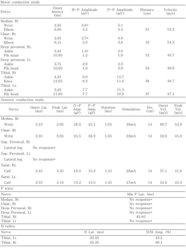
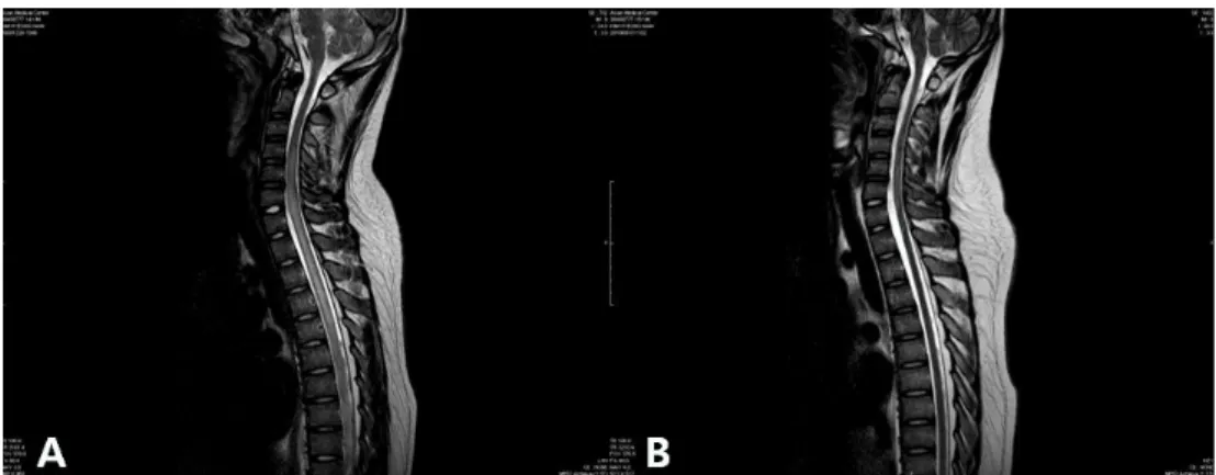
Neuropsychiatric symptoms are frequently associated in SLE, which is referred to as neurop- sychiatric SLE (NPSLE). NPSLE contains both central and peripheral nervous systems, which includes transverse myelitis, and Guillain-Barre syndrome (GBS). We report our experience of concurrent manifestation of transverse myelitis and GBS as an initial presentation of SLE, which suggests the common immune-mediated mechanisms of diseases. We here report the case of a 14-year-old boy with SLE who first presented with features of GBS. The patient developed ascending weakness starting from low extremities, experienced difficulty voiding, and had a facial rash. An initial diagnosis of GBS was made on the basis of clinical findings and nerve conduction studies. But he did not respond to intravenous immunoglobulin therapy and following spine MRI displayed T2 weighted high signal intensities from the cervical to thoracic region of the spinal cord, and serological analysis revealed the presence of anti- dsDNA, anti-smAb, anti nuclear antibody with decreased level of complements. The diagno- sis was revised to GBS and acute transverse myelitis resulting from SLE. Additional methyl- prednisolone pulse therapy led to rapid clinical improvement. This was followed by oral pred- nisolone and cyclophosphamide pulse therapy. This is the first case of concurrent manifesta- tion of GBS and transverse myelitis as initial presentation of SLE. The cross-reactivity of autoantibodies and increased susceptibility to infection owing to immunologic changes asso- ciated with lupus may form the basis of the association. Clinicians should consider a diagno- sis of SLE as an etiology of GBS or transverse myelitis.

Key Words : Guillain-Barre syndrome, Transverse myelitis, Systemic lupus erythematosus

(2)

causing typical pain, weakness, sensory chan- ges, and dysfunction of the bowel and/or blad- der

2)

.

Systemic lupus erythematosus (SLE) is an autoimmune disorder involving multiple organs.

To diagnose SLE, at least four of eleven patho- logic findings must be present in any given patient

3)

. One of these eleven pathologic fin- dings is a neurologic disorder, representing seizures and psychosis. Nonetheless, patients can also present with secondary phenomena involving the central nervous system, periphe- ral nervous system, and autonomic nervous system owing to treatment for complications or involvement of other organs.

Neuropsychiatric SLE (NPSLE) is used to describe the full range of possible symptoms experienced by patients with SLE. The pre- valence of NPSLE varies from 20% to 95%

among pediatric patients

4)

. Involvement of the CNS is more frequent than that of the PNS, with headache and cognitive dysfunction ac- counting for approximately 55% of NPSLE cases involving children

4)

. In the case of trans- verse myelitis, the prevalence is reported to be 1% to 2% in patients with SLE, including adults

2)

. However, the involvement of the PNS in SLE, as in GBS, is rare (less than 0.1%)

5)

. In spite of its low prevalence among patients with later stages of SLE, transverse myelitis is relatively common as an initial presentation in patients with SLE

2)

. In contrast, GBS is rarely reported as an initial feature of SLE. In this report, we describe what appears to be the first case of SLE, which initially manifested as GBS and transverse myelitis.

Case Report

A 14 year old male was admitted to the hos- pital with ascending weakness, starting from both lower extremities, as well as difficulty voiding, headache associated with back pain, and facial rash. The patient visited the emer- gency department four days before admission with symptoms of upper respiratory tract in- fection, including rhinorrhea, fever, and head- ache. However, neck stiffness was not definite at that time. He also had a history of diarrhea 1 week earlier, which lasted for only 1 day.

Neurologic examination indicated decreased motor power in the lower extremities bilate- rally. Grade II numbness was noted in the area under T1 and T2 of the dermatome. Examina- tion of the cranial nerve did not reveal any ab- normalities. However, both neck stiffness and a decreased deep tendon reflex were evident. As the patient complained of difficulties with both urination and defecation, a Foley catheter was inserted at the time of admission.

Laboratory tests revealed a hemoglobin le- vels of 15g/dL, a white blood cell count of 7,500/mm

3

, and a platelet count of 204,000/

mm

3

. Cell counts were within the respective reference limits, except for lymphopenia (15%

of leukocytes). Erythrocyte sedimentation rate (28 mm/hr) and levels of C-reactive protein (0.1 mg/dl) were within normal ranges. The serum concentrations of C3, C4 and CH50 was all decreased to 47.6 mg/dL (reference, 88- 201 mg/dl), 3.3 mg/dL (reference, 16-47 mg/

dL), and 15.4 mg/dL (reference 25-50 mg/dl),

respectively. Immunoglobulin G (IgG) level was

increased to 2,120 mg/dL (reference 700-

1,600 mg/dL), whereas IgA (217 mg/dl), and

IgM (113 mg/dL) levels were within the normal

ranges. The results of urinalysis were negative

for proteinuria or hematuria. Results of cereb-

(3)

Table 1. Nerve Conduction Study Motor conduction study

Nerve

Onset latency

(ms)

B-P Amplitude (mV)

P-P Amplitude (mV)

Distance (cm)

Velocity (m/s) Median, Rt

Wrist Elbow Ulnar, Rt

Wrist Elbow

Deep peroneal, Rt.

Ankle Fib head Deep peroneal, Lt

Ankle Fib head Tibial, Rt

Ankle Knee Tibial, Lt.

Ankle Fib head

2.85 6.80 2.65 6.15 3.95 10.80

3.75 10.60

4.25 12.05

3.95 11.80

3.6*

3.2 2.7*

2.6 1.4*

1.2 2.0 1.9 9.0 8.2 7.7 7.7

5.1 4.4 4.0 3.8 2.0 1.9 2.6 2.6 12.7 11.6 11.3 10.9

21

19

32

34

38

37

53.2

54.3

46.7

49.6

48.7

47.1 Sensory conduction study

Nerve Onset Lat.

(ms)

Peak Lat.

(ms)

O-P Amp.

( V) µ P-P Amp.

( V) µ

Duration

(ms) Stimulation Dis.

(cm) Onset

Vel.

(m/s) Peak

Vel.

(m/s) Median, Rt

Wrist 2.10 2.65 28.0 45.1 1.65 28mA 14 66.7 52.8

Ulnar, Rt

Wrist 2.35 3.05 24.5 38.9 1.65 33mA 14 59.6 45.9

Sup. Peroneal, Rt

Lateral leg No response*

Sup. Peroneal, Lt

Lateral leg No response*

Sural, Rt.

Calf 2.45 3.35 18.0 15.3 1.55 26mA 14 57.1 41.8

Sural, Lt.

Calf 2.55 3.10 13.2 13.5 1.35 27mA 14 54.9 45.2

F wave

Nerve Min F Lat. (ms)

Median, Rt Ulnar, Rt

Deep Peroneal, Rt Deep Peroneal, Lt Tibial, Rt Tibial, Lt

No response*

No response*

No response*

No response*

49.80 No response*

H reflex

Nerve H Lat. (ms) H/M Amp. (%)

Tibial, Lt Tibial, Rt

30.23 30.20

43.5

88.1

(4)

rospinal fluid (CSF) analysis was as follows:

total protein, 40.1 mg/dL; glucose, 49 mg/dL;

white blood cell count, 2/mm

3

; CSF albumin, 22.9 mg/dL; and CSF IgG, 8.9 mg/dL (IgG index: 0.75). An electrophysiologal examination study of the upper and lower extremities, which involved electromyography and assess- ment of nerve conduction velocity, revealed the absence of latency and decreased amplitude of complex muscle action potential in both lower extremities indicative of symmetric distal peri- pheral polyneuropathy (AMAN type), mainly axonal type (Table 1).

Although albumino-cytologic dissociation was not definite following CSF analysis, an initial diagnosis of Guillain-Barre syndrome (GBS) was made based on clinical findings and the electrophysiological study. Intravenous immu- noglobulin (IVIG) was administered after an initial survey for other neurological disorders and the weakness of both lower extremities was aggravated from motor power grade II to grade 0. However, given that injection of IVIG did not significantly improve clinical symptoms, brain and whole-spine MRI was done to identify whether other pathologic lesions might be present. The whole-spine MRI showed high signal intensity from the C3-4 to the T10-11 segments of the spinal cord in T2 weighted images with leptomeningeal enhancement, indi- cating transverse myelitis (Fig. 1A). No abnor- mality was apparent from the brain MRI.

The presence of facial rash, with a morpho- logy typical of malar rash, along with decrea- sed levels of components of the complement system, strongly implicated the possibility of an immunological disorder(s). Laboratory data for autoimmune serology were as follows: anti -nuclear factor antibody, 1:320 speckled (re-

ference<1:40); anti-dsDNA, 47.3I U/mL (refe- rence 0-7 I U/mL); anti-IgG for direct Coombs test, positive (1+) (reference negative); anti- Smith antigen antibody, positive (>120) (refe- rence <5); and platelet-associated antibody, positive (12.4) (reference negative). Two other markers, rheumatoid factor and anticar- diolipin antibody, were all negative. Together, anti-nuclear factor antibody, anti-dsDNA anti- body, malar rash, and decreased levels of com- plement components strongly implied the possi- bility of SLE.

Given that the effect of IVIG was not defini-

te, with the diagnosis of myelitis and presumed

diagnosis of SLE, pulse therapy involving deli-

very of 1 g of intravenous (IV) methylprednis-

olone was given for 3 consecutive days, star-

ting after the end of IVIG treatment. Motor

function of the lower extremities improved

after the steroid pulse therapy up to grade III,

and there was less difficulty voiding. Nonethe-

less, a sensory change was not definite, except

for a slight improvement in the level of numb-

ness experienced in both legs. Repeated analy-

sis of nerve conduction at this time revealed

symmetric distal peripheral polyneuropathy

(AMAN type), which was primarily of the axo-

nal type. Administration of oral steroids for

three weeks after admission failed to rectify

the decreased sensation, but dramatically imp-

roved motor function to the point that the pa-

tient could walk without any help. Despite im-

proved clinical manifestations, markers for SLE

activity did not show any improvement, with

C3, C4 and CH50 levels of 56.2 mg/dL, 3.9

mg/dL, and 15.4 U/mL, respectively. Monthly

IV administration of cyclophosphamide conti-

nued for five months until the time the patient

was discharged. Since then, the patient has

(5)

been treated with an oral steroid.

Motor and sensory function was much im- proved following a second dose of cyclophos- phamide. The grade of motor power appeared to be more than grade IV, and the patient no longer complained of sensory deficits. Repea- ted spine MRI showed improvement of lepto- meningeal enhancement and high signal inten- sity of myelitis involving the C3-4 to T10-11 segments of the spinal cord (Fig. 1B).

However, the levels of C3, C4, and CH50 were still decreased after five doses of cyclophos- phamide pulse therapy. An additional reduction in the dosage of methylprednisolone (500 mg of methylprednisolone weekly for a month, then biweekly for another month, and then 750 mg biweekly for 4 months), was made. Methyl- prednisolone therapy was followed by admini- stration of oral cyclosporine. The severity of the malar rash transiently got worse during methylprednisolone pulse therapy, but impro- ved without any additional medication. Photo- sensitivity developed as a new symptom during methylprednisolone pulse therapy. The patient is now exclusively on oral medication (cyclo- sporine and steroid). He has been stable

without further flare-ups for 15 months of follow up.

Discussion

The initial presentation in our patient was progressive, with ascending weakness begin- ning from both lower extremities, and a dec- reased deep tendon reflex. Initial diagnosis of GBS was made based on the patient s clinical ’ manifestations. Additional evaluation using MRI led to the diagnosis of transverse myelitis.

Although the patient fulfilled three SLE criteria malar rash, antibodies against dsDNA, and

antibodies against nuclear factor the low com — - plement levels detected suggested the possi- bility of SLE. Thus we suspected that the GBS and transverse myelitis might be neuropsychia- tric presentations of SLE. Steroid pulse the- rapy and cyclophosphamide therapy were given on the basis of our suspicion of SLE. Later the patient developed photosensitivity, which pro- mpted the formal diagnosis of SLE. The only current criteria for neurologic manifestations for SLE psychosis and seizure are both rela — — - tively infrequent, which decreases the sensi-

Fig. 1. (A) A high T2WI signal intensity from the C3-4 to the T10-11 segments of

spinal cord suggests myelitis. (B) A high T2WI signal intensity in the C3-4 to the

T10-11 segments of the spinal cord suggests improvement of myelitis.

(6)

tivity of diagnosis of the disease

6)

. The Ame- rican College of Rheumatology defined 19 po- tential neurologic manifestations of SLE in 1999

7)

. These include common complaints, such as cognitive impairment and headache, as well as relatively rare manifestations, such as myelitis and GBS, which are less frequently encountered in clinical practice. The group suggested that inclusion of above manifesta- tions should be considered in order to detect NPSLE during the early period

6)

.

Although the association of SLE with trans- verse myelitis is relatively well known, GBS is rarely reported as an initial presentation of SLE

8, 9)

. Furthermore, this appears to be the first report of GBS along with transverse mye- litis as an initial presentation of SLE. Patients with SLE may not necessarily fulfill the requi- site diagnostic criteria at the time of initial evaluation. Especially in the cases of patients with unusual neurologic manifestations, such as weakness of extremities, bladder dysfunction, or sensory dysfunction, MRI should be perfor- med to detect associated neurologic manifesta- tions such as GBS and/or transverse myelitis, even though these are rarely reported.

The pathogenic basis of SLE presenting as GBS and/or transverse myelitis is not well known. However, the generation of autoantibo- dies, along with other immunological changes, have been implicated

10, 11)

. In the case of GBS, about two-thirds of all patients have had infec- tion within the previous 6 weeks, most com- monly a flu-like illness with or without gastro- enteritis. The agents most likely to be respon- sible— Campylobacter jejuni , cytomegalovirus (CMV), Epstein-Barr virus (EBV), and Myco- plasma pneumoniae

1)

— express a lipooligosac - charide molecule that mimics various ganglio-

sides abundantly present in peripheral nerves.

This condition may result in typical signs of GBS

12)

. Like GBS, transverse myelitis is also associated with vulnerable infections, such as those by enterovirus, coxsackie virus, CMV, hepatitis A virus, M. pneumoniae , and Schisto- soma spp. Therefore, underlying changes of the immune system in SLE, along with cross- reactions of antibodies during infection, may be related to the presenting symptoms of GBS with transverse myelitis

10)

. In some cases, anticardiolipin antibodies are reported to be responsive to neuropathies, by causing myelin damage or cross-reaction with anti-myelin antibodies, with good responsiveness to ste- roids or IVIG

13)

. Additionally, recent study ex- plored the occurrence of aquaporinopathy in patients with SLE

14)

. This also should be in concern, although we did not carry out evalua- tions for NMO IgG or anti-aquaporin 4 anti- body.

Both GBS- and SLE-associated neuropathi-

es have been effectively treated with IVIG,

plasmapheresis, and corticosteroids

15)

. Impro-

vement of neurological manifestations with

these treatments may itself constitute further

evidence of autoimmune pathogenesis as a

common link between GBS and SLE

10)

. Curren-

tly, IVIG has replaced plasma exchange, mainly

because of its convenience. Several recent

studies indicated that plasma exchange follow-

ed by IVIG, or a combination of IVIG and me-

thylprednisolone, was not significantly more

effective than IVIG alone

16, 17)

. However, ano-

ther study reported that, after correcting some

known prognostic factors, the combination

treatment was only effective in the short

term

18)

. Our patient demonstrated rapid impro-

vement of motor function after combination

(7)

therapy involving IVIG and methylprednisolone, and he is still receiving immune-modulating therapy for SLE.

There has not been a large controlled study of therapy of SLE with neurologic manifesta- tion such as transverse myelitis. In particular, there have been no published treatment trials involving pediatric populations. Generally, cor- ticosteroids are the most commonly used medi- cation for SLE. They also constitute the stan- dard initial medication for SLE-associated transverse myelitis

2)

. Immune modulators such as cyclophosphamide may be given in instances when severe neuropsychiatric SLE is unres- ponsive to other treatment options. Plasmaphe- resis as a treatment option was reported in several severe cases, or rare cases, such as SLE-associated Devic s syndrome ’

2, 19)

. Our patient showed full recovery of sensory func- tion during cyclophosphamide pulse therapy. A small, randomized, controlled clinical trial that compared methylprednisolone and cyclophos- phamide reported better overall therapeutic control of SLE-related neurologic manifesta- tion, including peripheral neuropathy with monthly IV administration of cyclophospha- mide

20)

.

Not all patients fulfill the criteria of SLE at the time of diagnosis. Neurologic manifesta- tions may be the first symptoms of SLE that identify patients who will eventually meet the full criteria. Such manifestations may occasio- nally include GBS, other forms of peripheral neuropathy, myelitis, or any combination of these. We report the first case of combined GBS and transverse myelitis as an initial pre- sentation of SLE. It is important that physi- cians keep the option of SLE in mind whenever patients present atypical neurologic manifesta-

tions, or do not response to standard therapies.

Awareness of the possibility of SLE may be key to making the correct diagnosis and en- suring a better outcome.

국 문 요 약

길랑 바레 증후군과 횡단척수염으로 발현한 전신성 홍반성 루프스 1 례

울산대학교 의과대학 서울아산병원 소아청소년병원 소아청소년과학교실*

경희대학교 의과대학 소아청소년과학교실

강성한

*

ㆍ 염미선

*

ㆍ 이은혜

*, †

ㆍ 고태성

*

전신성 홍반성 루프스는 전신을 침범하는 자가 면

역 질환으로서 중추 신경과 말초 신경을 침범하는 ,

질환이 동반될 수 있다 대부분의 증례에서 두통 및 . 인지 장애와 같은 질환들이 보고 되고 있다 본 저자 . 들은 길랑 바레 증후군과 횡단성 척수염이 전신성

홍반성 루프스의 첫 증상으로 발현한 1 례를 경험하

였기에 보고하는 바이다.

References

1) van Sorge NM, van der Pol WL, Jansen MD, Geleijns KP, Kalmijn S, Hughes RA, et al. Se- verity of Guillain-Barre syndrome is associa- ted with Fc gamma Receptor III polymorphi- sms. J Neuroimmunol 2005;162:157-64.

2) Schulz SW, Shenin M, Mehta A, Kebede A, Fluerant M, Derk CT. Initial presentation of acute transverse myelitis in systemic lupus erythematosus: demographics, diagnosis, mana- gement and comparison to idiopathic cases.

Rheumatol Int 2011.

3) Tan EM, Cohen AS, Fries JF, Masi AT, Mc- Shane DJ, Rothfield NF, et al. The 1982 revised criteria for the classification of sys- temic lupus erythematosus. Arthritis Rheum 1982;25:1271-7.

4) Sibbitt WL, Brandt JR, Johnson CR, Maldonado

ME, Patel SR, Ford CC, et al. The incidence

and prevalence of neuropsychiatric syndromes

(8)

in pediatric onset systemic lupus erythema- tosus. J Rheumatol 2002;29:1536-42.

5) Unterman A, Nolte JE, Boaz M, Abady M, Sho- enfeld Y, Zandman-Goddard G. Neuropsychia- tric Syndromes in Systemic Lupus Erythema- tosus: A Meta-Analysis. Semin Arthritis Rhe- um 2010.

6) Petri M. Review of classification criteria for systemic lupus erythematosus. Rheum Dis Clin North Am 2005;31:245-54, vi.

7) The American College of Rheumatology no- menclature and case definitions for neuropsy- chiatric lupus syndromes. Arthritis Rheum 1999;42:599-608.

8) Ait Benhaddou E, Birouk N, El Alaoui-Faris M, Mzalek-Tazi Z, Aidi S, Belaidi H, et al. [Acute Guillain-Barre-like polyradiculoneuritis revea- ling acute systemic lupus erythematosus: two case studies and review of the literature]. Rev Neurol (Paris) 2003;159:300-6.

9) Hsu TY, Wang SH, Kuo CF, Chiu TF, Chang YC. Acute inflammatory demyelinating polyneu- ropathy as the initial presentation of lupus. Am J Emerg Med 2009;27:900 e3-5.

10) Vaidya S, Jasin HE, Logan J. Systemic lupus erythematosus and guillain-barre syndrome. J Clin Rheumatol 1999;5:349-53.

11) Muscal E, Brey RL. Neurologic manifestations of systemic lupus erythematosus in children and adults. Neurol Clin 2010;28:61-73.

12) Israeli E, Agmon-Levin N, Blank M, Chapman J, Shoenfeld Y. Guillain-Barre Syndrome-A Classical Autoimmune Disease Triggered by Infection or Vaccination. Clin Rev Allergy Im- munol 2010.

13) Nakajima H, Shinoda K, Doi Y, Tagami M, Fu- rutama D, Sugino M, et al. Clinical manifesta- tions of chronic inflammatory demyelinating polyneuropathy with anti-cardiolipin antibodies.

Acta Neurol Scand 2005;111:258-63.

14) Dellavance A, Alvarenga RR, Rodrigues SH, Kok F, de Souza AW, Andrade LE. Anti-aqua- porin-4 antibodies in the context of assorted immune-mediated diseases. Eur J Neurol 2012;19:248-52.

15) Lesprit P, Mouloud F, Bierling P, Schaeffer A, Cesaro P, Brun-Buisson C, et al. Prolonged remission of SLE-associated polyradiculoneu- ropathy after a single course of intravenous immunoglobulin. Scand J Rheumatol 1996;25:

177-9.

16) Randomised trial of plasma exchange, intrave- nous immunoglobulin, and combined treatments in Guillain-Barre syndrome. Plasma Exchange/

Sandoglobulin Guillain-Barre Syndrome Trial Group. Lancet 1997;349:225-30.

17) Hughes RA, Swan AV, Raphael JC, Annane D, van Koningsveld R, van Doorn PA. Immuno- therapy for Guillain-Barre syndrome: a syste- matic review. Brain 2007;130:2245-57.

18) van Koningsveld R, Schmitz PI, Meche FG, Visser LH, Meulstee J, van Doorn PA. Effect of methylprednisolone when added to standard treatment with intravenous immunoglobulin for Guillain-Barre syndrome: randomised trial.

Lancet 2004;363:192-6.

19) Polgar A, Rozsa C, Muller V, Matolcsi J, Poor G, Kiss EV. Devic's syndrome and SLE: chal- lenges in diagnosis and therapeutic possibilities based on two overlapping cases. Autoimmun Rev 2011;10:171-4.

20) Barile-Fabris L, Ariza-Andraca R, Olguin-Or-

tega L, Jara LJ, Fraga-Mouret A, Miranda-

Limon JM, et al. Controlled clinical trial of IV

cyclophosphamide versus IV methylpredniso-

lone in severe neurological manifestations in

systemic lupus erythematosus. Ann Rheum Dis

2005;64:620-5.

수치

Table 1. Nerve Conduction Study Motor conduction study
Fig. 1. (A) A high T2WI signal intensity from the C3-4 to the T10-11 segments of spinal cord suggests myelitis

참조

관련 문서

We describe a case of polymyositis/systemic lupus eryth- ematosus (SLE) overlap syndrome with severe generalized subcutaneous edema and interstitial lung

PURPOSE: This study examined the effects of core stability exercise on the strength, activation of the trunk muscle, and pulmonary function in a Guillain-Barre syndrome

Bronchiolitis obliterans organizing pneumonia as an initial manifestation in patients with

Acute left main coronary artery thrombosis as an initial presentation of systemic lupus erythematosus.. Kang Un