http://dx.doi.org/10.18325/jkmr.2016.26.3.1 Original Article
勝濕湯이 MIA로 유발된 골관절염 흰쥐에 미치는 영향
이종훈⋅우창훈⋅김영준⋅안희덕
대구한의대학교 한의과대학 한방재활의학과교실
Effects of Seungseup-tang on MIA-induced Osteoarthritis Rats
Jong-Hoon Lee, K.M.D., Chang-Hoon Woo, K.M.D., Young-Jun Kim, K.M.D., Hee-Duk Ahn, K.M.D.
Department of Rehabilitaion Medicine of Korean Medicine, College of Korean Medicine, Daegu Haany University
RECEIVED June 16, 2016 REVISED July 7, 2016 ACCEPTED July 8, 2016
CORRESPONDING TO Hee-Duk Ahn, Department of Rehabiliation Medicine of Korean Medicine, College of Korean Medicine, Daegu Hanny University, 136 Sincheondong-ro, Suseong-gu, Daegu 42158, Korea
TEL (053) 770-2116 FAX (053) 770-0566 E-mail [email protected]
Copyright © 2016 The Society of Korean Medicine Rehabilitation
Objectives This study was designed to evaluate the anti-inflammatory effects of Seungseup-tang (SST) on the monosodium iodoacetate (MIA)-induced osteoarthritis rats.
Methods Osteoarthritis was induced by injection of MIA (50 μl with 80 mg/ml) into knee joint cavity of rats. Rats were divided into 4 groups (normal group, control group, in- domethacin treated group, SST treated group, each n=6). Normal group was not injected with MIA and taken normal diet. Control group was injected with MIA and taken with dis- tilled water. Indomethacin treated group was injected with MIA and taken indomethacin 5 mg/kg by oral administration. SST treated group was injected with MIA and taken SST 200 mg/kg by oral administration. We examined the weight-bearing ability of hind paw, bio- markers related to oxidative stress in serum, inflammatory proteins and inflammatory medi- ators and cytokines. Moreover, histopathological examination of knee joint structure was also performed by Hematoxylin & Eosin (H&E), Safranin-O staining method.
Results In the present study, SST treated group showed a similar inhibitory effects alike indomethacin treated group, in most of the studied parameters of inflammation. The in- creased oxidative stress biomarker such as reactive oxygen species (ROS) and peroxy ni- trite (ONOO
−) in the serum were reduced with SST. Especially, the level of ONOO
−com- pared with control group significantly suppressed. Also, the expression of inflammatory mediators and cytokines induced by nuclear factor-kappa B (NF-κB) activation was modulated through inhibition of IkBaphosphorlation. In addition, histological analysis re- vealed that cartilage damage by MIA repaired markedly in SST treated group.
Conclusions According to the results, Seungseup-tang may be effective for preventing the progression of osteoarthritis. (J Korean Med Rehabil 2016;26(3):1-15)
Key words Seungseup-tang , Osteoarthritis, Anti-inflammative effect, Monosodium io- doacetate (MIA)
서론»»»
골관절염(osteoarthritis, OA)은 퇴행성관절염이라고도 불리며, 성인에서 가장 흔한 관절 질환이다. 주로 체중부 하 관절을 많이 침범하여 관절 연골의 국소적인 퇴행성 변화, 연골하골의 비대, 주변 골연골부의 과잉 골형성, 관
절의 변형 등을 특징으로 하며 염증변화는 소수에서 나타 나고 임상적으로는 반복적인 동통, 관절 강직감 및 점진 적인 운동 장애 등을 초래한다1).
골관절염 발생의 주요 위험인자는 나이와 성별이며, 과 체중이나 반복적인 스트레스가 가해지는 직업, 선천적 형 태이상과 염증성 질환도 위험인자이다. 현대에서는 노령
인구의 증가에 따라 그 유병률도 증가하는 추세이며 55세 이하에서는 남녀비가 유사하나, 나이가 많아질수록 여성 에게서 더 많이 나타난다2).
한의학에서 골관절염은 痺病證의 범주에 해당한다. 痺 症은 風寒濕熱의 邪氣가 經絡으로 침입하거나 關節에 응 체됨으로써 血氣運行을 저해하여 肌肉, 筋骨, 關節에 痲 木, 重着, 疼痛, 腫脹, 屈伸不利, 심하면 관절의 강직성 변 형을 초래하는 것으로, 치료원칙은 寒者는 溫之하고, 熱 者는 淸之하며, 濕痰瘀 등의 有形之邪가 있으면 去之하고 虛者는 補之하는 것이다1).
勝濕湯은 東醫寶鑑3)에 “治坐臥濕地, 或雨露所襲, 身 重脚弱, 大便泄瀉”라고 하여 습한 땅에 눕거나 앉는 것 혹은 비나 이슬을 맞아 몸이 무겁고 다리가 약하며 설사 하는 것을 치료한다고 하였다.
勝濕湯은 白朮, 人參, 乾薑, 白芍藥, 附子, 桂枝, 白茯 苓, 甘草로 구성된 처방으로 四君子湯에 乾薑, 白芍藥, 附 子, 桂枝를 가한 方劑로 寒濕이 주요 원인으로 작용한 골 관절염에 있어 효과가 기대된다. 또한 勝濕湯을 구성하고 있는 각각의 약재들은 항염증 또는 항산화에 대한 효과가 보고되어 있으나4-11) 복합제제인 勝濕湯의 효능에 대한 연 구는 없었기에 본 연구를 시작하게 되었다.
본 연구에서는 勝濕湯이 골관절염에 어떠한 영향을 미 치는지 알아보기 위하여 MIA를 사용하여 골관절염을 유 발시킨 Rat에 勝濕湯을 투여하고 뒷다리 체중 부하 검사, 혈액학적 분석, 혈청 내 염증 cytokine 및 매개인자 측정, 슬관절 조직의 cytokine 유전자 발현 분석과 연골의 병리 조직학적 검사를 시행하여 유의한 결과를 얻었기에 이를 보고하는 바이다.
재료 및 방법»»»
1. 재료
1) 시약
본 실험에 사용된 monosodium iodoacetate (MIA), phenylmethylsulfonyl fluoride (PMSF), dithiothreitol (DTT)는 Sigma Aldrich Co. Ltd. (St. Louis, MO, USA)에 서 구입하였다. 1차 항체 rabbit polyclonal antibodies against p22phox, nuclear factor-kappa B (NF-κBp65)와
goat polyclonal antibodies against tumor necrosis factor alpha (TNF-α), interleukin-6 (IL-6)와 mouse mono- clonal antibodies against Inducible nitric oxide synthase (iNOS), cyclooxygenase 2 (COX-2), phosphor-NF-κB inhibitor alpha (p-IκBα), histone, β-actin 및 2차 항체 는 Santa Cruz Biotechnology (Santa Cruz, CA, USA)로부 터 구입하였다. 또한, Rabbit polyclonal anti-reduced nic- otinamide adenine dinucleotide phosphate oxidase 4 (NOX4)는 LifeSpan BioSciences (Seattle, WA, USA)에서 구입하였다. 그리고 protease inhibitor cocktail, ethyl- enediaminetetraacetic acid (EDTA)는 Wako Pure Chemical Industries, Ltd. (Osaka, Japan)에서 구입하였으 며, 2΄,7΄-Dichlorofluorescein diacetate (DCF-DA)와 Dihydrorhodamine 123는 Molecular Probes (Eugene, OR, USA)에서 ECL Western Blotting Detection Reagents 와 nitrocellulose membranes는 Amersham GE Healthcare (Buckinghamshire, UK)에서 구입하여 사용하였다. 단백 질 정량을 위한 BCA protein assay kit는 Thermo Fisher Scientific (Waltham, MA, USA)에서 구입하였다.
2) 동물
대한바이오링크(충북, 한국)에서 공급하는 6주령의 수 컷 Sprague-Dawley rat (170∼200 g)을 공급받았고, 실 험 당일까지 고형 사료(삼양사료 Co., 한국)와 물을 충분 히 공급하고, 온도(22±2oC), 습도(55±5%)가 조절된 사 육실에서 12시간 light-dark cycle 환경을 유지 하며 1주 간 적응시킨 후 실험을 시작하였다. 그리고 동물실험의 윤리적, 과학적 타당성 검토 및 효율적인 관리를 위하여 대구한의대학교 동물실험윤리 위원회(Institutional Animal Care and Use Committee: IACUC)의 승인 (승인번호:
DHU2015-047)을 받았다.
3) 약재
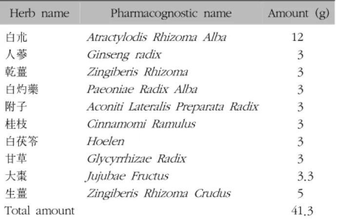
본 실험에서 사용한 처방의 약재들은 東醫寶鑑3)에 의거하여 옴니허브(영천, 한국)에서 구입, 정선 후 사용하 였으며 1錢은 4 g으로 계산하여 1첩 당 분량은 다음과 같 다(Table I).
勝濕湯 약재 82.6 g 분량에 증류수 826 ml를 가하여 열탕 추출기에서 2시간 추출하여 얻은 추출액을 감압 증 류장치로 농축하여, 이를 다시 동결 건조기를 이용하여
Herb name Pharmacognostic name Amount (g) 白朮 Atractylodis Rhizoma Alba 12
人蔘 Ginseng radix 3
乾薑 Zingiberis Rhizoma 3
白灼藥 Paeoniae Radix Alba 3 附子 Aconiti Lateralis Preparata Radix 3
桂枝 Cinnamomi Ramulus 3
白茯笭 Hoelen 3
甘草 Glycyrrhizae Radix 3
大棗 Jujubae Fructus 3.3
生薑 Zingiberis Rhizoma Crudus 5
Total amount 41.3
Table I. Composition of Seungseup-tang (SST)
완전 건조한 勝濕湯 분말(SST 24.36 g, 수율 31.7%)을 얻 었으며 勝濕湯 분말은 실험에 사용하기 직전까지 냉동보 관(−80oC)하였다.
4) 실험기기
본 실험에 사용된 기기는 열탕추출기(DWT-1800T, 대 웅, 한국), rotary vacuum evaporator (Buchi B-480, Switzerland), 동결건조기(FD5508, 일신, 한국), deep- freezer (Sanyo Co., Japan), vortex mixer, 냉장고속원심 분리기(Mega17R, 한일과학, 한국) 등을 사용하였다.
2. 방법
1) 약물투여와 군 분리
실험군은 골관절염을 유발시키지 않은 정상군(Normal), MIA로 골관절염 유발하고 증류수를 투여한 대조군 (Control), MIA로 골관절염 유발 후 indomethacin (J200, SK케미칼, 경기, 한국) 5 mg/kg 투여군(Indo), MIA로 골 관절염 유발 후 勝濕湯 200 mg/kg 투여군(STG)으로 총 4군으로 각각 6마리씩 군분리 하였다. 약물은 매일 증류 수에 녹여 매일 일정한 시각에 2주간 존대(Zonde)를 이 용하여 경구 투여하였다.
실험군은 각 군별로 난괴법에 의해 6마리씩 4군으로 나누었다.
① 정상군(Normal): 골관절염을 유발시키지 않은 정상군
② 대조군(Control): MIA로 골관절염 유발하고 증류수 를 투여한 대조군
③ Indomethacin 투여군(Indo): MIA로 골관절염 유발
후 indomethacin 5 mg/kg 투여군
④ 勝濕湯 투여군(STG): MIA로 골관절염 유발 후 勝濕 湯 200 mg/kg 투여군
2) MIA에 의한 골관절염 유발
정상군을 제외한 Rat에 마취제 Zoletile mixture (Vibrac, France)을 3.5 mg/kg를 복강에 투여하여 마취시킨다. 오 른쪽 무릎 주변을 깨끗이 제모한 후 골관절염 유발물질인 MIA (Sigma Chemical Co. Ltd, Cat. No. I2512)를 0.3 ml insulin 주사기(BD 31 G Ultra-Fine II, USA)를 사용하여 오른쪽 무릎 관절강 내에 50 μl (80 mg/kg/ml)씩 투여하 였다. MIA 희석 시에는 0.9% saline을 사용하였다. MIA 투 여 7 일 후에 관절염 유발 유무를 확인하여 관절염이 유발 된 동물만을 사용하였다.
3) 체중, 식이섭취량, 식이효율 측정
실험동물의 체중은 전자체중계(카스전자 저울, 한국)로 1회/2일으로 동일 시간 동일 조건에서 측정하였고, 실험 종료일 체중에서 실험 개시 전 체중을 빼서 체중증가량 (body weight gain)을 산출하였다. 식이섭취량은 2일 동 안 제공된 사료에서 2일간 섭취하고 남은 사료량을 제한 후 각 실험군의 하루 사료섭취량을 산출하였다. 한편, 각 Rat의 체중증가량을 동일 사육기간의 사료섭취량으로 나 누어 식이효율(FER; food efficiency ratio)을 구하였다.
(1) 체중(body weight)
① 체중변화: 1회/2일 오전 동일한 시간에 측정/기록
② 총 체중 증가량: Final body weight (g)−Initial body weight (g)
③ 1일 평균 체중 증가량
총 체중 증가량
×
(2) 식이섭취량(food intake)
① 1일 평균 식이섭취량
총 식이 섭취량
×
(3) 식이효율(food efficiency ratio, FER)
① 식이효율(%)
총 식이 섭취량 총 체중 증가량
×
284 304 324 344 364 384
0 day 7 days 15 days
Normal Control Indo
STG
Body weight change (g)
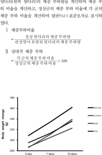
Fig. 1. Body weight changes.
All data are expressed means±S.E, n=6 rats per group.
There is no significant difference in the experimental groups.
Normal: Normal rats, Control: MIA-induced osteoarthritis rats, Indo: MIA-induced osteoarthritis rats administrated with Indomethacin 5 mg/kg body weight, STG: MIA-induced osteo- arthritis rats administrated with Seungseup-tang 200 mg/kg body weight.
4) 뒷다리 체중 부하 측정
시험물질은 오전 투여, 측정은 오후에 하는 것을 원칙 으로 하였다. 측정일은 시험물질을 경구투여 한 후 7일, 15일에 각각 시행하였다. Incapacitance tester (Ser No.
01/45/25, Linton instrument Co., UK)를 이용하여 오른 쪽, 왼쪽 발무게를 각각 측정하였다(Fig. 1). MIA에 의해 골관절염이 유발되면 Rat은 MIA를 투여하지 않은 발에 의지하여 tester의 holder 안에 서게 하였다. Rat의 배가 기기의 센서에 닿지 않은 상태에서 왼쪽, 오른쪽 각각의 발무게(g)를 측정하여 나타냈다. 실험결과는 관절염이 유 발된 뒷다리(오른쪽 뒷다리)의 체중 부하량에 대한 정상 뒷다리(왼쪽 뒷다리)의 체중 부하량을 계산하여 체중 부 하 비율을 계산하고, 정상군의 체중 부하 비율에 각 군의 체중 부하 비율을 계산하여 평균(%)±표준오차로 표시하 였다.
∫ 체중부하비율
=
관절염이 유발된 뒷다리의 체중 부하량
정상 뒷다리의 체중 부하량
∬ 상대적 체중 부하
=
정상군의 체중 부하 비율
각 군의 체중 부하 비율
×
5) 혈청 내 산화적 스트레스 바이오마커 측정
복대정맥에서 채혈한 혈액을 4,000 rpm 10분 원심 분 리하여 혈청을 얻었고 관절조직은 1 mM EDTA-50 mM sodium phosphate buffer (pH 7.4)를 이용하여 분쇄하였 다. Reactive Oxygen Species (ROS)를 측정하기 위하여 25 mM DCF-DA를 혼합한 후, 형광 광도계를 이용하여 0 분부터 매 10분씩 emission wavelength of 530 nm와 ex- citation wavelength of 486 nm를 이용하여 30분간 측정 한 산출 값을 계산하였다. 혈청에서 ROS와 Peroxy nitrite (ONOO−)를 측정하기 위하여, 각각 Kooy et al12)과 Mitsuru et al13)의 방법을 시행하였다.
6) 단백질 발현량 분석
관절조직의 세포질을 얻기 위해 100 mM Tris-HCl (pH 7.4), 5 mM Tris–HCl (pH 7.5), 2 mM MgCl2, 15 mM CaCl2, and 1.5 M sucrose, and then 0.1 M DTT and protease inhibitor cocktail을 첨가한 buffer A를 넣고 tis- sue grinder (Bio Spec Product, USA)로 분쇄한 후 10%
NP-40 용액을 첨가하였다. 아이스 위에서 20분간 정치시 킨 후 12,000 rpm으로 2분간 원심분리 하여 세포질을 포 함하고 있는 상층액을 분리하였다. 핵을 얻기 위해 10%
NP-40가 더해진 buffer A에 두 번 헹구고 100 μl의 buf- fer C (50 mM HEPES, 50 mM KCl, 0.3 mM NaCl, 0.1 mM EDTA, 1 mM DTT, 0.1 mM PMSF and 10% glycer- ol)를 첨가해 재부유 시킨 뒤 10분마다 vortex를 3번 하 였다. 4oC에서 12,000 rpm으로 10분간 원심 분리한 후 핵을 포함하고 있는 상층액을 얻어 −80oC에서 각각 냉 동 보관하였다. 관절조직의 세포질의 p-IκBα, NOX4, p22phox, COX-2, iNOS, TNF-α, IL-6, β-actin 및 핵의 NF-κBp65, Histone 단백질의 발현을 측정하기 위해 10 μg의 단백질을 8∼15% SDS-polyacrylamide gel을 이용 하여 전기연동 후, acrylamide gel을 nitrocellulose mem- brane으로 이동시켰다. 준비된 membrane에 각각의 1차 antibody를 처리하여 4oC에서 overnight 시킨 다음 PBS-T로 6분마다 5회 세척하고, 각각 처리된 1차 항체에 사용되는 2차 항체(PBS-T로 1:3000로 희석해서 사용)를 사용하여 상온에서 1시간 반응시킨 후, PBS-T로 6분마다 5회 세척하였다. 그리고 enhanced chemiluminescence (ECL) 용액을 GE Healthcare (Arlington Heights, IL,
Group Initial body weight (g)
Final body weight (g)
Body weight change (g)
Normal 312.7±8.3 365.7±9.4 53.0±4.1
Control 290.2±5.3 343.0±7.5 52.8±7.8
Indo 291.1±5.3 326.6±10.3 35.4±5.5
STG 299.3±7.6 339.6±11.2 40.4±3.8
All data are expressed means±S.E, n=6 rats per group.
There is no significant difference in the experimental groups.
Normal: Normal rats, Control: MIA-induced osteoarthritis rats, Indo: MIA-induced osteoarthritis rats administrated with Indomethacin 5 mg/kg body weight, STG: MIA-induced osteo- arthritis rats administrated with Seungseup-tang 200 mg/kg body weight.
Table II. Initial and Final Body Weight, Body Weight Change
Group Food intakes (g/day)
Body weight change (g/day)
Food efficiency ratio (FER, %)
Normal 17.37 3.53±0.27 20.35±1.57
Control 16.79 3.52±0.52 20.96±3.10
Indo 16.23 2.36±0.36 14.55±2.25
STG 16.41 2.69±0.25 16.40±1.53
All data are expressed means±S.E, n=6 rats per group.
There is no significant difference in the experimental groups.
Normal: Normal rats, Control: MIA-induced osteoarthritis rats, Indo: MIA-induced osteoarthritis rats administrated with Indomethacin 5 mg/kg body weight, STG: MIA-induced osteo- arthritis rats administrated with Seungseup-tang 200 mg/kg body weight.
Table III. Food Efficiency, Food Intakes and Body Weight Change
USA)에 노출시킨 후, Sensi-Q2000 Chemidoc (Lugen Sci Co., Ltd., 경기도, 한국)에 감광시켜 단백질 발현을 확인 한 후, 해당 band를 ATTO Densitograph Software (ATTO Corporation, Tokyo, Japan)프로그램을 사용하여 정량하였다.
7) 병리조직 분석
실험 종료 후 슬관절 부위를 절단하여 10% EDTA가 포함된 10% 포르말린(formalin) 용액에 넣어 관절을 탈회 (decalcification)시킨다. Radiographic technique을 이용 하여 탈회 유무를 확인한 후 parafin wax에 관절을 넣고 고정한 다음 coronal section을 실시하였다. 탈회 과정을 거쳐 파라핀으로 고정된 조직을 7 μm의 크기로 자른 뒤, H&E 및 Safranin-O 염색을 실시하여 조직의 상태를 관찰하였다. 염증 반응 발생 유무나 활막 세포의 증식, 염증 세포의 조직 침윤 여부는 H&E 염색 결과에서 확인 할 수 있으며, proteoglycan층을 염색하는 Safranin-O 염 색 결과에서는 연골 조직의 손상 여부를 확인할 수 있다.
8) 통계처리
모든 수치는 mean±standard error (Mean±S.E)로 표 시하였으며, SPSS program for windows version 22 (SPSS Inc., Chicago, USA)를 사용하여 one-way analysis of variance (ANOVA) 또는 Student T test로 각 자료의 통계적 유의성을 검증하였고, 대조군과 투여군 사이에 p-value<0.05 일 때 유의성이 있는 것으로 판정 하였다.
결과»»»
1. 체중, 식이섭취량 및 식이효율
적응기간을 제외한 실험기간 동안에 측정된 체중변화 는 정상군 53.0±4.1, 대조군 52.8±7.8, Indomethacin투 여군 35.4±5.5, 勝濕湯 투여군 40.4±3.8로 나타났다.
MIA 유발 후 15일간의 체중 측정에서 모든 군에서 체중 이 증가하였으며 각 군 간의 유의성은 없었다(Table II, Fig. 1).
식이효율은 정상군 20.35±1.57, 대조군 20.96±3.10, Indomethacin투여군 14.55±2.25, 勝濕湯 투여군 16.40±
1.53으로 나타났고 각 군 간의 유의성은 없었다(Table III, Fig. 2).
2. 뒷다리 체중 부하의 변화
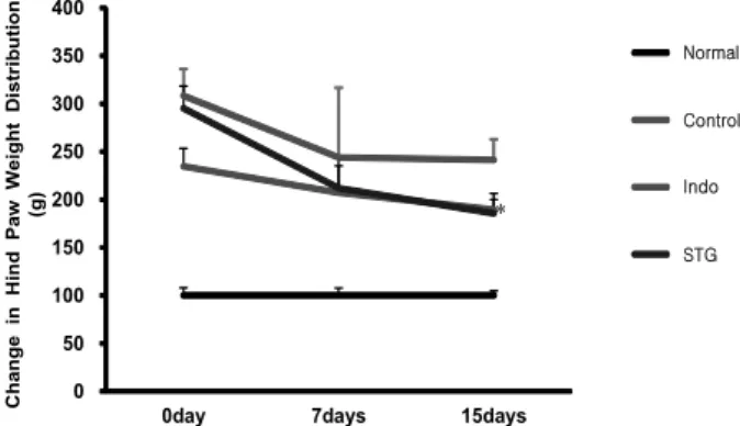
정상군의 뒷다리 체중 부하 비율을 100으로 하였을 때, 각 군의 상대적 뒷다리 체중 부하의 변화를 측정한 결과, MIA 투여 7일 후에는 대조군 243.92±72.75, Indomethacin 투여군 207.17±28.06, 勝濕湯 투여군 211.94±32.20으로 나타나 대조군에 비해 유의성은 없었다. 또한, 15일에는 대 조군 241.34±21.47, Indomethacin 투여군 189.58±18.87, 勝濕湯 투여군 185.65±14.26으로 나타나 뒷다리 체중 부 하가 Indomethacin 투여군과 勝濕湯 투여군 에서 개선되 었다. 특히, 勝濕湯 투여군에서 뒷다리 체중 부하가 대조군 에 비해 유의성 있게 감소(p<0.05)하였다(Fig. 3).
0 5 10 15 20 25
Normal Control Indo STG
Food efficiency ratio (%)
MIA-induced osteoathritis rats
Fig. 2. Food efficiency ratio (FER) in MIA-induced osteoarthritis rats.
All data are expressed means±S.E, n=6 rats per group.
There is no significant difference in the experimental groups.
Normal: Normal rats, Control: MIA-induced osteoarthritis rats, Indo: MIA-induced osteoarthritis rats administrated with Indomethacin 5 mg/kg body weight, STG: MIA-induced osteo- arthritis rats administrated with Seungseup-tang 200 mg/kg body weight.
0 50 100 150 200 250 300 350 400
0day 7days 15days
Normal Control Indo5 SST200
Change in Hind Paw Weight Distribution (g)
Control Normal
Indo
STG
Fig. 3. The changes in relative hind paw weight distribution in MIA-induced osteoarthritis rats.
All data are expressed means±S.E, n=6 rats per group.
Significance: *p<0.05 vs. MIA-induced osteoarthritis rats by student t test.
Normal: Normal rats, Control: MIA-induced osteoarthritis rats, Indo: MIA-induced osteoarthritis rats administrated with Indomethacin 5 mg/kg body weight, STG: MIA-induced osteo- arthritis rats administrated with Seungseup-tang 200 mg/kg body weight.
0 200 400 600 800 1000 1200 1400 1600
Indo STG ROS (fluorescence/min/mL)
Normal Control
MIA-induced osteoathritis rats
Fig. 4. The effects of SST on the ROS level in MIA-induced osteoarthritis rats.
All data are expressed means±S.E, n=6 rats per group.
There is no significant difference in the experimental groups.
Normal: Normal rats, Control: MIA-induced osteoarthritis rats, Indo: MIA-induced osteoarthritis rats administrated with Indomethacin 5 mg/kg body weight, STG: MIA-induced osteo- arthritis rats administrated with Seungseup-tang 200 mg/kg body weight.
3. 산화적 스트레스 관련 인자 분석
1) ROS level
혈청 내 ROS 생성량은 정상군 810±129, 대조군에서 1394±73, Indomethacin 투여군 1198±61, 勝濕湯 투여 군 1090±140으로 나타나 勝濕湯 투여군에서 대조군에
비하여 감소하였지만 유의성은 없었다(Fig. 4).
2) ONOO− level
혈청 내 ONOO− 생성량은 정상군 575.8±6.2, 대조군 600±2.7, Indomethacin 투여군 564.7±4.7, 勝濕湯 투여 군 546.8±4.9으로 나타나 勝濕湯 투여군에서 대조군에 비하여 유의성 있게 감소(p<0.001)하였다(Fig. 5).
4. 관절조직 내 산화적 스트레스 관련 단백질 분석
1) NOX4 분석
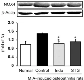
관절 조직에서 western blot을 실시 후 측정한 결과 NOX4의 발현은 정상군 1.00±0.13, 대조군 1.51±0.03, Indomethacin 투여군 1.03±0.09, 勝濕湯 투여군 1.12±0.12으로 나타나 勝濕湯 투여군에서 대조군에 비하 여 유의성 있게 감소(p<0.05)하였다(Fig. 6).
2) p22phox 분석
관절 조직에서 western blot을 실시 후 측정한 결과 p22phox의 발현은 정상군 1.00±0.36, 대조군 1.28±0.31, Indomethacin 투여군, 1.12±0.15, 勝濕湯 투여군 0.75±
0.11으로 勝濕湯 투여군에서 대조군에 비하여 감소하였지 만 유의성은 없었다(Fig. 7).
(fold of N)
-Actin NOX4
MIA-induced osteoathritis rats
Normal Control Indo STG
0 0.4 0.8 1.2 1.6 2.0
Fig. 6. The effects of SST on NOX4 in MIA-induced osteo- arthritis rats.
All data are expressed means±S.E, n=6 rats per group.
Significance: *p<0.05, **p<0.01 vs. MIA-induced osteoarthritis rats by student t test.
Normal: Normal rats, Control: MIA-induced osteoarthritis rats, Indo: MIA-induced osteoarthritis rats administrated with Indomethacin 5 mg/kg body weight, STG: MIA-induced osteo- arthritis rats administrated with Seungseup-tang 200 mg/kg body weight.
-Actin
(f old of N)
-Actin
(f old of N)
p22
phoxMIA-induced osteoathritis rats
Normal Control Indo STG
0 0.3 0.6 0.9 1.2 1.5 1.8
Fig. 7. The effects of SST on p22
phoxin MIA-induced osteo- arthritis rats.
All data are expressed means±S.E, n=6 rats per group.
There is no significant difference in the experimental groups.
Normal: Normal rats, Control: MIA-induced osteoarthritis rats, Indo: MIA-induced osteoarthritis rats administrated with Indomethacin 5 mg/kg body weight, STG: MIA-induced osteo- arthritis rats administrated with Seungseup-tang 200 mg/kg body weight.
500 520 540 560 580 600 620
Indo STG
ONOO- (fluorescence/mL)
Normal Control
MIA-induced osteoathritis rats
Fig. 5. The effects of SST on the ONOO
−level in MIA-in- duced osteoarthritis rats.
All data are expressed means±S.E, n=6 rats per group.
Significance: ***p<0.001 vs. MIA-induced osteoarthritis rats by student t test.
Normal: Normal rats, Control: MIA-induced osteoarthritis rats, Indo: MIA-induced osteoarthritis rats administrated with Indomethacin 5 mg/kg body weight, STG: MIA-induced osteo- arthritis rats administrated with Seungseup-tang 200 mg/kg body weight.
5. 관절조직 내 염증성 단백질 분석
1) p-IκBα 분석
관절 조직에서 western blot을 실시 후 측정한 결과 p-IκBα의 발현은 정상군 1.00±0.03, 대조군 1.79±0.06, Indomethacin 투여군 1.54±0.07, 勝濕湯 투여군 1.37±
0.06으로 나타나 勝濕湯 투여군에서 대조군에 비하여 유 의성 있게 감소(p<0.01)하였다(Fig. 8).
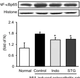
2) NF-κBp65 분석
관절 조직에서 western blot을 실시 후 측정한 결과 NF- κBp65의 발현은 정상군 1.00±0.28, 대조군 1.77±0.10, Indomethacin 투여군 1.45±0.12, 勝濕湯 투여군 1.48±
0.09으로 나타나 勝濕湯 투여군에서 대조군에 비하여 유의 성 있게 감소(p<0.05)하였다(Fig. 9).
6. 염증성 매개인자에 미치는 영향
1) COX-2 분석
관절 조직에서 western blot을 실시 후 측정한 결과 COX-2의 발현은 정상군 1.00±0.11, 대조군 1.87±0.17,
(fold of N)
-Actin p-IB
MIA-induced osteoathritis rats Normal Control Indo STG 0
0.5 1.0 1.5 2.0 2.5
Fig. 8. The effects of SST on the p-IκBα protein expression in MIA-induced osteoarthritis rats.
All data are expressed means±S.E, n=6 rats per group.
Significance: *p<0.05, **p<0.01 vs. MIA-induced osteoarthritis rats by student t test.
Normal: Normal rats, Control: MIA-induced osteoarthritis rats, Indo: MIA-induced osteoarthritis rats administrated with Indomethacin 5 mg/kg body weight, STG: MIA-induced osteo- arthritis rats administrated with Seungseup-tang 200 mg/kg body weight.
(fold of N)
Histone NF-Bp65
0 0.6 1.2 1.8 2.4
MIA-induced osteoathritis rats Normal Control Indo STG
Fig. 9. The effects of SST on the NF-κBp65 protein ex- pression in MIA-induced osteoarthritis rats.
All data are expressed means±S.E, n=6 rats per group.
Significance: *p<0.05 vs. MIA-induced osteoarthritis rats by student t test.
Normal: Normal rats, Control: MIA-induced osteoarthritis rats, Indo: MIA-induced osteoarthritis rats administrated with Indomethacin 5 mg/kg body weight, STG: MIA-induced osteo- arthritis rats administrated with Seungseup-tang 200 mg/kg body weight.
(fold of N)
-Actin COX-2
0 0.5 1.0 1.5 2.0 2.5
MIA-induced osteoathritis rats
Normal Control Indo STG
Fig. 10. The effects of SST on the COX-2 protein expression in MIA-induced osteoarthritis rats.
All data are expressed means±S.E, n=6 rats per group.
Significance: *p<0.05, **p<0.01 vs. MIA-induced osteoarthritis rats by student t test.
Normal: Normal rats, Control: MIA-induced osteoarthritis rats, Indo: MIA-induced osteoarthritis rats administrated with Indomethacin 5 mg/kg body weight, STG: MIA-induced osteo- arthritis rats administrated with Seungseup-tang 200 mg/kg body weight.
Indomethacin 투여군 1.18±0.13, 勝濕湯 투여군 1.26±
0.14으로 나타나 勝濕湯 투여군에서 대조군에 비하여 유 의성 있게 감소(p<0.05)하였다(Fig. 10).
2) iNOS 분석
관절 조직에서 western blot을 실시 후 측정한 결과 iNOS의 발현은 정상군 1.00±0.15, 대조군 1.86±0.14, Indomethacin 투여군 1.36±0.11, 勝濕湯 투여군 1.28±
0.18으로 나타나 勝濕湯 투여군에서 대조군에 비하여 유 의성 있게 감소(p<0.05)하였다(Fig. 11).
7. 염증성 cytokine 발현에 미치는 영향
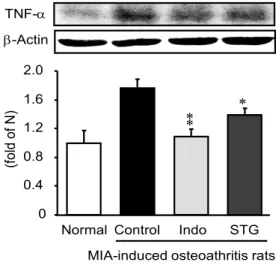
1) TNF-α 분석
관절 조직에서 western blot을 실시 후 측정한 결과 TNF-α의 발현은 정상군 1.00±0.17, 대조군 1.76±0.13, Indomethacin 투여군 1.08±0.11, 勝濕湯 투여군 1.39±
0.09으로 나타나 勝濕湯 투여군에서 대조군에 비하여 유 의성 있게 감소(p<0.05)하였다(Fig. 12).
-Actin
(f old of N )
iNOS
-Actin
(f old of N )
0 0.5 1.0 1.5 2.0 2.5
MIA-induced osteoathritis rats Normal Control Indo STG 0
0.5 1.0 1.5 2.0 2.5
Fig. 11. The effects of SST on the iNOS protein expression in MIA-induced osteoarthritis rats.
All data are expressed means±S.E, n=6 rats per group.
Significance: *p<0.05 vs. MIA-induced osteoarthritis rats by student t test.
Normal: Normal rats, Control: MIA-induced osteoarthritis rats, Indo: MIA-induced osteoarthritis rats administrated with Indomethacin 5 mg/kg body weight, STG: MIA-induced osteo- arthritis rats administrated with Seungseup-tang 200 mg/kg body weight.
MIA-induced osteoathritis rats
(f old of N)
-Actin TNF-
0 0.4 0.8 1.2 1.6 2.0
Normal Control Indo STG
Fig. 12. The effects of SST on the TNF-α protein expression in MIA-induced osteoarthritis rats.
All data are expressed means±S.E, n=6 rats per group.
Significance: *p<0.05, **p<0.01 vs. MIA-induced osteoarthritis rats by student t test.
Normal: Normal rats, Control: MIA-induced osteoarthritis rats, Indo: MIA-induced osteoarthritis rats administrated with Indomethacin 5 mg/kg body weight, STG: MIA-induced osteo- arthritis rats administrated with Seungseup-tang 200 mg/kg body weight.
-Actin IL-6
(f ol d of N )
0 0.4 0.8 1.2 1.6 2.0
MIA-induced osteoathritis rats
Normal Control Indo STG
Fig. 13. The effects of SST on the IL-6 protein expression in MIA-induced osteoarthritis rats.
All data are expressed means±S.E, n=6 rats per group.
Significance: *p<0.05 vs. MIA-induced osteoarthritis rats by student t test.
Normal: Normal rats, Control: MIA-induced osteoarthritis rats, Indo: MIA-induced osteoarthritis rats administrated with Indomethacin 5 mg/kg body weight, STG: MIA-induced osteo- arthritis rats administrated with Seungseup-tang 200 mg/kg body weight.
2) IL-6 분석
관절 조직에서 western blot을 실시 후 측정한 결과 IL-6의 발현은 정상군 1.00±0.11, 대조군 1.51±0.15, Indomethacin 투여군 1.02±0.13, 勝濕湯 투여군 1.02±
0.15으로 나타나 勝濕湯 투여군에서 대조군에 비하여 유 의성 있게 감소(p<0.05)하였다(Fig. 13).
8. 조직병리학적 변화에 미치는 영향
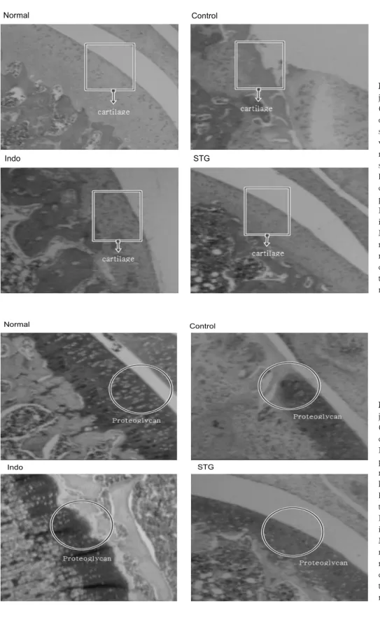
1) H&E stain
정상군의 활막 조직, 연골 등은 정상이었고, 대조군에 서는 골관절염이 유발되어 활막 조직과 연골의 변성 및 변형이 현저하게 나타났다. Indomethacin 투여군와 勝濕 湯 투여군의 활막 조직 및 연골 등은 대조군과 비교하여 정상에 가까워 조직 손상이 효과적으로 억제되는 것으로 나타났다(Fig. 14).
2) Safranin-O stain
정상군 관절조직은 proteoglycan은 정상적으로 관찰되 었으나 대조군에서는 골관절염이 유발되어 관절조직이
Control Normal
STG Indo
Fig. 15. The effects of SST on joint in pathological observation (Safranin-O staining) in MIA-in- duced osteoarthritis rats. In the Normal, there are much of proteoglycan. In the Control, most of proteoglycan was bro- ken down. The proteoglycans of Indo and STG, were well main- tained compared with Control.
Normal: Normal rats, Control: MIA- induced osteoarthritis rats, Indo:
MIA-induced osteoarthritis rats ad- ministrated with Indomethacin 5 mg/kg body weight, STG: MIA-in- duced osteoarthritis rats adminis- trated with Seungseup-tang 200 mg/kg body weight.
STG Control Normal
Indo
Fig. 14. The effects of SST on joint in pathological observation (H&E staining) in MIA-induced osteoarthritis rats. In the Normal, synovial tissue and cartilage were well observed. In the Control, most of aricular cartilage and synovial tissue were destructed.
In the Indo and STG, articular cartilage and synovial tissue were preserved, compared with Control.
Normal: Normal rats, Control: MIA- induced osteoarthritis rats, Indo:
MIA-induced osteoarthritis rats ad- ministrated with Indomethacin 5 mg/kg body weight, STG: MIA-in- duced osteoarthritis rats adminis- trated with Seungseup-tang 200 mg/kg body weight.
변형되고, proteoglycan의 대부분이 파괴되었다. 반면, Indomethacin 투여군과 勝濕湯 투여군에서는 대조군에 비해 관절조직의 변형이 억제되고 proteoglycan의 소실도
억제되는 것으로 나타났다(Fig. 15).
고찰»»»
골관절염은 퇴행성 관절염이라고도 불리며, 국소적인 관절 부위의 연골이 점차 소실되고 그와 관젼된 2차적인 변화를 일으키는 질환으로2) 55세 이상의 성인에서 가장 흔히 발병하며, 그중 슬관절은 골관절염의 호발 부위라고 알려져 있다14). 골관절염을 일으키는 정확한 원인은 아직 알려지지 않았으나 골관절염은 노화로 인한 관절의 마모 가 원인이 되고 그 이후에는 연골의 변성을 일으키는 다 양한 생물학적 인자와 기계적인 요인으로 인해 발생되는 것으로 보인다. 골관절염의 치료의 목표는 연골의 변성을 유발하는 인자들을 최대한으로 억제함으로써 통증을 억 제하고 연골의 재생과 기능을 회복시키는 것이다15).
관절염 초기에 지속적인 외부자극이 주어지게 되면 역 학적, 생화학적 변화로 불균형이 오면서 연골세포와 조직 에 영향을 주게 된다16). 연골의 항상성 유지에 중요한 역 할을 하는 연골세포의 변화와 사멸로 인한 collagen, pro- teoglycan, aggrecan 등 세포외기질의 생산이 감소하는 동시에 분해가 촉진되면서, 연골의 기본구조가 파괴되어 관절부하를 견디지 못하게 된다17). 이러한 연골 파괴 과 정에 TNF-α, IL-6 등과 같은 cytokines이 작용하여 중요 한 역할을 하는 것으로 알려져 있다18).
한의학에서 골관절염은 痺證의 범주에 해당한다. 痺症 은 風寒濕熱의 邪氣가 인체가 허약한 틈을 타고 經絡으로 침입하거나 關節에 응체됨으로써 血氣運行을 저해하여 肌 肉, 筋骨, 關節에 痲木, 重着, 疼痛, 腫脹, 屈伸不利, 심하면 관절의 강직성 변형을 초래한다, 痺證의 치료원칙은 宣通 하는 것으로 寒者는 溫之하고, 熱者는 淸之하며, 濕痰瘀 등 의 有形之邪가 있으면 去之하고 虛者는 補之하는 것이다1). 한의학에서 골관절염 연구는 약물, 침구, 약침, 물리적 요 법 등 다방면으로 이루어지고 있으며, 연골세포의 재생 및 연골 보호, 통증 및 관절 운동 개선 등의 효과가 있는 것으 로 보고되고 있다19).
본 연구에 사용된 勝濕湯은 白朮, 人參, 乾薑, 白芍藥, 附子, 桂枝, 白茯苓, 甘草로 구성된 方으로 四君子湯에 乾 薑, 白芍藥, 附子, 桂枝를 가한 方劑이다. 方 중의 四君子 湯은 補氣補脾하고, 白朮, 附子, 乾薑, 桂枝의 배합으로 寒濕을 제거하며, 白朮, 茯苓에 白芍藥의 배합으로 止瀉 하여 身重脚弱, 大便泄의 濕症, 中濕이 스스로 치유되도 록 한다20). 이중 白朮은 補氣藥으로 健脾益氣하고 燥濕利
水하는 효능이 있으며21), 白朮의 에탄올추출물은 콜라겐 유도 관절염 마우스의 혈청 내 항-콜라겐 항체와 TNF-α 의 증가를 유의적으로 감소시켜 콜라겐 유도 관절염 마우 스에서 관절염 개선 효과가 있는 것으로 보고되었다4). 人 蔘은 補氣藥으로 大補元氣 하고 固脫生津 하는 효능이 있 으며21), 관절염 유도 생쥐 모델에서 TNF-α, IL-1β 등의 cytokine 감소와 항산화 효과가 있는 것으로 보고되었다5-6). 乾薑은 溫裏藥이며 溫中逐寒, 回陽通脈 하는 효능이 있으 며21), 실험적으로 쥐의 면역세포에 대한 항염증 효과가 보고되었다7). 白芍藥은 補血藥으로 養血柔肝, 緩中止痛의 효능이 있으며, 甘草는 補氣藥으로 和中緩急, 解毒의 효 능이 있다21). 白芍藥과 甘草의 조합으로 이루어진 芍藥甘 草湯이 생체 내에서 전염증성 cytokine들의 생산에 영향 을 주어 염증억제효과가 보고되었다8). 附子는 溫裏藥으로 回陽補火, 散寒除濕의 효능이 있으며21), 附子 藥鍼으로 IgG, IgM, IL-6, IL-1β, TNF-α를 억제하여 관점염에 효 과가 있다고 보고되었다9). 桂枝는 發散風寒藥으로 發汗解 肌하며 溫經通脈하며21), collagen유발된 골관절염에서 소 염효과가 보고되었다10). 白茯苓은 利水退腫藥으로 利水 滲濕하며 健脾寧心하며21), 항염증 효과 외에도 면역 반응 조절, 혈관 생성 억제 효능이 보고되었다11).
이처럼 勝濕湯을 구성하고 있는 각각의 약재는 항산화 효과, 항염증효과, 면역반응 조절 등의 효과를 가지고 있 음이 실험적으로 확인되어 있으므로, 각 약재의 복합제인 勝濕湯 역시 골관절염에 효과가 있을 것으로 예상되었다.
이에 본 연구에서는 MIA를 주사하여 골관절염을 유발한 Rat 모델에서 勝濕湯의 경구투여 하여 체중변화, 식이효 율, 뒷다리 체중부하, 산화적 스트레스 관련인자, 염증성 단백질, 염증성 매개인자, 염증성 cytokine, 병리조직학적 변화에 대해 알아보고자 하였다.
MIA로 골관절염을 유발한 동물실험은 Kalbhen and Blum에 의해서 최초로 확립되었다22). MIA는 관절연골의 손상, 기능장애, 통증 등의 양상을 사람의 골관절염과 유 사하게 발현시킬 수 있고, 주입농도에 따라 다양한 관절 퇴행정도를 가진 동물 모델을 만들 수 있을 뿐 아니라 질 환의 진행정도와 통증수준을 정확하게 평가할 수 있다는 장점이 있다23-24).
약물의 안정성을 알아보기 위하여 체중변화와 식이효 율 변화를 측정하였다. 각 군의 체중 변화는 대조군과 비 교하여 모든 군에서 체중이 증가하였고(Fig. 1), 식이효율
에서도 유의성 있는 차이는 관찰 되지 않았다(Fig. 2). 이 러한 점으로 보아 勝濕湯은 실험에 사용된 Rat의 식이에 부작용을 줄만한 영향이 없었다고 할 수 있다.
본 실험에서는 골관절염이 유발된 Rat의 통증 변화를 확인하기 위하여 뒷다리 체중부하검사를 실시하였다. 체 중부하검사는 염증이 나타난 관절을 보호하기 위한 자세 의 변형, 움츠림, 핥기 등의 자발통, 행동 관찰보다 더 정 량적이고 객관적으로 통증의 정도를 측정할 수 있다는 장 점이 있다25). MIA투여 7일 후에는 대조군에 비하여 유의 성 있는 감소는 없었으나 15일 이후에는 勝濕湯 투여군과 Indomethacin 투여군에서 감소가 나타났으며, 특히 勝濕 湯 투여군에서는 유의성 있는 감소가 나타났다(Fig. 3).
이러한 결과로 보아 勝濕湯은 골관절염이 유발된 Rat에서 통증 감소 효과가 있다고 사료된다.
ROS에는 superoxide radical, hydrogen peroxide, hy- droxyl radical, hypochlorous acid 등이 속하며, 연골조직 을 파괴하는 물질 중 하나로 추측되고 있다. superoxide radical와 hydrogen peroxide는 조직을 직접 손상시키지 는 않지만 여러 기전에 의해서 반응성 높은 hydroxyl radi- cal로 전환되어 조직에 손상을 주는 것으로 알려져 있다26). ONOO−는 단백질, 아미노산, DNA등과 반응하여 세포 및 조직손상을 야기하고 염증반응 및 동맥경화에 관여할 뿐 만 아니라, NO signaling을 저해하여 여러 만성 질환에 발 생에 관여하는 것으로 알려져 있다27). 본 실험에서 측정 한 혈청 ROS는 勝濕湯 투여군과 Indomethacin 투여군에 서 대조군에 비하여 감소하였으나 유의성 있는 변화를 나 타내지는 못하였다(Fig. 4). 혈청 ONOO−측정 결과는 勝 濕湯 투여군과 Indomethacin 투여군이 모두 대조군에 비 하여 유의성 있는 감소를 나타내었다(Fig. 5). 이러한 결 과는 勝濕湯이 혈액 내 산화적 스트레스 인자중 ONOO− 를 조절하여 골관절염에 효과가 있다고 할 수 있겠다.
O2−
을 생산하는 효소인 NADPH oxidase는 NOX4, p22phox, p47phox, p67phox 등으로 구성 되어있고, 이중 p22phox는 혈관에서의 주된 산화 스트레스인 과산화음이 온을 생성시킨다28). NOX4는 세포막에 위치한 p22phox의 조절에 의해 H2O2를 과잉 생성하며, 이 과정에서 미토콘 드리아의 전자전달계에 의해 과잉 생산된 O2−
에 전자를 운반하여 H2O2로 전환시켜 염증반응에 관여한다고 알려 져 있다29). 본 실험에서 관절 조직에서 western blot을 실 시 후 측정한 결과 NOX4의 발현은 勝濕湯 투여군에서
대조군에 비하여 유의성 있게 감소하였으며(Fig. 6), p22phox의 발현은 勝濕湯 투여군에서 대조군에 감소하였 으나 유의성은 없었다(Fig. 7). 이러한 점으로 미루어 보 아 勝濕湯은 관절 조직내 산화적 스트레스 인자중 NOX4 를 억제하여 항산화 효과를 나타내어 골관절염에 효과가 있을 것으로 사료된다.
NF-κB는 일반적인 상태에서는 inhibitory kappa B (IκB)와 결합한 상태로 세포질 내에서 불활성 상태로 존 재하지만, TNF-α 등의 자극을 통해 비활성 상태의 in- hibitory kappa B kinase (IKK)가 활성화되고 이로인해 인산화된 IκBα가 분해되면, p65로 대표되는 단백질들 이 세포핵 내로 이동하여 전사인자로서의 역할을 수행하 게 된다30-31). 이때 핵에서 전사가 이루어지면서 염증성 매개인자인 COX-2와 iNOS, 염증성 cytokine인 TNF-α와 IL-6을 분비하게 된다. 본 실험에서 p-IκBα와 NF-κB 의 발현은 勝濕湯 투여군에서 대조군에 비하여 유의성 있 게 감소하였다(Fig. 8, 9).
Cyclooxygenase (COX)는 COX-1과 COX-2로 나뉘어지 며, 아라키돈산(arachidonic acid)을 프로스타글란딘 (prostaglandin)으로 전환하는 효소이다. COX-1은 보호 작용을 하는 효소로서 혈소판에서 발견되며 다양한 생리 적 반응을 매개한다. COX-2는 주로 염증과 관계되는 대 식세포, 단핵구세포, 활액막세포에서 높은 비율로 발현되 고, COX-2를 선택적으로 억제할 수 있다면 이론적으로 위장관 점막의 세포보호 및 혈소판 기능을 저해하지 않으 면서 항염증작용을 한다고 본다32). iNOS는 NO를 과량 생성해 골관절염 연골세포에서 염증성 cytokine의 분비와 이화작용을 촉진하는 효소로, iNOS에 의해 과량 생성된 NO는 proteoglycan과 collagen의 합성을 모두 억제하며 기질분해효소로 작용하여 연골세포의 사멸과 염증 반응 을 조장한다33-34). 또한 iNOS에 의해 NO가 다량 생성되면 관절 조직에 산화적 스트레스를 주는 ROS도 같이 생성된 다35). 본 실험에서 COX-2와 iNOS의 발현은 勝濕湯 투여 군에서 대조군에 비하여 유의성 있게 감소하였다(Fig. 10, 11).
TNF-α와 IL-6은 염증성 cytokine의 일종으로 TNF-α 는 IL-1, IL-6, IL-8, IL-10, Matrix Metalloproteinases (MMPs) 등의 하위염증 매개물질 생성을 유발하여 염증을 증폭시키고, 관절연골기질로부터의 proteoglycan 소실율 을 증가시킴과 동시에 재합성을 억제하여 관절연골을 손
상시키고 골관절염의 진행을 유도한다36). IL-6은 만성 염 증 면역반응의 영구화와 급성 염증의 시작에 있어 중요한 역할을 하며 병인과 급성 스트레스에서 숙주 방어에 중요 한 역할을 한다37-38). 본 실험에서 TNF-α와 IL-6의 발현 은 勝濕湯 투여군에서 대조군에 비하여 유의성 있게 감소 하였다(Fig. 12, 13). 이러한 점으로 미루어 보아 勝濕湯 은 관절조직에서 염증매개 인자들의 생성을 억제함과 동 시에 염증성 cytokine 분비를 억제하여 항염증 효과를 낼 수 있을 것으로 사료된다.
골관절염의 병리조직학적 관찰을 위하여 H&E와 Safranin-O 염색을 실시하였다. H&E 염색은 호염기성인 Hematoxylin과 호산성인 Eosin이 핵, proteoglycan, 세포 질 등을 염색하는 방법이며, Safranin-O 염색은 양이온성 염색소로서 keratan sulfate나 chondroitin sulfate 등의 음 이온과 결합해 연골에 분포하는 proteoglycan의 양에 비례 하여 적색 또는 오렌지색으로 염색하는 방법으로 연골의 proteoglycan 농도 변화를 개략적으로 추정할 수 있다39).
H&E 염색 결과에서 MIA로 관절염을 유발한 대조군은 활막 조직과 연골의 변성 및 변형이 현저하게 나타났으나, 勝濕湯 투여군과 Indomethacin 투여군에서는 활막 조직 및 연골 등은 대조군과 비교하여 정상에 가까워 조직 손 상이 효과적으로 억제되는 것으로 나타났다(Fig. 14).
Safranin-O 염색 결과에서 대조군은 골관절염이 유발되 어 관절조직이 변형되고, proteoglycan의 대부분이 파괴 되었으나, 勝濕湯 투여군과 Indomethacin 투여군에서는 대조군에 비해 관절조직의 변형이 억제되고 proteoglycan 의 소실도 억제되는 것으로 나타났다(Fig. 15).
본 실험에서 사용한 indomethacin은 비스테로이드성 소 염진통제 (Nonsteroidal anti-infalmmatory drugs, nSAIDs) 의 일종으로 prostaglandin의 생성억제 기전이 확실히 밝 혀져 있는 약물로서 비교적 널리 사용되고 있다. 그러나 indomethacin은 비선택적 소염진통제 중 하나로 prosta- glandin을 생성하는 COX효소를 억제하는데 생리작용을 담당하고 있는 COX-1 마저 억제하여 그로 인한 위장관 합 병증이 흔히 발생한다41). 이외에 청각부작용, 피부발진 등 의 부작용이 나타날 수 있어42) 신중을 기하여 투여하는 약 물중의 하나이다.
勝濕湯과 indomethacin의 효능을 실험결과를 통해 비 교해 보았을 때 뒷다리 체중부하검사에서 두 약물은 모두 통증 감소효과를 유의하게 나타내었으며, 혈청 내 산화적
스트레스 관련인자인 ONOO−와 관절 조직 내 산화적 스 트레스 인자인 NOX4 또한 유의하게 감소시켰다. 관절 조직 내 염증성 단백질인 p-IκBα, NF-κBp65 염증성 매개인자인 NOX4, iNOS 염증성 cytokine인 TNF-α의 수치를 유의성 있게 감소시켰다. 또한 H&E와 Safranin-O 염색에서도 두 약물은 모두 조직 손상을 억제하여 관절 보호 효과가 있음을 알 수 있었다.
이상의 결과들을 종합해 볼 때, 勝濕湯은 MIA로 유발 된 Rat의 골관절염에 작용하여 진통작용과 항염증 작용을 나타내었으며 염증 세포로부터 연골과 뼈를 보호하고 활 막 주변의 proteoglycan을 보존 하는 효과가 있으므로 추 가적인 연구를 통해 향후 골관절염 치료 약물로서 임상에 서 활용 될 수 있을 것이라 사료된다.
결론»»»
勝濕湯이 골관절염에 미치는 영향을 확인하기 위하여 MIA로 유발된 Rat에 본 연구에서는 체중변화, 식이효율, 뒷다리 체중부하, 산화적 스트레스 관련인자, 염증성 단 백질, 염증성 매개인자, 염증성 cytokine, 병리조직학적 변화를 관찰하여 다음과 같은 결과를 얻었다.
1. 뒷다리 체중부하 검사에서 통증 감소가 나타났다.
특히 MIA투여 15일 후에는 유의한 감소가 나타났다.
2. 혈청 내 ONOO− 생성량은 유의하게 감소하였다.
3. 관절 조직에서 NOX4의 발현량은 유의하게 감소하 였다.
4. p-IκBα, NF-κBp65, COX-2, iNOS, TNF-α, IL-6 의 발현은 유의하게 감소하였다.
5. 병리조직검사에서 활막 조직 및 연골의 손상이 효과 적으로 억제되었으며 proteoglycan의 소실도 억제되었다.
이상의 결과로 보아 勝濕湯은 MIA로 유발된 Rat의 골 관절염을 진행시키는 인자를 효과적으로 억제하며, 관절 조직과 연골을 보호하여 골관절염 치료에 유의할 것으로 사료된다.