ABSTRACT
This case report illustrates a successful nonsurgical orthodontic treatment of two patients with a bilateral or unilateral scissors bite on the posterior teeth. The first patient was a 15-year-old boy who was diagnosed with Class II malocclusion with bilateral scissors bite. A mandibular lingual arch and a modified transpalatal arch (TPA) with preadjusted edgewise appliances were used to correct the bilateral scissors bite. After active treatment of 38 months, the facial profile was improved and good interdigitation with ideal incisor relationship was achieved. The other was a 20-year-old man who was diagnosed with Class I malocclusion with scissors bite on the right posterior teeth. Glass ionomer bite blocks were bonded on the left mandibular posterior teeth, and two miniscrews were inserted in the right mandibular buccal shelf for intrusion and uprighting of the posterior teeth. Intrusion and contraction of the maxillary arch were performed using modified TPA and miniscrews. After active treatment of 14 months, the unilateral scissors bite on the right side was corrected and appropriate occlusion was achieved. The treatment results suggest that lower lingual arch, modified TPA, miniscrews and bite blocks were effective for correction of the scissors bite on multiple posterior teeth without surgical intervention. (Clin J Korean Assoc Orthod 2020;10(3):177-191)
Key words Scissors bite, Nonsurgical orthodontic treatment, Lingual arch, Modified transpalatal arch, Temporary anchorage devices
다수의 구치부 가위교합에 대한 비수술적 치료
박진아,
1차봉근
2춘천예치과,
1강릉원주대학교 치과대학 치과교정학교실
2Non-Surgical Treatment of Multiple Posterior Scissors Bite
Jina Park,
1Bong-Kuen Cha
21
Chuncheon YE Dental Clinic, Chuncheon, Korea
2
Department of Orthodontics, College of Dentistry, Gangneung-Wonju National University, Gangneung, Korea
Dr. 차 봉 근 Dr. 박 진 아
Corresponding author: Jina Park
Chuncheon YE Dental Clinic, Topclinic Building, 2025 Yeongseo-ro, Chuncheon 24415, Korea
Tel: +82-33-262-2078 E-mail: [email protected]
Received: July 16, 2020 / Revised: August 13, 2020 / Accepted: August 13, 2020
여러 형태의 반대교합 중에서 상악 구치의 구개측 교 두의 설면이 대합되는 하악 구치의 협측 교두의 협면과 교합이 되는 관계를 상악 제1대구치를 중심으로 하악 이 설측에 위치한다고 하여 설측 반대교합이라고 분류 한다.
1또한 하악궁 전체가 상악궁에 포개어지는 모양 을 표현하기 위해 이러한 형태의 반대교합을 가위교합 (scissors bite)이라고 명명하기도 한다.
2이러한 다수의 구치부 치아에서 양측성으로 나타나는 교차교합의 원 인에 대해 Moyers
3는 상·하악골의 골격적 부조화가 원 인인 경우가 많다고 하였고 이는 하악골의 기능적 후퇴 를 동반하며, 편측으로 발생하는 경우에는 교합 평면의 경사와 하악의 편측 변위를 유발할 수 있다고 하였다.
3,4구치의 양측성 혹은 편측성 가위교합은 높은 빈도의 부정교합 형태는 아니지만 이환측의 저작 장애를 유발 시킬 수 있다.
3또 조기에 발생된 구치부 가위교합의 경 우 성장기 동안 하악의 정상적인 성장을 방해하여 하악 의 비대칭과 같은 골격성 부정교합을 야기할 수 있다.
골격성 부조화가 심한 경우 악교정 수술 또는 횡적 골 신장술(transverse distraction osteogenesis)을 동반한 치료가 필요한 경우도 있다.
5,6그러나 악교정 수 술 또는 골신장술을 이용한 하악궁의 확장은 큰 비용 이 따르고 전신 마취와 수술에 따른 위험 요인이 증가 하여 환자의 입장에서는 섣불리 선택하기가 어려운 측 면이 있다. 본 증례 보고는 청소년기 잔여 성장이 기대 되는 환자에서 양측성 혹은 편측성으로 다수의 구치에 서 보이는 교차교합을 비수술적 접근으로 하악궁 확장 및 상악궁 축소를 성공적으로 치료한 두 치험례를 소개 하고자 한다.
증례 1 진단
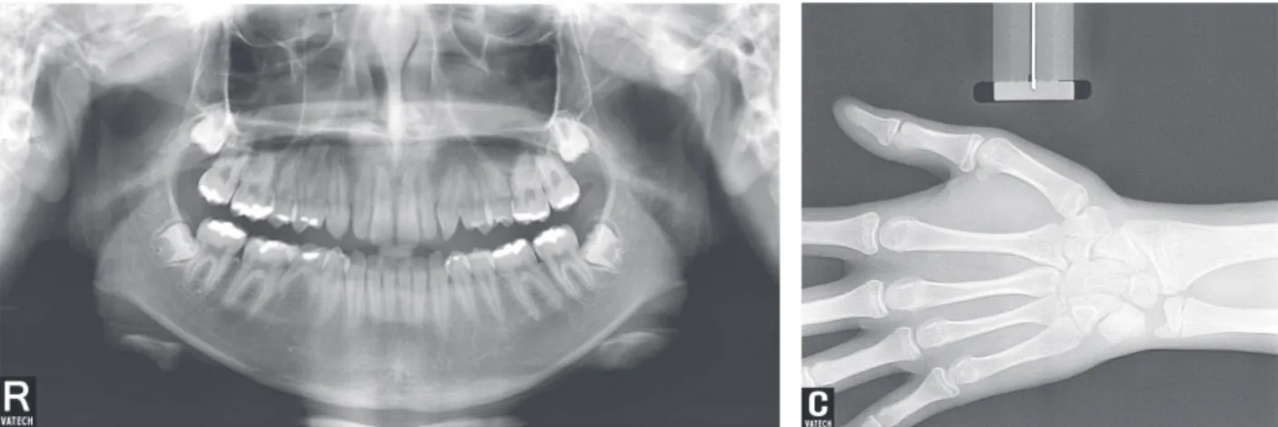
경, 깊은 이순구 및 비교적 잘 발달된 이부를, 측모에서 는 직선형 안모를 보이고 있었다. 구내사진 및 모델 분 석에서 전치부의 다소 깊은 수직피개(4.8 mm), 하악 우측 제2유구치의 만기 잔존 및 양측 II급 구치 관계 를 보였다(Figure 1, Table 1). 상악 전치부 및 견치 후 방에 치간 공극이 관찰되었고, 상악 치아 중심선에 대 해 하악 치아 중심선이 약 0.5 mm 좌측으로 편위되 어 있었다. 양측 소구치 및 대구치 부위에서 넓은 상악 궁, 좁고 설측 경사된 하악 치열궁으로 인한 설측 교차 교합을 가지고 있었다(Table 2). 심한 양측성 가위교합 임에도 불구하고 환자나 보호자는 이를 인지하지 못하 고 있었고, 상악 좌우측 측절치가 왜소치로 전치부 볼 턴 비(Bolton anterior ratio)의 부조화를 보이고 있었 다. 파노라마방사선사진상에서 하악 우측 제2유구치의 만기 잔존 및 계승 영구치 결손을 보였고, 미맹출된 하 악 제3대구치의 근심 경사가 관찰되었다(Figure 2). 측 면 두부규격방사선사진 분석 결과 상·하악골의 전후방 적 위치는 골격성 I급 관계를 가지며, 상·하악 전치의 설측 경사 및 수평 성장 양상의 단안모를 가지고 있었 다(Figure 3A, Table 1). 정면 두부규격방사선사진상 의 Cg-ANS 기준선에 대해 이부(menton)가 좌측으로 1.5° 내의 변위를 보여, 비대칭 정도는 미약하다고 판단 되었고(Figure 3B), 수완부 방사선사진상에서 요골의 골단부 골화가 진행 중으로 악골의 잔여 성장이 남아있 는 것으로 추측되었다(Figure 2).
치료계획
양측성 구치부 가위교합 개선 및 전치부 공간 폐쇄,
II급 구치 관계 개선 및 심피개교합 개선을 치료목표로
하였다. 하안면 고경의 증가와 양측 구치부의 텔레스코
픽 교합(telescopic occlusion)을 개선하기 위해 상악
의 분절 르포르 I 골절단술, 하악의 횡적 골 신장술 등
의 수술적 방법이 사용되기도 하지만, 본 증례에서는
Figure 1. Facial and intraoral photos before treatment.
Table 1. Comparison of the cephalometric measurements at pretreatment, posttreatment, and retention Pretreatment
(15 years 3 months old)
Posttreatment (18 years 7 months old)
Retention (19 years 9 months old)
SNA (°) 82.8 82.3 83.5
SNB (°) 79.9 81.1 82.2
ANB (°) 2.9 1.2 1.3
Mandibular plane angle (°) 29.7 26.8 25.1
Gonial angle (°) 125.5 123.2 124.6
Mx. 1 to FH plane (°) 109.3 117.1 116.6
Mn. 1 to mandibular plane (°) 89.4 95.6 95.8
Overbite (mm) 4.8 2.2 1.6
Overjet (mm) 5.1 2.5 2.7
Mandibular length (mm) 126.2 135.4 137.2
A to N⊥FH plane (mm) -3.4 -3.6 -1.6
Lower anterior facial height (mm) 76.0 78.9 78.8
Facial axis (°) 87.8 88.6 89.7
Figure 3. Lateral and posteroanterior cephalometric radiograms before treatment.
Table 2. Dental cast measurements
Variable (mm) Pretreatment
(15 years 3 months old)
Posttreatment (18 years 7 months old)
Retention (19 years 9 months old)
Maxillary intercanine width 34.6 34.2 34.4
Mandibular intercanine width 26.2 25.0 26.2
Maxillary inter-first molar width 62.6 56.2 58.6
Mandibular inter-first molar width 41.4 50.6 51.4
Maxillary inter-second molar width 71.0 67.0 68.6
Mandibular inter-second molar width 50.6 60.2 60.4
The intercanine width: the distance between the cusps of the bilateral canines. The intermolar width: the distance between the mesiobuccal cusps of the bilateral molars.
Figure 2. Panorama and hand-wrist radiograms before treatment.
가 강하여 고정식 교정장치와 설측 호선을 이용한 절충 적 교정치료, 즉 하악 구치부 직립 및 상악 구치부 치열 궁의 축소, 왜소치인 상악 좌우측 측절치의 보철적 수 복을 시행하기로 하였다.
치료경과 및 결과
상악 치열에 0.022-inch 슬롯 Damon MX
Ⓡ브라켓 (Ormco, Glendora, CA)을 부착하여 치아 배열을 시 작하였고, 하악에는 제1대구치의 설측 호선을 확장하여 하악 대구치부를 직립시켰다(Figure 4A). 하악 설측 호선 조절 및 상악궁 레벨링 4개월 만에 제1대구치 가 위교합이 개선되어 하악 치열에도 브라켓을 부착하였다 (Figure 4B). 이후 상악궁 축소를 위한 변형된 구개 호
선을 삽입하여 상악 소구치부와 구치부의 폭경을 조절 하였고(Figure 4C), 상악 전치부 공간을 닫기 위해 상 악 6전치를 견인하고(Figure 4D), 구치부 정출을 통해 교합을 거상하였다. 좌측 견치 및 구치 II급 관계를 개 선하기 위해 좌측 상악 구치부에 미니 임플랜트를 식립 하였다(Figure 4E). 3년 2개월 후 치료가 종료되었고 왜소치인 상악 좌우측 측절치는 레진으로 수복하였다.
고찰
교정치료 종료 후 하악골의 전방 성장과 함께 환자의 측모의 심미적 개선을 보였고, 양측성 가위교합 및 구 치, 견치간 교합관계, 심피개교합이 개선되었다(Figure 5). 전치부 공간 폐쇄 기간 동안 환자의 불성실한 내
Figure 4. Intraoral photos at 3 months (A), 4 months (B), 9 months (C), 13 months (D), and 35 months (E).
A
B
C
D
E
원 등으로 치료기간이 장기화되어 아쉬움이 남는다. 측 면 두부규격방사선사진 중첩 결과 하악골이 매우 폭 발적인 성장이 보이는데 유효 하악골 길이(effective mandibular length; Co-Pog) 변화가 9 mm 정도로 같은 기간 한국인 남자의 표준 성장량이 약 1.4 mm
7인 것을 고려하면 임상적으로 매우 흥미로운 관찰이었 다(Figures 6, 7, Table 1). 이는 상·하악궁의 폭경 부
조화로 지연되었던 하악골의 잠재적인 성장이 폭경 부 조화의 개선과 함께 자발적으로 개선될 수 있다는 점을 시사한다고 볼 수 있다.
8하악골의 전하방 성장과 구치 부 가위교합의 개선이 이루어졌으나 좌측 편위를 보였 던 하악골의 비대칭 정도는 비슷한 정도로 유지되었다.
교정치료 약 1년 후에도 구치부의 교합은 긴밀하게 유지 되고 있었다(Figure 8).
Figure 5. Facial and intraoral photos after treatment.
Figure 6. Radiographic examination after treatment.
Figure 7. Superimposition of cephalometric tracings before (black line) and after (red line) treatment.
증례 2
진단
20세 8개월의 남자 환자로 우측 어금니가 반대로 물 린다는 것을 주소로 내원하였다. 타 병원에서 교정 상 담 시 양악 수술을 동반한 교정치료 설명을 듣고 수 술에 대한 부담감으로 치료를 포기하였다고 하였으 며, 내원 당시 환자는 절충적 교정치료에 대한 강한 의 사를 표명하였다. 안정 시 입술은 이개되어 있고(lip incompetency), 특히 하순이 건조하고 약간 전방으로 말려(everted) 있어 구호흡이 의심되었다(Figure 9).
의도적으로 입술을 다물 때는 상순과 하순이 약간 돌 출되는 양상을 보이고 있었다. 구내 소견에서도 구개편 도가 비대되어 있고 구강위생이 매우 불량하며, 특히 상·하악 전치 치경부에는 띠 형태의 백색 반점이 관찰 되었다. 상악 전치부는 임상 치관의 길이가 매우 짧아 지속적인 마모의 진행이 의심되었다.
측면 두부규격방사선사진상에서 ANB 각도는 약 1°
로 골격적으로 전후방적인 문제는 크지 않았으며, 상·
하악 전치의 각도도 정상 범주에 속하나 단안모의 수
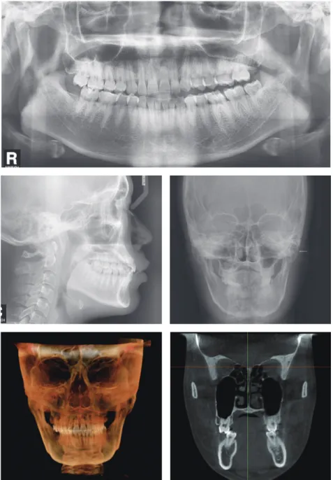
의도적으로 입술을 다물 때 상·하순이 돌출되는 현상 은 이러한 수직 성장의 결여에 의한 것으로 판단되었으 며 이는 두부규격방사선사진상에서 보이는 후순 공간 (retrolabial space)에 의해 확인될 수 있었다(Figure 10). 상·하악의 구치부 폭경 분석 결과 협착된 하악 구 치 폭경을 보였으며, 우측 소구치 및 대구치부의 가위교 합이 관찰되었다(Table 4). 파노라마방사선사진상에서 상악 좌측 중절치의 치근 흡수(rounding root apex) 가 관찰되었다(Figure 10).
치료계획
우측 하악 소구치와 구치의 직립 및 정출된 상악 우 측 구치의 함입, 상악 치열궁 축소를 통한 구치부 가위 교합의 개선과 I급 구치 및 견치 관계 획득을 목표로 한 비발치 교정치료를 계획하였다.
치료경과 및 결과 치료 경과
상악 좌측 제1대구치의 근관치료 시행 후 교정치료를
시작하였다. 상악에 변형된 형태의 횡구개 호선 및 하
Figure 8. Intraoral photos at 14 months of retention.
Figure 9. Facial and intraoral photos before treatment.
Table 3. Comparison of the cephalometric measurements at pretreatment, posttreatment, and retention
Pretreatment
(20 years 8 months old)
Posttreatment (22 years 11 months old)
Retention (23 years 11 months old)
SNA (°) 80.8 81.7 80.6
SNB (°) 79.9 81.3 79.5
ANB (°) 0.9 0.4 1.1
Mandibular plane angle (°) 18.3 19.0 19.4
Gonial angle (°) 115.9 111.9 111.9
Mx. 1 to FH plane (°) 113.0 118.1 117.5
Mn. 1 to mandibular plane (°) 94.8 96.7 94.3
Overbite (mm) 2.0 2.4 1.8
Overjet (mm) 3.8 3.8 3.4
Mandibular length (mm) 134.5 133.0 134.5
A to N⊥FH plane (mm) 2.7 0.6 0.9
Lower anterior facial height (mm) 78.9 76.4 77.6
Facial axis (°) 88.8 90.1 88.8
Figure 10. Radiographic examination before treatment.
해 협측에 미니 임플랜트를 식립하였다. 한편 상악 우 측 구치부는 구개측에 미니 임플랜트를 식립하여 탄성 체인을 이용하여 상악궁 축소 및 대구치와 소구치의 압 하를 시행하였다. 하악 좌측 구치부에는 교합을 이개하 기 위한 컴포짓 레진 교합 블록을 부착하였다(Figure 11A). 약 6개월 경과 후 우측 구치부 및 소구치부의 가 위교합이 해소되었고(Figure 11B), 치료 9개월째 점 진적으로 삭제해 가던 교합 블록을 완전히 제거하였다 (Figure 11C). 환자의 개인 사정으로 약 3개월 정도 치 료가 연장되었고, 동적 치료기간은 14개월 정도 소요되 었다.
치료 결과
교정치료 후 우측 구치부 가위교합이 성공적으로 개 선되었고, I급 구치 및 견치 관계를 보였으며, 악골의 비 대칭 정도는 유지되었다(Figure 12). 비발치 교정치료 를 시행하였으므로 입술 돌출도는 치료 전과 유사하였 고 치료 전 관찰되었던 치경부의 백색 탈회 부위는 지 속적인 구강위생 지도에도 불구하고 대부분 와동 형성 으로 진행된 것이 아쉬움으로 남았다. 해당 부위는 교 정장치 제거 후 보존적 수복치료를 시행하였다. 측모 두부규격방사선사진 중첩 결과 구치의 압하를 동반한 가위교합의 개선으로 전안면 고경이 약간 감소하였다 (Figures 13 and 14, Table 3). 약 1년의 유지 기간 이 Figure 11. Intraoral photos at 0 month (A), 6 months (B), and 9 months (C).
A
B
C
Table 4. Dental cast measurements
Variable (mm) Pre-treatment
(20 years 8 month)
Post-treatment (22 years 11 months)
Retention (23 years 11 months)
Maxillary intercanine width 36.2 34.8 34.6
Mandibular intercanine width 26.2 26.8 26.0
Maxillary inter-first molar width 55.6 52.4 53.4
Mandibular inter-first molar width 41.6 50.0 47.6
Maxillary inter-second molar width 62.2 58.2 58.6
Mandibular inter-second molar width 45.8 52.0 51.8
The intercanine width: the distance between the cusps of the bilateral canines. The intermolar width: the distance between the mesiobuccal cusps of the bilateral
molars.
후 관찰에서 교합은 양호하게 유지되었다(Figure 15).
고찰
구치부 측방치 군의 협설측 교합 이상에 대해서는 전 통적으로 협측 반대교합(buccal cross bite)과 설측 반 대교합(lingual cross bite)
1으로 표현해 왔다. 상·하악 궁의 폭경 부조화로 인한 구치부의 교차교합은 협측이 냐 설측이냐에 따라 치료계획이 정반대이므로 정확히 구별해야 한다. 이 분류는 단순하고 간편하긴 하지만, 그 정도를 정량적으로 표현하지는 못한다. 좀 더 정량적 으로 표현하고 어느 악궁에 문제가 있는지를 평가하기 위해서는 콘빔 영상화단층촬영이 도움이 될 수 있을 것 이다(Figures 10, 13).
가위교합 증례를 분절 골절단술과 구치부 치근단 하 악 수술을 이용해서 치료한 증례들
9이 발표되고 있고
그 안정성도 매우 좋은 것으로 보고되고 있다. 아마도 이전 병원에서 수술을 권유한 것도 이러한 이유일 것으 로 생각된다. 그러나 수술적 접근은 비수술적 접근에 비해 비용과 위험 요인이 상대적으로 크므로 교정치료 현장에서 가장 적정한 치료를 선정하는 것도 교정 의사 의 몫이라 생각하여 다양한 선택지를 제시하는 것도 매 우 중요한 업무라 생각된다.
결론
가위교합은 구강악안면계의 근신경계의 부조화 및 측두하악관절질환의 발생과도 유의한 관계가 있다.
10따 라서 이러한 부정교합은 가능한 발견 즉시 특히 조기에 개선하는 것이 바람직하다고 생각한다.
Figure 12. Facial and intraoral photos after treatment.
Figure 13. Radiographic examination after treatment.
Figure 14. Superimposition of cephalometric tracings before (black line) and after (red line) treatment.
Figure 15. Intraoral photos at 12 months of retention.
REFERENCES