• 검색 결과가 없습니다.

A pattern of cell death induced by 40 kHz ultrasound in yeast cell model

N/A
N/A
Protected

Academic year: 2021

Share "A pattern of cell death induced by 40 kHz ultrasound in yeast cell model"

Copied!
7
0
0

로드 중.... (전체 텍스트 보기)

전체 글

(1)

A pattern of cell death induced by 40 kHz ultrasound in yeast cell model

40 kHz 초음파에 의해 유도된 효모세포 모델에서 세포사멸 패턴

Ji Wook Kim, 1 Hee Jeong Kong, 2 Young H. Kim, 3 and Kwang Il Kang 1†

(김지욱,

1

공희정,

2

김영환,

3

강광일

1†

)

1

Department of Chemistry and Biology, Korea Science Academy of KAIST,

2

Biotechnology Research Division, National Institute of Fisheries Research,

3

Applied Acoustics Laboratory, Korea Science Academy of KAIST

(Received January 25, 2017; revised March 8, 2017; accepted May 30, 2017)

ABSTRACT: Ultrasound has been widely used for biological and medical applications including induction of cell death, but a precise mechanism of induced cell death by ultrasound is controversial. In this study, an irradiation system with 40 kHz ultrasound was developed for a suitable cell death test of a representative unicellular organism, yeast, and used to study the biological effect of ultrasound on inducing cell death. Potassium Iodide (KI) dosimetry was used to devise an optimal system that successfully delivers 40 kHz ultrasound and produces reactive oxygen species in a 1.5 ml Eppendorf tube. Cell death was observed in an ultrasound transmission time-dependent fashion in this system. Thermal effect during irradiation was not observable in ultrasound induced cell death. Co-treatment of 40 kHz ultrasound and hydrogen peroxide showed a synergistic effect in inducing cell death. This finding suggests that 40 kHz ultrasound is related to reactive oxygen species formation. However, NAC (N-acetyl-L-cyste- ine) oxygen scavenger slightly inhibited the cell death by 40 kHz ultrasound. It was also found that 40 kHz ultrasound induced cell death was slightly inhibited by inhibitors of necrosis or apoptosis (glycyrrhizin or zVAD-fmk). This study suggests that cell death induced by 40 kHz ultrasound may not be exclusively related to reactive oxygen species formation and thermal effects in irradiated yeast cells.

Keywords: Cell death, 40 kHz ultrasound, yeast, Reactive oxygen species

PACS numbers: 43.35.Wa., 43.80.Gx

초 록: 초음파는 세포사멸을 포함하여 의학 및 생물학분야에 널리 응용되고 있으나 그 정확한 기작에 대해선 논쟁의 여지가 있다. 본 연구에서는 40 kH 초음파 조사시스템을 단세포 효모에 적합하게 개발하고 세포사멸 유도시 40 kH 초음 파의 생물학적 현상을 살펴보았다. 아이오딘화 칼륨 선량 측정법을 이용하여 1.5 ml 실험튜브에 40 kH 초음파 조사 시스 템의 최적 조건을 맞추어 세포사멸을 시간 의존적 방식으로 연구하였고 초음파 조사과정동안 온열효과와는 별개로 세포 사멸이 관찰되었다. 40 kH 초음파와 과산화수소의 동시 처리는 세포사멸에 상조적인 효과가 관찰되어 활성산소가 40 kH 초음파사멸에 관련이 있었다. 그러나 활성산소 저해제, NAC(N-acetyl-Lcysteine)는 초음파에 의한 세포사멸에 약 한 영향만을 미쳤고 다른 세포사멸, 괴사억제제[글리실리진(glycyrrhizin) 또는 zVAD-fmk] 역시도 세포사멸을 완전히 억제하진 못하였다. 본 연구를 통하여 40 kH 초음파에 의한 세포사멸에는 온열효과나 활성산소만으로 사멸이 유도되지 는 않는 것으로 보인다.

핵심용어: 세포사멸, 40 kHz 초음파, 효모, 활성산소

†Corresponding author: Kwang Il Kang (kikangos@kaist.ac.kr) Department of Chemistry and Biology, Korea Science Academy of KAIST, 105-47, Baeqyanggwanmun-ro, Busanjin-gu, Busan 47162, Republic of Korea

(Tel: 82-51-606-2224, Fax: 82-51-894-0280)

(2)

Fig. 1. Irradiation system of 40 kHz ultrasound applied to the yeast cell culture of the 1.5 ml Eppendorf tube.

I. Introduction

Many applications of low frequency ultrasound from 20 to 200 kHz have been reported in physical, medical and biological domains. Low frequency ultrasound has used for diverse cleaning systems and medical therapeutic methods.

[1]

When blood vessels are clogged by thrombus or fibrin, 40 kHz ultrasound accelerates the thrombolysis or fibrinolysis.

[2]

Drug transport is enhanced by low frequency ultrasound, as it creates circulating eddies and acoustic pressure around oscillating bubbles that are formed by stable cavitation.

[3]

Ultrasound increases the membrane permeability and enhances the drug uptake of certain target cells such as HL-60 cells in vitro.

[3]

Low frequency ultrasound is also used for breaking kidney stones and for tearing fat tissue to assist liposuction.

[1]

It has been also reported that ultrasound irradiation with low frequency could be applied to induce chronic wound healing.

[4]

For the studies of cell death, high frequency ultrasound over MHz has been widely applied to diverse cancer cells to induce cell death in cancer cells.

[5,6]

However, ultrasound is also used in monitoring of cell death images and could induce cell death.

[7,8]

Physical effects of low frequency ultrasound such as sonoporation of cell membranes are known as ways cells die under transmission. The biological effect of low frequency ultrasound via cavitation and ROS (Reactive Oxygen Species) formation is suggested as another way cell death occurs, but the mechanism is not fully understood. The precise mechanism and efficacious method of ultrasound induced-cell death remains to be elucidated.

Ultrasound at a frequency of 40 kHz is easily applicable and widely used in medical and biological devices. Therefore, we first developed the irradiation system by applying a 40 kHz ultrasound to the unicellular representative organism, yeast, Saccharomyces cerevisiae. The yeast cells are easy to manipulate and have many advantages including applica- bility to human cancer cells. Then, we examined the effect of 40 kHz ultrasound on the cell death and analyzed the thermal effect, radicals effects and other possible effects.

We used the 50 W output of ultrasound generator and had

worked in condition of standing wave. Therefore, the intensity and acoustic pressure are spatially not uniform and were not measured. In our study, we considered cell death dependent on the irradiated time instead of the intensity.

II. Materials and method

2.1 Yeast strain

The yeast Saccharomyces cerevisiae BY4743 was purchased from EUROSCARF (Germany). The obtained results are applicable to human cells. The strain was incubated in 5 ml YPD medium (50 g/l, GIBCO, BRL) at 25 °C shaking incubator of 200 r/min.

2.2 Ultrasound transmission system A pyrex beaker of outer diameter 60 mm was used as an ultrasonic transmission cell. A BLT (Bolt-clamped Langevin Transducer) with a 40 kHz nominal frequency was attached at the bottom of the transmission cell. The BLT was excited by an ultrasonic generator (50 W Power, Kodo Technical Research Co., Korea). Two vertical height adjustors were placed 15 cm apart. At the middle of the two adjustors, the transmission cell was fixed using a bolt.

Distilled water was poured into the beaker, and ultrasound was transmitted using an ultrasound producer.

In order to transmit a 40 kHz ultrasound into the yeast

cell culture of the 1.5 ml Eppendorf tube, a stable Eppendorf

(3)

Fig. 2. Ab sorb ance at wavelength 355 nm of KI solution with different distilled water level in irradiation system. Absorbance was measured after 1 min irradiation with 40 kHz ultrasound.

tube fixing system was devised using acrylic. (Fig. 1) A U-shaped acrylic extension was added to the middle of 160 mm × 38 mm acrylic. In the middle of the U-shaped extension, a hole of 10 mm was made to fix the Eppendorf tube.

2.3 Potassium Iodide (KI) dosimetry The OH radicals attack Potassium Iodide (KI) and liberate iodine. When liberated iodine accumulates, I

3-

forms in the solution. I

3-

of ultrasound irradiation at certain level of water surface was estimated by measuring the absorbance of KI solution at 355 nm wavelength.

2.4 Viability spotting assay

The yeast cell culture was prepared and adjusted with optical density 0.1 at 660 nm wavelength (OD660 = 0.1).

The solution was diluted one fifth for four times to prepare solutions with concentration of 1/5, 1/25, 1/125, 1/625 of the original solution. 10 ㎕of series of diluted solutions are spotted on the YPD-agar plate (15 g/l) in a row, and incubated for 24 h before colony was counted.

2.5 Heat shock assay

The yeast cell was plated on YPD-agar media (15 g/l) and incubated with 5 min at normal condition at 25 °C. It was also incubated for 5 min under heat shock conditions at 30

°C, 40 °C, 50 °C in a waterbath. Temperature was controlled within ± 1 °C. After heat shock, the yeast cells were returned to the normal condition and incubated overnight.

2.6 ROS (Reactive Oxygen Species) Hydrogen peroxide (Sigma-Aldrich, USA) of 0.1 mM, 0.5 mM and 1 mM were treated to each yeast cell culture (O.D.660 = 0.1) to induced condition of Reactive Oxygen Species. ROS was inhibited by adding different concentrations of NAC (N-acetyl-L-cysteine; ENZO Life Sciences) to cells right before ultrasound transmission.

2.7 ROS detection

ROS formed due to ultrasound irradition can be viewed under a fluorescence microscope when stained with ROS reaction dyes. Oxygen radicals were induced by adding pyocyanin to negative control cell. ROS was detected by adding 10 g/ml 2`,7'-dichlorohydrouorescein diacetate (Sigma- Aldrich, USA) to yeast cell cultures (O.D.660 ≈ 0.4). Cells were left to grow for an additional two h and then viewed under Zeiss Axiovert 40 Fluorescence microscope (Zeiss, Swiss).

III. Results

3.1 Cell death is induced by the optimized ultrasound condition

An irradiation system with ultrasound at 40 kHz frequency was developed (Fig. 1) and used to test the KI ionization for optimal position. Fig. 2 shows the optical density values of KI solution at 355 nm after 1 min of 40 kHz ultrasound transmission at different water heights.

The two peaks at 4.3 cm and 6.4 cm mark the water height

at which oxygen radicals formed the most.

[9]

This indicates

that at these water height (4.3 cm and 6.4 cm), the 40 kHz

ultrasound is successfully transmitted to the Eppendorf

tube placed at the water surface level. Another point worth

noting is that the distance between two antinodes (2 cm) is

similar to the theoretically calculated distance between

antinodes of 40 kHz ultrasound in water. Among the two

(4)

Fig. 3. For cell viability test, overnight cultured yeast cells were treated with 40 kHz ultrasound (US) at various times, incubated for 24 h (left), and then measured at absorbance of 660 nm with different time intervals (right) for cell death detection.

Fig. 4. The growth of yeast cells was measured with exposure to various heat shock environments for 5 min. Spt dilution assay (left) and viability test (right) after 5 min exposure of indicated temperature.

peaks, 4.3 cm water height was chosen as the optimal condition for 40 kHz ultrasound transmission, since absor- bance is higher than that of 6.4 cm.

The cell viability of yeast exposed to 40 kHz ultrasound for different durations was detected in Fig. 3. Cell death is induced from 3 min to 5 min of ultrasound transmission. This result implies that our optimized ultrasound irradiation system successfully transmits ultrasound to yeast cells (diameter 5 ∼10 µm) and induces cell death.

3.2 Heat shock effects on cell death During the ultrasound transmission, hyperthermia occurred under the tested conditions. The temperature of the system increased a maximum temperature of 50 °C when the system was irradiated by the 40 kHz ultrasound for 5 min.

Since hyperthermia has an important role in ultrasound effects, we checked whether exposing yeast cells to high temperatures for 5 min would induce cell death or not. In

this condition, the experiment failed to show that heat shock induced cell death (Fig. 4) This result suggests that hyperthermia of the irradiation system is not relevant to 40 kHz ultrasound induced cell death.

3.3 Reactive oxygen species on cell death It is well known that ultrasound stimulates inertial cavitation and oxygen radials. ROS accumulated in the yeast cells irradiated with 40kHz ultrasound for 3 min and 5 min (Fig. 5). This results correspond with that of cell viability for 3 min and 5 min. Thus, the result suggests that reactive oxygen species may be involved in 40 kHz ultrasound using this transmission system.

If ultrasound induced cell death is closely related to

reactive oxygen species formation, extraneous oxygen

radicals will be promoted by the effect of ultrasound

transmission. To check this, yeast cells were co-treated

with hydrogen peroxide and ultrasound irradiation. Interes-

(5)

Fig. 5. Reactive oxygen species levels of yeast cells irradiated with 40 kHz ultrasound irradiation at various times were assessed by dichlorohydrouorescein diacetate staining under fluorescence microscope. A: control, B:

0.5 min, C: 1 min, D: 3 min, and E: 5min.

Fig. 6. Synergistic cell death with cotreatment of ultrasound and hydrogen peroxide is concentration dependent manner. Yeast cell was treated with various concentration of hydrogen peroxide and then irradiated with 40 kHz ultrasound. After irradiation, viability of yeast cells were analyzed.

Fig. 7. NAC treatment with ultrasound irradiation slightly increased cell viability. The 40 kHz ultrasound irradiated for 3 min to yeast cells with 0, 0.5 mM, 5 mM, 20 mM NAC treatment. After irradiation, viability of yeast cells were analyzed.

tingly cell death is synergistically induced in ultrasound irradiated cells with hydrogen peroxides (Fig. 6). It must be noted that the amount of cell death induced by co-treat- ment is larger than the sum of cell death induced by two treatments independently. If ultrasound is one of the reactive oxygen sources which can trigger cell death, the inhibition of reactive oxygen species will suppress the cell death by ultrasound. During 3-min ultrasound irradiation test, yeast cells were treated with different concentrations of NAC as a well known reactive oxygen scavenger. NAC treatment with yeast cells slightly inhibited cell death induced by 40 kHz ultrasound (Fig. 7). These results suggest another way of cell death induced by ultrasound beyond that of reactive oxygen species generated by ultrasound.

3.4 Biological mechanism of ultrasound induced cell death

The biological patterns of cell death are necrosis and

apoptosis which can be induced by various stress agents

including physical and chemical stresses.

[10,11]

When yeast

cells containing glycyrrhizin (necrosis inhibitor) or z-vad-fmk

(apoptosis inhibitor) are treated with 40 kHz ultrasound,

(6)

Fig. 8. Inhibitors of cell death with ultrasound irradiation slightly increase cell viability. The 40 kHz ultrasound irradiated for 3 min to yeast cells with glycyrrhizin (Glz) 200 µM and z-VAD-fmk (zvad) 20 µM. After irradiation, viability of yeast cells were analyzed.

cell viability is slightly increased (Fig. 8). This result suggests that either necrosis and apoptosis are not major types of cell death or chemical reagents cannot inhibit both types of cell death in this transmission system.

IV. Conclusions

In this study, a system that transmits 40 kHz ultrasound was developed and optimized to study yeast as a representative unicellular organism. Using an ultrasound transmission system, yeast cell death was examined. We found that cell death is dependent with irradiation time. We used the 50 W output of ultrasound generator and had worked in condition of standing wave. Therefore, the intensity and acoustic pressure are spatially not uniform and were not measured. In our study, we considered cell death dependent on the irradiated time instead of the intensity. Large amount of cell death was observed after 3 min of ultrasound irradiation.

After the optimization of the ultrasound transmission system, a mechanism of cell death was examined. The thermal effect of low frequency ultrasound was not plausible to ultrasound induced cell death because the viability of heat shocked cells was similar to that of control cells (Fig. 4). A reactive oxygen species was suggested as one of the cause of cell death induced by ultrasound, as hydrogen peroxide and ultrasound showed synergistic effects in inducing cell death. Also, the amount of reactive oxygen species formed

corresponded with the amount of cell death occurred as ultrasound transmission time increased. Therefore, it was assumed that ultrasound induced yeast cell death might be involved in reactive oxygen species generation. However, the treatment of NAC did inhibit slightly the ultrasound induced cell death. Necrotic and apoptotic cell death were also slightly inhibited by each specific cell death inhibitor.

Taken together, this study suggests that ultrasound induced cell death may be not exclusively related to reactive oxygen species formation in the cell and that other cell death pathways other than apoptosis and necrosis must be carried out.

It shoud be noted that applying these results to cell deaths studies may be difficult because of non-linear effects of low frequency ultrasound.

Acknowledgement

This work was supported by the Korea Science Academy of KAIST with funds from the Ministry of Science, ICT and Future Planning.

References

1. F. Ahmadi and I. McLoughlin, “A new mechanical index for gauging the human bioeffects of low frequency ultrasound,” Conf. Proc. IEEE Eng. Med.

Biol. Soc. 1964-1967 (2013).

2. V. Suchkova, F. N. Siddiqi, E. L. Carstensen, D.

Dalecki, S. Child, and C. W. Francis, “Enhancement of fibrinolysis with 40-kHz ultrasound,” Circulation, 98, 1030-1035 (1998).

3. W. G. Pitt, G. A. Husseini, and B. J. Staples, “Ultrasonic drug delivery - A general review,” Expert Opin. Drug Deliv. 1, 37-56 (2004).

4. J. Voigt, M. Wendelken, V. Driver, and O. M. Alvarez,

“Low-frequency ultrasound (20-40 kHz) as an adjunctive therapy for chronic wound healing: a systematic review of the literature and meta-analysis of eight randomized controlled trials,” Int. J. Low Extrem Wounds, 10, 190-199 (2011).

5. H. Ashush, L. A. Rozenszajn, M. Blass, M. Barda-Saad, D. Azimov, J. Radnay, D. Zipori, and U. Rosenschein,

“Apoptosis induction of human myeloid leukemic

cells by ultrasound exposure,” Cancer Res. 60, 1014-

(7)

1020 (2000).

6. S. Brand, B. Solanki, D. B Foster, G. J. Czarnota, and M. C. Kolios, “Monitoring of cell death in epithelial cells using high frequency ultrasound spectroscopy,”

Ultrasound Med. Biol. 35, 482-493 (2009).

7. R. M. Vlad, M. C. Kolios, and G. J. Czarnota, “Ultrasound imaging of apoptosis: spectroscopic detection of DNA- damage effects at high and low frequencies,” Methods Mol. Biol. 682, 165-187 (2011).

8. B. A. Scheven, J. L. Millard, P. R. Cooper, S. C. Lea, A. D. Walmsley, and A. J. Smith, “Short-term in vitro effects of low frequency ultrasound on odontoblast-like cells,” Ultrasound Med. Biol. 33, 1475-1482 (2007).

9. E. J. Hart and A. Henglein, “Free radical and free atom reactions in the sonolysis of aqueous iodide and formation solutions,” J. Phys. Chem. 89, 4342-4347, (1985).

10. S. W. Ryter, H. P. Kim, A. Hoetzel, J. W. Park, K.

Nakahira, X. Wang, and A. M. Choi, “Mechanisms of cell death in oxidative stress,” Antioxid Redox Signal, 9, 49-89 (2007).

11. G. Kroemer, L. Galluzzi, P. Vandenabeele, J. Abrams, E. S. Alnemri, E. H. Baehrecke, M. V. Blagosklonny, W. S. El-Deiry, P. Golstein, D. R. Green, M. Hengartner, R. A. Knight, S. Kumar, S. A. Lipton, W. Malorni, G.

Nuez, M. E. Peter, J. Tschopp, J. Yuan, M. Piacentini, B.

Zhivotovsky, and G. Melino, “Classication of cell death:

recommendations of the Nomenclature Committee on Cell Death 2009,” Cell Death Differ. 16, 3-11 (2009).

Profile

▸Ji Wook Kim (김지욱)

He graduated Korea Science Academy of KAIST in 2015. Currently, he is student of Seoul National University, majoring in Biological Sciences and minoring in Brain-Mind-Behavior program.

▸Hee Jeong Kong (공희정)

She received her PhD in Molecular Biology from Pusan National University, Republic of Korea in 2001. Then, she was trained at the National Institutes of Health, USA as a postdoctoral fellow from 2001 to 2006. She has been working for Biotechnology Research Division, National Institute of Fisheries Science, Republic of Korea as a researcher since 2006. Her research interest includes transcriptional gene regulation in innate immunity of marine organisms, and devel- opment and risk assessment of transgenic fish.

▸ Young H. Kim (김영환)

He graduated with a BSc in Science Education from Seoul National University in 1979. He took the MSc degree (1981) and PhD (1990) in Phys ics at Korea Advanced Institute of Science and Technology. He worked at Korea Research Institute of Standards and Science, Korea Inspection Engineering Corporation and Sungkyunkwan University. He has been working for Korea Science Academy of KAIST since 2005.

His main research interest are applied acoustics such as wave propagation, high intensity ultrasound and musical instruments.

▸Kwang Il Kang (강광일)

He was graduated from Pusan National University with BSc majored in Science Education in 1983. He received his Msc degree in Seoul National University in 1985 and PhD in Molecular Cell Biology at University of PARIS VI in France in 1995.

He has been working for Korea Science

Academy of KAIST since 2005. His main

research interest is mechanism of cell

death by environmental stresses including

ultrasound irradiation.

수치

Fig. 1. Irradiation system of 40 kHz ultrasound applied to the yeast cell culture of the 1.5 ml Eppendorf tube.
Fig.  2.  Ab sorb ance  at  wavelength  355  nm  of  KI  solution with different distilled water level in irradiation  system
Fig. 3. For cell viability test, overnight cultured yeast cells were treated with 40 kHz ultrasound (US) at various  times, incubated for 24 h (left), and then measured at absorbance of 660 nm with different time intervals (right)  for cell death detection
Fig. 5. Reactive oxygen species levels of yeast cells irradiated with 40 kHz ultrasound irradiation at various times were assessed by dichlorohydrouorescein diacetate staining under fluorescence microscope
+2

참조

관련 문서

interaction with anti-apoptotic Bcl-2 family members, we investigated whether Mcl-1 can inhibit the Noxa-induced cell death.. As expected, Mcl-1 significantly inhibits

– Embryonic stem cell : Pluripotent cells that can give rise to all tissue types. – Adult stem cell : Multipotent cells

Therefore, in this paper, the solar array of HST is modeled as a simple model with beams and membranes, and the phenomenon of thermal induced vibration is verified and

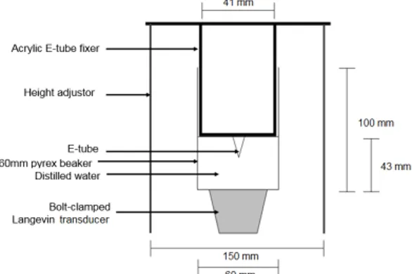
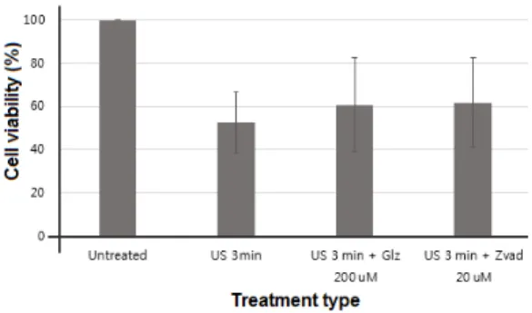
Low intensity pulsed ultrasound used in this

Objective: The purpose of this study was to analyze recent trend in incidence of basal cell carcinoma and squamous cell carcinoma in patients from the Gwangju City

Glutamate excitotoxicity induced by excessive activation of NMDA receptor causes various damage to cells, which leads to cell death.. In previous studies, increased ROS

These results suggest that the bilobalide inhibits the cell proliferation and induces the apoptotic cell death in FaDu human pharyngeal squamous cell carcinoma via both

Taken together, these results suggested that latex containing the ficin inhibited the cell growth and induced apoptosis by caspase and Bcl-2 family signaling pathway in