Received: September 19, 2018 Revised: December 11, 2018 Accepted: December 25, 2018 CliniCAl
neurophysiology
Correspondence to Byung-Nam Yoon
Department of Neurology, Seoul Paik Hos- pital, Inje University College of Medicine, 9 Mareunnae-ro, Jung-gu, Seoul 04551, Korea Tel: +82-2-2270-0946
Fax: +82-2-2270-0045 E-mail: [email protected]
Motor dominant polyradiculopathy with Primary Sjögren’s syndrome mimicking motor neuron disease
Suk-Won Ahn1 and Byung-Nam Yoon2
1Department of Neurology, Chung-Ang University Hospital, Chung-Ang University College of Medicine, Seoul, Korea
2Department of Neurology, Seoul Paik Hospital, Inje University College of Medicine, Seoul, Korea
Sjögren’s syndrome (SS)-associated polyradiculopathy is rarely reported. A 51-year-old woman presented with a history of gradual weakness in all four extremities for several months. Based on electrophysiological studies, spinal magnetic resonance imaging and cerebrospinal fluid examination, inflammatory polyradiculopathy was confirmed. During a search for the aetiol- ogy, the patient was ultimately diagnosed with SS. This study introduces SS-associated poly- radiculopathy that primarily presented with motor symptoms, thus mimicking motor neuron disease.
Key words: Polyradiculopathy; Sjögren’s syndrome; Motor neuron disease
Sjögren’s syndrome (SS) is an autoimmune epithelitis with lymphocytic infiltration of exocrine glands.1 At least one-third of patients present with systemic extraglandular symptoms.2 Peripheral nervous system involvement is the most common neurological symptom and may be the first symptom observed before diagnosis of SS. Symmetric sen- sory polyneuropathy infiltrates are most commonly observed; multiple neuropathy, small fiber neuropathy and cranial neuropathy are also involved.3 Among these disorders, SS-as- sociated polyradiculopathy is rarely observed. Here, we describe a patient with SS who presented with inflammatory polyradiculopathy mimicking motor neuron disease (MND) as the first symptom, as well as slowly progressive weakness and muscular atrophy.
CASE
A 51-year-old female patient visited the hospital with a main complaint of weakness in both upper limbs for 9 months. The symptoms started with neck stiffness and difficulty
ORCID Suk-Won Ahn
http://orcid.org/0000-0002-9979-4589 Byung-Nam Yoon
http://orcid.org/0000-0003-0946-0276
in lifting her arm over her shoulder. Five months later, the strength in her left arm had decreased, and her legs began to lose strength. She had visited the department of neurol- ogy at our hospital due to suspected MND based on exten- sive denervation changes observed in an electromyography (EMG) examination performed at another hospital. She did not have any past medical history. She complained of stiff- ness of the neck and a sense of weakness in both shoulders, hands and legs. She showed no fasciculation or atrophy of the tongue. Atrophy was observed in the proximal upper extremities. Regarding the Medical Research Council grade, her bilateral shoulders received grade 3. Her elbows showed an extension of grade 5 and a flexion of grade 3 for the right elbow and grade 4 for the left elbow. In the lower extrem- ities, flexion and extension of the bilateral hips, knees and ankles were grade 4. Her sensory examinations were normal.
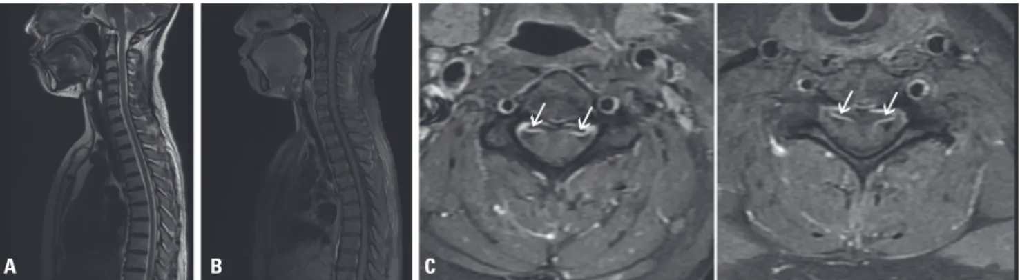
The deep tendon reflexes of the brachioradialis, biceps bra- chii, and triceps muscles were hypoactive. Bilateral Hoffman signs were positive. Plantar reflex signs and ankle clonus were negative. Whole spine MRI showed the enhancement of multiple ventral nerve rootlets in the cervical and lumbar spinal cord (Fig. 1). In the nerve conduction test, the com- pound motor action potential of the right median ulnar, axil- lary, and musculocutaneous nerves was reduced by approx- imately 50-70% compared to that of the left side (Table 1).
EMG confirmed active denervation and reinnervation chang- es in the bilateral abductor pollicis brevis; flexor carpi radialis;
first dorsal interosseous; biceps brachii; triceps brachii; tibialis anterior; vastus lateralis; and cervical, thoracic and lumbar paraspinal muscles (C6-T1, T6, and L4-5). EMG also revealed
giant motor unit potential and delayed recruitment (Table 2).
A cerebrospinal fluid (CSF) test showed normal results with red blood cell 17/mm3, white blood cell 1/mm3 and glucose 58 mg/dL, whereas CSF protein levels were elevated to 65 mg/dL. No evidence of malignancy was detected in the CSF culture test, and herpes virus, zoster virus and JCV PCR were negative. Antinuclear antibodies were 1:20 speckled, anti-Ro antibodies were positive, and anti-La antibodies and other vasculitis-related test results were negative. Anti-HCV, anti-HIV, anti-GM1, anti-GD1a, anti-GD1b, anti-GT1a and an- ti-GQ1b were negative. Creatine phosphokinase and VDRL levels were normal. We started steroid pulse therapy (dose: 1 g solumedrol for 5 days) as an empirical treatment based on the possibility of unspecified inflammatory polyradiculopa- thy, due to spinal MRI findings and elevated CSF protein lev- els. Three days after starting the steroid, the patient showed improvements in neck stiffness and overall weakness. We confirmed that she had symptoms of dry mouth and dry eyes over the past 2 years. In her salivary gland scan test, the absorption of the contrast agent was decreased in the pa- rotid and submandibular glands. According to SS diagnostic criteria, the patient exhibited 2 symptoms of the eyes and 2 symptoms of the mouth.4 The non-stimulated salivary flow- rate test showed a rate of 0.31 g/0.3 mL (<1.5 mL), the wafer test result was 8 minutes and 25 seconds (>2 minutes), and the Schirmer test results were 8 mm on the right side and 7 mm on the left side (<5 mm over 5 minutes). Salivary scin- tigraphy showed delayed uptake, reduced concentration and delayed excretion of the tracer. As she satisfied 4 of the 6 criteria, she was diagnosed with SS.
Fig. 1. Cervical and thoracic spinal MRI. A T2 sagittal image (A) and T1 gadolinium-enhanced sagittal image (B) show unremarkable findings. T1 gadolini- um-enhanced axial images at the (C) C4 level and (D) C6 level show enhancement of the ventral nerve rootletsof the ventral nerve rootlets (white arrows). MRI, magnetic resonance imaging.
A B C
She was discharged with a maintenance treatment of oral steroids. Based on the results of the muscle strength assess-
ment performed at the 2-week follow-up visit, an improve- ment in overall strength was observed. According to the fol- Table 1. The results of nerve conduction study
Nerve Stimulation Latency (msec) Amp. Velocity (m/sec) F-latency (msec)
Motor
Lt. median Wrist 3.13 (<3.6) 11.3 (>5.0) 24.7
Elbow 10.7 58.5 (>50.0)
Axilla 10.4 60.1 (>56.0)
Rt. median Wrist 3.17 (<3.6) 5.4 (>5.0) 26.6
Elbow 5.3 53.7 (>50.0)
Axilla 5.3 64.5 (>50.0)
Lt. ulnar Wrist 2.46 (<2.5) 8.8 (>5.0) 21.4
Elbow 8.8 62.7 (>50.6)
Axilla 8.5 75.2 (>52.7)
Rt. ulnar Wrist 2.29 (<2.5) 6.1 (>5.0) 20.8
Elbow 5.6 54.9 (>50.6)
Axilla 5.4 59.9 (>52.7)
Lt. Axillary Erb 3.05 (<5.4) 8.9 (>4.6)
Rt. Axillary Erb 4.01 (<5.4) 5.4 (>4.6)
Lt. MC Erb 4.11 (<5.6) 5.4 (>4.0)
Rt. MC Erb 4.23 (<5.6) 4.2 (>4.0)
Lt. peroneal Ankle 3.25 (<4.8) 5.1 (>4.0) 42.5
Popliteal fossa 5.1 46.4 (>41.9)
Rt. peroneal Ankle 4.04 (<4.8) 5.3 (>4.0) 45.0
Popliteal fossa 4.4 45.9 (>41.9)
Lt. tibial Ankle 3.21 (<5.1) 12.9 (>5.0) 46.1
Fibular neck 8.7 45.3 (>40.6)
Rt. tibial Ankle 3.96 (<5.1) 10.9 (>5.0) 45.0
Fibular neck 6.9 46.2 (>40.6)
Sensory
Lt. median Digit 2 2.48 27.2 (>10.0) 44.4 (>41.3)
Rt. median Digit 2 2.54 21.3 (>10.0) 43.3 (>41.3)
Lt. ulnar Digit 5 1.98 10.4 (>10.0) 45.5 (>39.3)
Rt. ulnar Digit 5 1.85 34.3 (>10.0) 48.6 (>39.3)
Lt. sural Calf 2.71 9.9 (>6.0) 51.7 (>34.7)
Rt. sural Calf 2.85 21.0 (>6.0) 49.1 (>34.7)
H reflex Lt 26.6
Rt 29.3
Latencies are in milliseconds, amplitudes of compound muscle action potentials in millivolts. amplitudes of sensory nerve action potentials in microvolts.
velocities in m/sec.
Amp, amplitude; Lt, left; Rt, right; MC, musculocutaneous.
low-up spinal MRI results, the initially observed ventral nerve root enhancement had disappeared. After 2 years, the pa- tient was under prognostic observation without recurrence and had maintained a low-dose prednisolone treatment.
DISSCUSION
Our patient showed a pattern of gradual progression of weakness and atrophy with no definite sensory symptoms over 9 months. In addition, because of EMG findings with a widespread denervation pattern, she was referred to our
hospital with suspicion of early-stage MND. We focused on the discrimination of MND. In her spinal MRI, only the ventral roots were contrast enhanced, and the dorsal roots were not enhanced. Spinal MRI results and increased CSF protein lev- els were more consistent with polyradiculopathy than with MND. In the differential diagnosis, we also considered myop- athies. As observed in this patient, if sensory symptoms are rarely accompanied by muscle weakness, we should consid- er myopathy, such as inflammatory myositis and inclusion body myopathy. However, we considered the possibility of myopathies low and did not perform muscle biopsy be- cause the patient had normal creatine phosphokinase, and Table 2. The findings of electromyography
Muscle Spontaneous activity Voluntary contraction
IA Fibrillation PSWs Amplitude Duration Recruitment IP
Lt. ABP ↑ +1 +1 NL NL NL NL
Rt. ABP ↑ 0 0 ↑ ↑ ↓ ↓
Lt. FDI ↑ +1 +1 NL NL NL NL
Rt. FDI ↑ +1 +1 NL NL NL NL
Lt. FCR ↑ 0 0 NL NL NL NL
Rt. FCR ↑ +1 +1 ↑ ↑ ↓ ↓
Lt. biceps brachii ↑ +1 +2 ↑ ↑ ↓ ↓
Rt. biceps barchii ↑ +2 +2 ↑ ↑ ↓ ↓
Lt. triceps brahii ↑ +1 +2 ↑ ↑ ↓ ↓
Rt. triceps brachii ↑ +2 +2 ↑ ↑ ↓ ↓
Lt. vastus lateralis ↑ +1 +1 ↑ ↑ ↓ ↓
Rt. vastus lateralis ↑ 0 +1 ↑ ↑ ↓ ↓
Lt. TA ↑ 0 0 ↑ ↑ ↓ ↓
Rt. TA ↑ 0 0 ↑ ↑ ↓ ↓
Lt. GCM ↑ 0 0 ↑ ↑ ↓ ↓
Rt. GCM ↑ 0 0 ↑ ↑ ↓ ↓
Lt. paraspinal C5-C8, T1, T6 ↑ +1 +1
Rt. paraspinal C5-C8, T1, T6 ↑ +1 +1
Lt. paraspinal L4-5 ↑ +1 +1
Rt. paraspinal L4-5 ↑ +1 +1
Lt. paraspinal S1 NL 0 0
Rt. paraspinal S1 NL 0 0
Lt. masseter NL 0 0 NL NL NL NL
Lt. tongue NL 0 0 NL NL NL NL
IA, insertional activity; PSW, positive sharp wave; IP, interference pattern; Lt, left; ABP, abductor pollicis brevis; NL, normal; Rt, right; FDI, first dorsal interosse- ous; FCR, flexor carpi radialis; TA, tibialis anterior; GCM, gastrocnemius medialis.
the EMG finding was compatible with neuropathy.
Peripheral nervous system involvement in SS varies in the literature, ranging from 2% to 23% and 60%.3,5,6 In two large-scale studies, four of 92 patients and six of 54 patients showed symptoms of polyradiculopathy.7,8 Most of these patients exhibited a subacute pattern (2 weeks to a few months) and mainly described sensory symptoms, and dor- sal root involvement was identified upon neuroimaging.7 A case of subacute inflammatory polyradiculopathy associated with SS was previously reported in 2008.9 The patient com- plained of simultaneous sensory discomfort and weakness.
However, in contrast to our patient, this patient exhibited sensory dominant symptoms.
Inflammatory polyradiculopathy and SS may be comorbid conditions. However, SS can share a pathological mecha- nism as it causes autoimmune inflammatory responses, such as lymphocytic infiltration of the self-organ. If an autoim- mune response occurs in the spinal nerve, specifically in the motor root, then it may cause the same symptoms reported in the present case. Moreover, steroid use improved our pa- tient’s condition; thus, our patient likely had an inflammatory lesion.
Based on the results of this case study, primary SS may in- duce inflammatory polyradiculopathy, primarily presenting with motor rather than sensory symptoms. A rapid, accurate diagnosis is needed for proper treatment with steroids and an appropriate immunosuppressive agent.
REFERENCES
1. Venables P. Sjögren’s syndrome. Best Pract Res Clin Rheumatol 2004;18:313-329.
2. Asmussen K, Andersen V, Bendixen G, Schiødt M, Oxholm P. A new model for classification of disease manifestations in primary Sjögren’s syndrome: evaluation in a retrospective long-term study.
J Intern Med 1996;239:475-482.
3. Pavlakis PP, Alexopoulos H, Kosmidis ML, Mamali I, Moutsopoulos HM, Tzioufas AG, et al. Peripheral neuropathies in Sjögren’s syn- drome: a critical update on clinical features and pathogenetic mechanisms. J Autoimmun 2012;39:27-33.
4. Vitali C, Bombardieri S, Jonsson R, Moutsopoulos HM, Alexander EL, Carsons SE, et al. Classification criteria for Sjögren’s syndrome:
a revised version of the European criteria proposed by the Amer- ican-European Consensus Group. Ann Rheum Dis 2002;61:554- 558.
5. Lafitte C. Neurological manifestations in Sjögren’s syndrome. Arch Neurol 2000;57:411-413.
6. Delalande S, de Seze J, Fauchais AL, Hachulla E, Stojkovic T, Ferriby D, et al. Neurologic manifestations in primary Sjögren syndrome.
Medicine (Baltimore) 2004;83:280-291.
7. Mori K, Iijima M, Koike H, Hattori N, Tanaka F, Watanabe H, et al. The wide spectrum of clinical manifestations in Sjögren’s syndrome-as- sociated neuropathy. Brain 2005;128(Pt 11):2518-2534.
8. Grant IA, Hunder GG, Homburger HA, Dyck PJ. Peripheral neuropa- thy associated with sicca complex. Neurology 1997;48:855-862.
9. Rigamonti A, Lauria G, Balgera R, Agostoni E. Subacute inflamma- tory polyradiculopathy associated with Sjögren’s syndrome. Mus- cle Nerve 2009;39:855-857.