Clinical significance of T cell receptor excision circle (TREC) quantitation after allogenic HSCT
Neveen Lewis Mikhael
1, Manal Elsorady
21Clinical Pathology Department, Alexandria Faculty of Medicine, 2Clinical Hematology Department, Head of BMT Unit, Alexandria Faculty of Medicine, Alexandria, Egypt
p-ISSN 2287-979X / e-ISSN 2288-0011 https://doi.org/10.5045/br.2019.54.4.274 Blood Res 2019;54:274-281.
Received on August 28, 2019 Revised on October 15, 2019 Accepted on November 5, 2019
Background
Hematopoietic stem cell transplantation (HSCT) is a well-established treatment modality for a variety of diseases. Immune reconstitution is an important event that determines outcomes. The immune recovery of T cells relies on peripheral expansion of mature graft cells, followed by differentiation of donor-derived hematopoietic stem cells. The for- mation of new T cells occurs in the thymus and as a byproduct, T cell receptor excision circles (TRECs) are released. Detection of TRECs by PCR is a reliable method for estimating the amount of newly formed T cells in the circulation and, indirectly, for estimating thymic function. The aim of this study was to determine the role of TREC quantitation in predicting outcomes of human leucocyte antigen (HLA) identical allogenic HSCT.
Methods
The study was conducted on 100 patients receiving allogenic HSCT from an HLA identical sibling. TREC quantification was done by real time PCR using a standard curve.
Results
TREC levels were inversely related to age (P=0.005) and were significantly lower in pa- tients with malignant diseases than in those with benign diseases (P=0.038). TREC levels could predict relapse as an outcome but not graft versus host disease (GvHD) and infections.
Conclusion
Age and nature of disease determine the TREC levels, which are related to relapse.
Key Words TRECs, Immune, Allogenic, HSCT, Outcomes
Correspondence to Neveen Lewis Mikhael, M.D.
Clinical Pathology Department, Alexandria Faculty of Medicine, Elkhartoum Square, Alexandria, Egypt E-mail: [email protected]
Ⓒ 2019 Korean Society of Hematology
INTRODUCTION
Allogenic hematopoietic stem cell transplantation (allo- HSCT) is widely used as a mode of treatment in a variety of benign and malignant disorders. Despite being lifesaving in some situations, it is not without severe drawbacks, such as failure of engraftment, graft-versus-host disease (GvHD), relapse, and profound and long-lasting immunodeficiency with fatal infections [1].
Reconstitution of the different lymphocyte populations and myeloid cells is an important event after allo-HSCT, routinely tested with absolute lymphocyte and lymphocyte subset counts, as well as antibody titers. The thymus has an important role in long-term reconstitution which may provide a chance of targeting it therapeutically [2]. T cell
reconstitution occurs either by peripheral expansion of donor and recipient T cells that survived conditioning, or by de novo production of naive T cells in the recipient thymus.
This T cell repertoire is vital for the development of a strong adaptive immune response against pathogens and tumors, without leading to GvHD [3-5].
T cell receptor excision circles (TRECs) are proposed to be quantitative markers of thymic output which is not yet routine in transplantation procedures [6]. TRECs are circular DNA by-products generated from double-stranded interven- ing sequences during the V(D)J recombination process that joins the TCR gene segments. TRECs seem to be stable throughout the life of a T-lymphocyte. The population of TRECs is diluted by cell proliferation. At the double-positive
-TCR/CD3- stage of thymocyte development, most TCR-
gene loci first undergo a rearrangement that deletes much
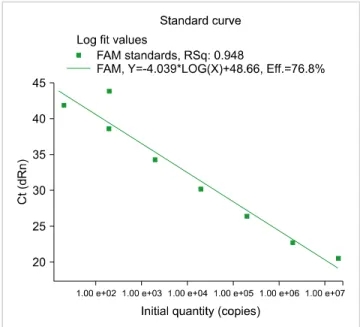
Fig. 1. TREC standard curve.
of the TCR- gene locus, which is located between clusters of V and J segments. This rearrangement forms a signal joint (sj) between the Rec segment and the downstream
J segment. sjTREC is the segment that contains the deleted D, J, and C segments [7].
Studies on the clinical utility of TRECs were initiated by screening programs for severe combined immunodeficiency (SCID) [8]. This was followed by research on the role of TREC measurement in various diseases and infections like T cell lymphoma, and HIV and retroviral infections [9-12].
The role of TREC quantification has evolved in both HSCT and solid organ transplantation. Some researchers have re- ported that pre-transplant TREC predicts acute rejection in renal transplant patients [10]. Others reported increased TREC levels during rejection episodes of cardiac transplants [13].
In the context of HSCT, studies were done in different settings and time points, and correlated with outcomes [14-16]. However, correlations between TREC levels and HSCT outcomes remain to be elucidated. Given the simplicity of the test and the provisional value in the evaluation of different outcomes of transplant, we aimed at analyzing the role of measurement of TRECs in a number of allogenic HSCT transplant recipients. We compared TREC levels to their age-matched sibling healthy donors, to different param- eters, and to different transplant outcomes. We focused on early single-point measurements to emphasize the role of the test in predicting outcomes which, in turn, may facilitate therapeutic interventions.
MATERIALS AND METHODS
PatientsÊ data
The study was conducted on 200 subjects, 100 patients receiving allogenic HSCT from an HLA-identical sibling and 100 donors taken as controls. The cases were collected from both BMT units in Alexandria and Nasser Institute, Cairo over a period of two years.
TREC analysis
DNA extraction was done using ABIOpure extraction kit (Cat No: M501DP100, Alliance Bio Inc., Bothell, WA, USA).
In some patients, T cells were separated by rosette selection technique (StemCell Technologies, Vancouver, BC, Canada) and DNA extracted. Samples were collected from controls once and compared to both pretransplant and day 28 samples from patients.
Detection of TREC values was done by real time PCR using standard curve method for target gene amplification.
Primers and probes were supplied by Applied Biosystems (ThermoFisher Scientific, California, CA, USA) with the fol- lowing sequences: CACATCCCTTTCAACCATGCT (forward primer); GCCAGCTGCAGGGTTTAGG (reverse primer);
and FAM-ACACCTCTGGTTTTTGTAAAGGTGCCCACT- TAMRA (TaqMan probe). The PCR mixture contained 10
L of mastermix (containing 0.125 L; Ampli Taq, 2.5 L;
Buffer, 1.75 L of 50 mM Mg, 0.5 L of 10 mM dNTP), 1 L of 12.5 M of each forward and reverse primer and 1 L of 5 M probe, 5 L of template DNA (or standard plasmid dilution), and 3 L water. The experiment was done on Stratagene thermocycler (ThermoFisher Scientific) with cycles as follows: 95oC for 5 min to activate the Platinum Taq, followed by 50 cycles of 95oC for 30 s and 60oC for 1 min. The standard curve was constructed using a plasmid generously provided by Dr. Daniel Douek, Vaccine Research Center, National Institute of Allergy and Infectious Diseases, Bethesda. The concentration of the plasmid as determined by its absorbance was 1.4 mg/mL. Following the instructions of the providing laboratory 994 L water was added to 6
L plasmid to yield 1 mL of 2×10⁹ plasmid copies/L. The standard curve was constructed with the following serial dilu- tions: 2×10⁷, 2×10⁶, 2×10⁵, 2×10⁴, 2×10³, 2×10², 20, and 2 plas- mid copies/L. The curve efficiency was 76.8%, RSq was 0.948, and Y intercept (slope) was -4.039 (Fig. 1).
TRECs were calculated as TREC copies/g DNA, and TRECs/mL.
TREC copies/mL blood were calculated using the following equation:
(
Total DNA (g in L WDNA (g amplified in RQ-PCR
)
× (No. of TREC)×(
1000300)
Where (WB—whole blood; RQ-PCR—real time quantita- tive PCR; No. of TREC is absolute value derived from RQ-PCR standard curve) [16].
GvHD prophylaxis
GvHD prophylaxis in patients was done using cyclosporine A from day 1 till 1 year after transplantation, as guided by trough levels. In addition, methotrexate was administered
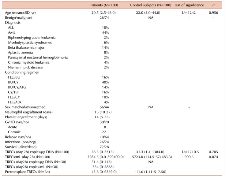
Table 1. Patient characteristics.
Patients (N=100) Control subjects (N=100) Test of significance P
Age (mean±SD, yr) 20.5 (2.5–48.0) 22.0 (3.0–44.0) U=1242 0.956
Benign/malignant 26/74 NA - -
Diagnosis
ALL 18%
AML 44%
Biphenotyping acute leukemia 2%
Myelodysplastic syndromes 6%
Beta thalassemia major 14%
Aplastic anemia 8%
Paroxysmal nocturnal hemoglobinuria 2%
Chronic myeloid leukemia 4%
Niemann pick disease 2%
Conditioning regimen
FLU/BU 16%
BU/CY 40%
BU/CY/ATG 14%
CY/TBI 16%
FLU/CY 10%
FLU/ALK 4%
Sex matched/mismatched 56/44 NA -
Neutrophil engraftment (days) 15 (10–27) Platelet engraftment (days) 14 (5–33)
GvHD (yes/no) 30/70
Acute 8
Chronic 22
Relapse (yes/no) 10/64
Infections (pos/neg) 26/74
Survival (alive/dead) 72/28
TRECs (day 28) copies/g DNA (N=100) 28.3 (0–2215) 31.3 (1.4–1384.0) U=1210.5 0.785 TRECs/mL (day 28) (N=100) 2984.5 (0.0–399400.0) 5723.0 (114.5–571483.3) 990.5 0.074
TRECs (day28) copies/g DNA (N=30) 51.4 (0–448) NA
TRECs (day28) copies/mL (N=30) 534 (0–5888)
Pretransplant TRECs (N=34) 43.6 (0–6359.0) 111.0 (1.41–517.50)
on days 1, 3, 6, 11, and 15. For B thalassemia patients, metho- trexate was replaced with steroids from day 7 to +4.
CD34 dose and source of stem cells
All cases received mobilized peripheral blood stem cells as a source of the graft. The mean and median dose of CD34+
cells was 9.1±5.7×10⁶/kg BW (range of 3.4–36.5×10⁶ cells/kg BW) and 8.55×10⁶/kg BW, respectively.
Statistical analysis
Collected data were analyzed using IBM SPSS software version 20.0 (IBM Corp, Armonk, NY, USA). Qualitative data were described using number and percent. Quantitative data were described using range, mean, standard deviation, and median values. Significance of the obtained results was judged at the 5% level.
RESULTS
Patient characteristics
Two hundred subjects were involved in this study─ 100 patients undergoing allogenic HSCT and 100 age matched controls who were their donors. Patients’ ages ranged from 2.5 to 48 years with a mean of 20.5 years, (72% adults and 28% pediatric patients). Twenty-six and 74% of cases were transplanted for benign and malignant diseases, respectively.
Additionally, 56 and 44% of cases were sex matched and mismatched, respectively. Follow-up of patients was done for a median of 2 years. Outcomes were measured as days to neutrophil and platelet engraftment, development of in- fections, and GvHD, as well as survival rates during the follow-up period. Neutrophil engraftment occurred at a me- dian of 15 days (range, 10–27) and failed in one patient, while platelet engraftment occurred at a median of 14 days (range, 5–33) and failed in three patients. Twenty-six percent of patients developed infections, while 30% developed GvHD (10 of them acute GvHD and 20 chronic GvHD). Out of
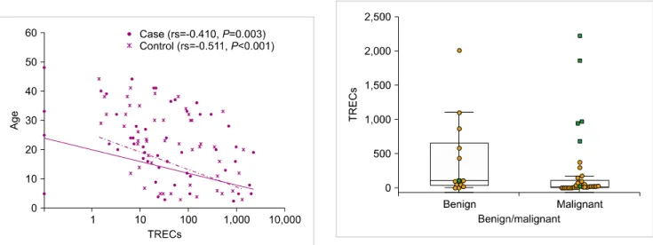
Fig. 2. Correlation between TRECs and age in patients and controls. Fig. 3. Comparison between TREC levels at day 28 in patients with benign and malignant disease.
the 74 malignant cases, 10 relapsed (13.5%). Twenty-eight percent of patients died during the follow-up period due to infections, relapse, and failure of engraftment (Table 1).
Day 28 whole blood TRECs
TRECs were measured at a single point in all patients- at day 28 post-transplant.
Median TREC levels was 28.3 copies/g DNA in patients (range, 0–2215) versus 31.3 copies/g DNA in control samples (range, 1.4–1384), while median TREC/mL blood was 2984.5 (0.0–399400.0) in patients versus 5723 (114.5–571483.3) in control samples. Therefore, no significant difference in TREC copies/g DNA at day 28 was detected between patients and controls (P=0.785). TREC levels/mL blood in patients was significantly lower than that in control (P=0.074).
Day 28 TREC levels and patient characteristics
There was a negative correlation between patients’ age and both TRECs and TRECs/mL (P=0.002 and 0.003, re- spectively). We further tested this correlation in healthy controls and found a similar decrease in TREC levels with age in healthy controls (P=0.001) (Fig. 2). No difference was detected between males and females regarding TREC and TREC/mL levels. (P=0.334 and 0.633, respectively).
There was a significant difference between TREC copies/g DNA at day 28 between patients with malignant diseases (21.0; range was 0–2215.0) and benign diseases (108.2; range was 0–1997.0) (P=0.045). Similarly, TRECs/mL value was 12178.9 (0–399400.0) and 2730.0 (0–370600.0) in benign and malignant diseases, respectively (P=0.054) (Fig. 3). Among the 38 patients aged below 18 years, 22 were transplanted for benign diseases versus 6/62 in cases above 18 years, but median levels of TRECs in benign diseases were higher in both age groups as compared to those in malignant diseases within the same age group.
Day 28 TREC levels and patient outcomes
There was no correlation between TRECs and TRECs/mL and days to neutrophil and platelet engraftment. There was
no significant difference in TREC levels between patients who developed infections and GvHD. However, levels of TRECs at day 28 were lower in patients who later developed infections, and higher in those who developed GvHD.
Median TREC level values were 52.9 copies/g DNA (range, 0–2215.0) in patients who developed GvHD versus 23.4 cop- ies/g DNA (0–1853.0) in those who didn’t (P=0.330).
Additionally, median values were 23.4 copies/g DNA (0–
2215.0) in patients with infections versus 35.0 copies/g DNA (0–1997.0) in patients without infections (P=0.626). Infectious complications were cytomegalovirus (CMV) reactivation in six patients, Herpes simplex virus in two, and severe bacterial sepsis and pneumonia in 18 patients. We could not study TREC levels in patients according to the type of infections due to the small number of patients in each group.
We compared TREC levels at day 28 between patients who showed a relapse (N=10) and other patients who suffered malignant diseases but did not show a relapse (N=64). Patients who showed a relapse had lower median levels though with no significant difference [21.0 (0–175.2) vs. 44.821.0 (0–2215.0) copies/g DNA]. No correlation was detected between TREC levels and survival during the follow-up period (Fig. 4).
We further divided our cases into low or high levels ac- cording to the median TREC levels, which were 28.3 and 2894.5 for TRECs and TRECs/mL, respectively. We found a statistically significant difference between TREC levels in patients with benign and malignant diseases─75.6% of malignant patients had low TREC levels, whereas 24.4%
had high TREC levels (P=0.02). Additionally, none of the patients with high TRECs/mL showed a relapse, while 15%
of patients with low TRECs/mL showed a relapse (P=0.009).
No other differences were detected between the patients with low and high TREC levels (Table 2).
Multivariate analysis
Multivariate analysis was conducted to study the different variables in correlation to TREC levels. Multivariate analysis showed significance with age (P=0.026) and relapse (P=
Fig. 4. TREC levels at day 28 and outcomes of transplantation.
Table 2. Relationship between outcomes and patient groups divided as having high or low TREC/mL.
TRECs/mL High
(≥2894.5) Low
(<2894.5) P
Malignant (N=74) 24.4% 75.6% 0.021
GvHD (N=30) 53% 47% 0.216
Infections (N=26) 30.8% 69.2% 1.000
Relapse (N=10) 0% 100% 0.009
Death (N=28) 26.6% 71.4% 0.7
-0.037) and an approaching significance regarding survival (P=0.075).
T cell TRECs
TRECs were measured from DNA isolated from T cells at day 28 in 30 cases. Measurement of TRECs from T cell DNA did not seem to add any value to the test. T cell TRECs were 51.4 copies/g DNA (0–448) while T cell TREC/mL was 534 copies/mL blood (0–5888).T cell TRECs did not correlate with whole blood TRECs in these thirty cases (rs=0.064 and P=0.820 for TRECs, and rs=0.075 and P=0.791 for TRECs/mL).
Pre-transplant TRECs
TRECs were measured in 34 patients pre-transplant. No difference was detected between pre-transplant patient and control samples. The median levels in pre-transplant patients were 43.61 copies/g DNA (0–635) versus 111.0 copies/g DNA (1.41–517.50) in the control samples. There was no significant difference between the pre-transplant and post-transplant levels of TRECs. Mean TREC/g DNA levels were 487.2 versus 309.9 copies, while mean TREC/mL values were 7569 versus 7098.8 copies (P=0.177 and 0.501, re- spectively). The levels of pre-transplant TRECs seemed to correlate with post-transplant levels (r=0.473; P=0.055) but did not seem to predict outcomes. This shows that the level of TRECs pre-transplant is an important determinant of post-transplant levels.
DISCUSSION
TRECs have been studied as markers of immune recon- stitution following HSCT. However, the role of this test as a predictor of outcome in HSCT remains to be elucidated.
The aim of our work was to study TREC levels at a single point post-transplantation, correlate it with different patient parameters, and test whether the levels would predict out- comes of transplant. The study was conducted on 200 sub-
jects−100 patients undergoing HSCT from an HLA identical sibling and their donors were taken as controls.
We chose the measurement at day 28 for sampling feasi- bility, as this is the point when samples are withdrawn for chimerism testing. Additionally, this time point is early, so their role in prediction of outcomes could also be studied.
Initially, we had collected the samples at engraftment, but this would have varied from patient to patient. Previously, studies have used many different points for measurement of TRECs, from day 15 to one-year post-transplant [17, 18].
At day 28, there was no statistically significant difference in TRECs/g DNA levels between the patients and controls.
Alternatively, the TREC copies/mL values were lower in patients than in controls, with a P-value approaching sig- nificance (0.07). Previous studies have not measured TREC levels before three months post-transplant, nor have they reported the full recovery of TRECs before 3–9 months [17-19]. Considering our data regarding TREC/g DNA, it seems that patients reached the same level of TRECs as their controls. One possible reason for this relatively rapid recov- ery of TRECs is that all our patients had a completely matched sibling donor. In a study by Fu et al. [19] TREC recovery was the most rapid in patients with a matched sibling donor compared to haploidentical sibling or matched unrelated donors. We should rely more on TRECs/mL blood as the number of TRECs/mL is independent of cell count, cell death, and dilution by peripheral expansion of naive T cells [16].
In a study by Ringhoffer et al. [20] the effect of cell pro- liferation causing dilution of TRECs was overcome by consid- ering the ratio of sjTREC and TREC.
In the current study the three factors that affected TREC levels post-transplant were age, nature of the disease, ie, whether benign or malignant, and TREC levels pre-transplant.
In accordance with published data, TRECs had a strong negative correlation with age in controls and patients at day 28. Age has also been inversely correlated with thymopoiesis. This has been reported in normal individuals and in many clinical situations, including recipients of high-dose chemotherapy, HSCT recipients, and patients in- fected with human immunodeficiency virus [21]. This is attributed to the thymic involution occurring with age and contributes to superior outcomes of HSCT in younger patients [22, 23]. This indicates another reason for the patient and control TREC levels being comparable. Controls were patient siblings, mostly in the same age category. Since age affects TREC levels the most, it seems reasonable that the difference between patients and controls would not be significant.
Patients transplanted for malignant diseases showed sig- nificantly lower TREC levels than benign diseases. One study states that thymopoiesis was markedly reduced both in newly diagnosed and chemotherapy-treated patients with acute lymphocyte leukemia. Others have reported decreased TREC numbers and poorer thymic function following treatment in patients with hematological diseases [1, 24-26]. Moreover, a recent study has shown that TBI used in conditioning of some of malignant cases compromises cortical and medul- lary thymic epithelial cells (TEC), a critical population for
thymic renewal and thymopoiesis [27].
Pre-transplant thymic function is perhaps one of the least studied possible effects on TREC levels. One study used pre-transplant TREC levels as a prognostic factor for trans- plant outcome. They found that patients with high TREC levels have better OS, and decreased incidence of severe GvHD and opportunistic infections. Another study found similar results, with a correlation between high levels of sjTRECs before HSCT and a reduced risk of death, mainly due to a reduction in the incidence of relapse. Unfortunately, in both studies pre-transplant TREC levels were not corre- lated to TREC levels post HSCT or other thymic activity parameters [28, 29]. In our study pre-transplant TRECs corre- lated with post-transplant values, though none of them corre- lated with clinical outcomes.
We did not find any correlation between day 28 levels of TRECs and either development of infection or GvHD.
However, we observed that patients who developed in- fections had lower TRECs and those who developed GvHD had higher TRECs at this time point. A number of studies have referred to decreased levels of TRECs at the onset of acute GvHD and during chronic GvHD. They have gone further to include the thymus as one of the organs involved in GvHD damage [30-32]. None have, however, used this single point measurement to predict occurrence of GvHD.
None of our patients had actually developed GvHD at the time of sampling. We assume that increased level of TRECs in patients is one mechanism of host naive T cell proliferation, leading to GvHD. Similarly, a study conducted in renal trans- plant patients reported increased levels of recent thymic emigrants (RTE) before renal graft rejection [33]. Skert et al. [34] reported that the level of TRECs was increased at day 28 in patients who developed chronic GvHD, among many other changes in immunological variables. This would further support our data and may urge an extension of this single point measurement study on larger numbers of patients.
Although statistically insignificant, we found lower levels of TRECs in patients who developed infections. This is con- tradictory to most of the published data, which report sig- nificantly lower levels of TRECs in patients who develop infections [35]. In a study by da Rocha et al. [36] a 3-fold lower risk of developing severe infections was observed in those patients who had effective sjTREC+ T-cell recovery at 6 months, and a 9-fold lower risk at 12 months. In another study, lower TREC levels at 9 months were related to CMV episodes, but not to other infections [36]. We did not segre- gate based on types of infection in our study.
None of the patients exhibiting high TREC levels/mL showed a relapse, while 15% of patients with low TREC/mL showed a relapse and the result was statistically significant (P=0.009). A study by Uzunel et al. [37] reported that in acute myeloid leukemia (AML) patients, low TREC level 2 months post-transplantation was correlated to high relapse incidence at 5 years, while in patients with chronic leukemia and myelodysplastic syndrome (MDS), high TREC levels were correlated with improved relapse-free survival. This,
together with our results, strongly suggests the utility of TREC levels to predict relapse. Sairafi et al. [38] also support the use of TREC measurement as part of the standard reper- toire of immunological monitoring after autologous stem-cell transplantation (ASCT).
The published data on TRECs in the field of transplantation show great variation. This is probably due to many factors.
One of them is the variability in method design, since some use the absolute quantification of TREC, while others use relative quantification by the delta-CT method. Another is the quantification of TREC in different subpopulations, like CD3+, CD4+ or CD8+ T cells. In addition, TREC results have been expressed in different ways, such as TREC/cell count, TREC/mL or L of blood, or even TREC/g of DNA. sjTREC levels are also influenced by many factors, such as longevity of naïve T cells, peripheral expansion or apoptosis of T cells, and intracellular degradation [14, 16].
We aimed at studying whole TREC levels at a single point and to detect its utility as a cost-effective test. Limitations of the study include heterogeneity of the patients, though this is the case in many studies involving HSCT. Another limitation is that different T cell subset counts were not recorded and we did not follow up TREC levels at different time points, but we aimed at detecting the utility of a simple and cost-effective test and its predictive value at a single point.
We present some preliminary data in this study that should be further investigated. We aim at extending this research to larger groups of HSCT recipients and stratifying them into subgroups according to the different patient-related and transplant-related variables. The simplicity of the test and the predictive value would make it a likely candidate for routine testing post-transplantation.
ACKNOWLEDGMENTS
We would like to acknowledge Dr. Gamal Eldin Mohammed Fathy for helping us in the collection of patient samples and data from Nasser Institute, Cairo.
The study protocol was approved by the ethics committee at Alexandria Faculty of Medicine and informed consent was obtained from all patients or caregivers of the included children, since participation was voluntary.
AuthorsÊ Disclosures of Potential Conflicts of Interest
No potential conflicts of interest relevant to this article were reported.
REFERENCES
1. Toubert A, Glauzy S, Douay C, Clave E. Thymus and immune reconstitution after allogeneic hematopoietic stem cell transplantation in humans: never say never again. Tissue Antigens
2012;79:83-9.
2. van den Brink MR, Velardi E, Perales MA. Immune reconstitution following stem cell transplantation. Hematology Am Soc Hematol Educ Program 2015;2015:215-9.
3. Ogonek J, Kralj Juric M, Ghimire S, et al. Immune reconstitution after allogeneic hematopoietic stem cell transplantation. Front Immunol 2016;7:507.
4. Chaudhry MS, Velardi E, Malard F, van den Brink MR. Immune reconstitution after allogeneic hematopoietic stem cell transplantation: time to t up the thymus. J Immunol 2017;198:
40-6.
5. de Koning C, Plantinga M, Besseling P, Boelens JJ, Nierkens S.
Immune reconstitution after allogeneic hematopoietic cell transplantation in children. Biol Blood Marrow Transplant 2016;22:195-206.
6. van der Spek J, Groenwold RH, van der Burg M, van Montfrans JM. TREC based newborn screening for severe combined immunodeficiency disease: a systematic review. J Clin Immunol 2015;35:416-30.
7. Yamanaka K, Yawalkar N, Jones DA, et al. Decreased T-cell receptor excision circles in cutaneous T-cell lymphoma. Clin Cancer Res 2005;11:5748-55.
8. Serana F, Chiarini M, Zanotti C, et al. Use of V(D)J recombination excision circles to identify T- and B-cell defects and to monitor the treatment in primary and acquired immunodeficiencies. J Transl Med 2013;11:119.
9. Drylewicz J, Vrisekoop N, Mugwagwa T, et al. Reconciling longitudinal naive T-cell and TREC dynamics during HIV-1 infection. PLoS One 2016;11:e0152513.
10. Bamoulid J, Courivaud C, Crepin T, et al. Pretransplant thymic function predicts acute rejection in antithymocyte globulin- treated renal transplant recipients. Kidney Int 2016;89:1136-43.
11. Morgun A, Shulzhenko N, Socorro-Silva A, Diniz RV, Almeida DR, Gerbase-Delima M. T cell receptor excision circles (TRECs) in relation to acute cardiac allograft rejection. J Clin Immunol 2004;24:612-6.
12. Weinberg K, Blazar BR, Wagner JE, et al. Factors affecting thymic function after allogeneic hematopoietic stem cell transplantation.
Blood 2001;97:1458-66.
13. Przybylski GK, Kreuzer KA, Siegert W, Schmidt CA. No recovery of T-cell receptor excision circles (TRECs) after non-myeloablative allogeneic hematopoietic stem cell transplantation is correlated with the onset of GvHD. J Appl Genet 2007;48:397-404.
14. Gaballa A, Sundin M, Stikvoort A, et al. T cell receptor excision circle (TREC) monitoring after allogeneic stem cell transplantation;
a predictive marker for complications and clinical outcome. Int J Mol Sci 2016;17:E1705.
15. Gaballa A, Norberg A, Stikvoort A, et al. Assessment of TREC, KREC and telomere length in long-term survivors after allogeneic HSCT: the role of GvHD and graft source and evidence for telomere homeostasis in young recipients. Bone Marrow Transplant 2018;53:69-77.
16. Lorenzi AR, Patterson AM, Pratt A, et al. Determination of thymic function directly from peripheral blood: a validated modification to an established method. J Immunol Methods 2008;339:185-94.
17. Eyrich M, Wollny G, Tzaribaschev N, et al. Onset of thymic recovery and plateau of thymic output are differentially regulated
after stem cell transplantation in children. Biol Blood Marrow Transplant 2005;11:194-205.
18. Jiménez M, Martínez C, Ercilla G, et al. Reduced-intensity conditioning regimen preserves thymic function in the early period after hematopoietic stem cell transplantation. Exp Hematol 2005;33:1240-8.
19. Fu YW, Wu DP, Cen JN, et al. Patterns of T-cell reconstitution by assessment of T-cell receptor excision circle and T-cell receptor clonal repertoire after allogeneic hematopoietic stem cell transplantation in leukemia patients--a study in Chinese patients.
Eur J Haematol 2007;79:138-45.
20. Ringhoffer S, Rojewski M, Döhner H, Bunjes D, Ringhoffer M.
T-cell reconstitution after allogeneic stem cell transplantation:
assessment by measurement of the sjTREC/TREC ratio and thymic naive T cells. Haematologica 2013;98:1600-8.
21. Weinberg K, Blazar BR, Wagner JE, et al. Factors affecting thymic function after allogeneic hematopoietic stem cell transplantation.
Blood 2001;97:1458-66.
22. Velardi E, Dudakov JA, van den Brink MR. Clinical strategies to enhance thymic recovery after allogeneic hematopoietic stem cell transplantation. Immunol Lett 2013;155:31-5.
23. Krenger W, Blazar BR, Holländer GA. Thymic T-cell development in allogeneic stem cell transplantation. Blood 2011;117:6768-76.
24. Svaldi M, Lanthaler AJ, Dugas M, et al. T-cell receptor excision circles: a novel prognostic parameter for the outcome of transplantation in multiple myeloma patients. Br J Haematol 2003;122:795-801.
25. Li Y, Yin Q, Yang L, et al. Reduced levels of recent thymic emigrants in acute myeloid leukemia patients. Cancer Immunol Immunother 2009;58:1047-55.
26. Haining WN, Neuberg DS, Keczkemethy HL, et al. Antigen- specific T-cell memory is preserved in children treated for acute lymphoblastic leukemia. Blood 2005;106:1749-54.
27. Williams KM, Mella H, Lucas PJ, Williams JA, Telford W, Gress RE. Single cell analysis of complex thymus stromal cell populations: rapid thymic epithelia preparation characterizes radiation injury. Clin Transl Sci 2009;2:279-85.
28. Clave E, Rocha V, Talvensaari K, et al. Prognostic value of pretransplantation host thymic function in HLA-identical sibling hematopoietic stem cell transplantation. Blood 2005;105:2608-13.
29. Saglio F, Cena S, Berger M, et al. Association between thymic function and allogeneic hematopoietic stem cell transplantation outcome: results of a pediatric study. Biol Blood Marrow Transplant 2015;21:1099-105.
30. Törlén J, Gaballa A, Remberger M, et al. Effect of graft-versus-host disease prophylaxis regimens on T and B cell reconstitution after allogeneic hematopoietic stem cell transplantation. Biol Blood Marrow Transplant 2019;25:1260-8.
31. Krenger W, Schmidlin H, Cavadini G, Holländer GA. On the relevance of TCR rearrangement circles as molecular markers for thymic output during experimental graft-versus-host disease. J Immunol 2004;172:7359-67.
32. Wu X, Zhu K, Du X, et al. Frequency analysis of TRBV subfamily sjTRECs to characterize T-cell reconstitution in acute leukemia patients after allogeneic hematopoietic stem cell transplantation.
J Hematol Oncol 2011;4:19.
33. Bamoulid J, Courivaud C, Crepin T, et al. Pretransplant thymic function predicts acute rejection in antithymocyte globulin- treated renal transplant recipients. Kidney Int 2016;89:1136-43.
34. Skert C, Perucca S, Chiarini M, et al. Sequential monitoring of lymphocyte subsets and of T-and-B cell neogenesis indexes to identify time-varying immunologic profiles in relation to graft-versus-host disease and relapse after allogeneic stem cell transplantation. PLoS One 2017;12:e0175337.
35. Wils EJ, van der Holt B, Broers AE, et al. Insufficient recovery of thymopoiesis predicts for opportunistic infections in allogeneic hematopoietic stem cell transplant recipients. Haematologica 2011;96:1846-54.
36. da Rocha LKA, Freschi de Barros S, Bandeira F, et al . Thymopoiesis in pre- and post-hematopoietic stem cell transplantation. Front Immunol 2018;9:1889.
37. Uzunel M, Sairafi D, Remberger M, Mattsson J, Uhlin M. T-cell receptor excision circle levels after allogeneic stem cell transplantation are predictive of relapse in patients with acute myeloid leukemia and myelodysplastic syndrome. Stem Cells Dev 2014;23:1559-67.
38. Sairafi D, Mattsson J, Uhlin M, Uzunel M. Thymic function after allogeneic stem cell transplantation is dependent on graft source and predictive of long term survival. Clin Immunol 2012;142:
343-50.