Influence of genetic polymorphisms in the folate pathway on toxicity after high-dose methotrexate treatment in pediatric osteosarcoma
Jeong A Park
1, Hee Young Shin
21Department of Pediatrics, Haeundae Paik Hospital, Inje University College of Medicine, Busan, 2Department of Pediatrics, Cancer Research Institute, Seoul National University Children’s Hospital, Seoul National University College of Medicine, Seoul, Korea
p-ISSN 2287-979X / e-ISSN 2288-0011 http://dx.doi.org/10.5045/br.2016.51.1.50 Blood Res 2016;51:50-7.
Received on January 29, 2016 Revised on March 14, 2016 Accepted on March 22, 2016
Background
Methotrexate (MTX), one of the main drugs used to treat osteosarcoma, is a representative folic acid antagonist. Polymorphisms of various enzymes involved in the metabolism of MTX could contribute to differences in response to MTX in pediatric osteosarcoma patients.
Methods
Blood and tissue samples were obtained from 37 pediatric osteosarcoma patients who were treated with high-dose MTX therapy. The following 4 single nucleotide poly- morphisms (SNPs) were analyzed: ATIC 347C>G, MTHFR 677C>T, MTHFR 1298A>C and SLC19A1 80G>A. Serial plasma MTX concentrations after high-dose MTX therapy and MTX-induced toxicities were evaluated. Correlations among polymorphisms, MTX concentrations and treatment-induced toxicities were assessed.
Results
Plasma MTX levels at 48 hours after high-dose MTX infusion were significantly associated with SLC19A1 80G>A (P=0.031). Higher plasma levels of MTX at 48 and 72 hours were significantly associated with MTX-induced mucositis (P=0.007 and P=0.046) and renal toxicity (P=0.002), respectively. SNP of SLC19A1 gene was associated with development of severe mucositis (P=0.026).
Conclusion
This study suggests that plasma levels of MTX are associated with GI and renal toxicities after high-dose MTX therapy, and genetic polymorphisms that affect the metabolism of MTX may influence drug concentrations and development of significant side effects in pediatric patients treated with high-dose MTX.
Key Words Pediatric, Osteosarcoma, Methotrexate, Toxicity, Single nucleotide polymorphism
*This research was supported by a grant from the Alumni of Pediatrics, Seoul National University Children’s Hospital, Seoul, Korea.
Correspondence to Hee Young Shin, M.D., Ph.D.
Department of Pediatrics, Cancer Research Institute, Seoul National University Children’s Hospital, Seoul National University College of Medicine, 101 Daehak-ro, Jongno-gu, Seoul 03080, Korea E-mail: [email protected]
Ⓒ 2016 Korean Society of Hematology
INTRODUCTION
High-dose methotrexate (MTX) with leucovorin (5-formyl- tetrahydrofolic acid) rescue in combination with doxorubicin and a platinum agent has served as a cornerstone of neo- adjuvant chemotherapy for osteosarcoma. High-dose MTX treatment is associated with various toxicities, including tox- icities of central nervous system (CNS), liver, kidney, bone marrow and gastrointestinal system, particularly oral mucosa.
Various studies have reported that several pharmacokinetic parameters, including a high plasma MTX concentration and
prolonged exposure to high levels of MTX, were associated with the development of high-dose MTX-induced toxicities [1-3]. However, MTX levels exhibit significant inter-in- dividual variability, and acute toxicity after high-dose MTX is often unexpected and not typically dose dependent; thus, it is difficult to predict who will develop a more serious adverse response to high-dose MTX. Folate pathway genes are involved in the metabolism of MTX and are very polymorphic. Numerous pharmacogenetic studies have re- ported that single nucleotide polymorphisms (SNPs) alter the activity or expression of folate pathway enzymes, which may influence the response to and toxicity caused by MTX
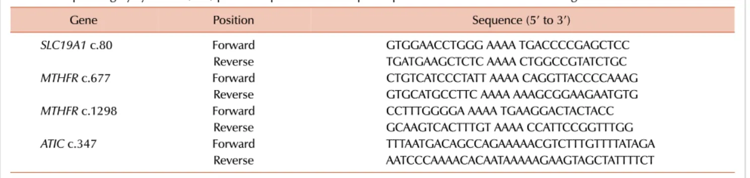
Table 1. Sequencing by synthesis (SBS) primer sequences for multiple amplification of methotrexate related genes.
Gene Position Sequence (5’ to 3’)
SLC19A1 c.80 Forward GTGGAACCTGGG AAAA TGACCCCGAGCTCC
Reverse TGATGAAGCTCTC AAAA CTGGCCGTATCTGC
MTHFR c.677 Forward CTGTCATCCCTATT AAAA CAGGTTACCCCAAAG
Reverse GTGCATGCCTTC AAAA AAAGCGGAAGAATGTG
MTHFR c.1298 Forward CCTTTGGGGA AAAA TGAAGGACTACTACC
Reverse GCAAGTCACTTTGT AAAA CCATTCCGGTTTGG
ATIC c.347 Forward TTTAATGACAGCCAGAAAAACGTCTTTGTTTTATAGA
Reverse AATCCCAAAACACAATAAAAAGAAGTAGCTATTTTCT
in various malignancies and autoimmune diseases [4-6].
However, comparatively few data are available regarding associations between various genetic polymorphisms of the folate pathway genes and pediatric osteosarcoma. We hy- pothesized that polymorphisms of the folate pathway genes may influence plasma concentrations of MTX and MTX-in- duced toxicity in pediatric patients with osteosarcoma.
Among various candidate SNPs in folate pathway genes, SNPs in solute carrier family 19, member1 (SLC19A1), meth- ylenetetrahydrofolate reductase (MTHFR) and 5-amino- imidazole-4-carboxamide ribonucleotide formyltransferase (ATIC) are potential biomarkers that confer susceptibility to MTX. In this study, we retrospectively examined whether SLC19A1 80G>A, MTHFR 677C>T, MTHFR 1298A>C, and ATIC 347C>G polymorphisms affected plasma MTX concentrations and susceptibility to high-dose MTX-induced toxicity in Korean pediatric patients with osteosarcoma.
MATERIALS AND METHODS
Study population
The patients included in this study were Korean children with osteosarcoma who were treated with high-dose MTX-containing chemotherapeutic protocols between 1986 and 2010. This study was approved by the Ethics Committee of Institutional Review Board (IRB No. H-0906-067-284).
In all cases, informed consent was obtained from the patients, their parents or both. The patients received high-dose MTX according to the following treatment protocols: CCG 7921A, CCG 7921B and COG AOST 0331 [7, 8]. High-dose MTX at 12 g/m2 was administered separately by an interval of 1 week. Each infusion lasted for 4 hours. Intravenous hydra- tion and alkalization were achieved 12 hours prior to the start of MTX therapy. High-dose MTX was started at a urine pH>7.0 to provide protection against MTX-induced renal dysfunction. Leucovorin rescue was initiated 24 hours after the start of MTX infusion at a dose rate of 15 mg/m2, and the dosages were adjusted based on plasma MTX concen- trations and continued until plasma MTX levels reached
<0.1 mol/L. The patient characteristics and clinical data used in this study were collected retrospectively.
MTX concentration and toxicity
Therapeutic drug monitoring was performed at 24, 48 and 72 hours from the beginning of the infusion of high-dose MTX. Plasma MTX concentrations were measured by a fluo- rescence polarization immunoassay on a TDx system (Abbot Laboratories, Abbot Park, IL). The highest plasma levels of total bilirubin, aspartate aminotransferase (AST), alanine aminotransferase (ALT), blood urea nitrogen (BUN) and crea- tinine were used as criteria for liver and kidney toxicity.
Grades of mucositis, liver, kidney and neurologic toxicities were assessed according to the CTCAE 3.0 [9].
DNA extraction and genotyping
Genomic DNA from peripheral blood and formalin-fixed, paraffin-embedded (FFPE) tumor tissues from the 37 patients were analyzed. Genomic DNA was extracted from peripheral blood lymphocytes and tumor tissues using a QIAamp DNA Mini Kit and a QIAamp DNA FFPE Tissue kit (QIAGEN, Hilden, Germany), respectively, according to the manu- facturer’s instructions. DNA yield, integrity and protein con- tamination were determined by spectrophotometry.
Candidate SNPs of genes involved in the folate pathway and the transport of MTX included SLC19A1 80G>A, MTHFR 677C>T and 1298A>C, and ATIC 347C>G.
Gene amplification
PCR amplification was performed in a total volume of 20 L with a mixture containing 5 M forward and reverse sequencing by synthesis (SBS) primers (Table 1), 0.25 mM deoxynucleotide (dNTP) mix, 112.5 mM Tris HCl (pH 9.0), 3 mM MgCl2, 75 mM KCl, 30 mM (NH4)2SO4, and 1.5 U of Taq polymerase (Biotools, Madrid, Spain). The reactions were preheated to 94oC for 2 minutes, followed by 10 amplifi- cation cycles, which consisted of the following: denaturation at 94oC for 15 seconds, annealing at 50oC for 30 seconds and extension at 72oC for 1 minute. This protocol was fol- lowed by 40 amplification cycles using following parameters:
denaturation at 94oC for 15 seconds, annealing at 60oC for 230 seconds and extension at 72oC for 40 seconds in a 96-well thermal cycler (Applied Biosystems, CA, USA). Sixteen mi- croliters of PCR product was mixed with 4 L of an activation solution (YeBT, Seoul, Korea), and the resultant mixture was incubated at 37oC for 60 minutes and 85oC for 15 minutes.
Table 2. Allele-specific primer extension (ASPE) primer sequences.
Gene Taq sequence Sequence (5’ to 3’)
SLC19A1 80G TCAAAATCTCAAATACTCAAATCA AAAGGTAGCACACGAGGC
SLC19A1 80A CAATTCAAATCACAATAATCAATC AAAGGTAGCACACGAGGT
MTHFR 677C CTAACTAACAATAATCTAACTAAC TGCGTGATGATGAAATCGG
MTHFR 677T AATCTTACTACAAATCCTTTCTTT TGCGTGATGATGAAATCGA
MTHFR 1298A CTACAAACAAACAAACATTATCAA GAGCTGACCAGTGAAGA
MTHFR 1298C CAATATACCAATATCATCATTTAC GAGCTGACCAGTGAAGC
ATIC 347C TCAATTACCTTTTCAATACAATAC CACAGCCTCCTCAACAG
ATIC 347G TTACTCAAAATCTACACTTTTTCA CACAGCCTCCTCAACAC
Table 3. Characteristics of patients.
Characteristics No. of Patients
Total number of patients 37
Age at diagnosis (yr) 7.2–15.7 (median 11.8)
Gender (male/female) 21/17
Primary site
Femur 18 (48.6%)
Tibia/fibula 10 (27.0%)
Humerus/radius 6 (16.2%)
Axial 3 (8.1%)
Metastasis at diagnosis
Absent 24 (64.9%)
Present 13 (35.1%)
Histological subtype
Osteoblastic 32 (86.5%)
Chondroblastic 3 (8.1%)
Telangiectatic 2 (5.4%)
Total number of high-dose MTX therapy 393
No. of high-dose MTX therapy per person 2–20 (median 10)
Labeling of activation products
Five L of activated PCR product was added to 20 L of an allele-specific primer extension (ASPE) reaction mix- ture containing 75 mM Tris HCl (pH 9.0), 2 mM MgCl2, 50 mM KCl, 20 mM (NH4)2SO4, 25 nM ASPE primer mix (Table 2), 6 mM biotin dCTP, 50 mM of dATP, dGTP, dTTP mix and 1.0 U of Taq polymerase (Biotools, Spain). The reactions were denatured at 94oC for 5 minutes, followed by 35 amplification cycles to label the amplicons: 94oC for 30 seconds, 55oC for 1 minute, 72oC for 2 minutes and a final extension at 72oC for 7 minutes.
Hybridization of amplicons
Microspheres (FlexMAP beads, Tm Bioscience, Toronto, Canada) with anti-tags for each allele-specific primer were added to 22 L of the ASPE reaction product to a final volume of 42 L in 2× Hybrisol (YeBT, Seoul, Korea). The mixtures were denatured for 10 min at 95oC, and then in- cubated for 30 min at 37oC. The microspheres were washed thrice in 160 L of TM hybridization buffer (0.2 M NaCl, 0.1 M Tris (pH 8.0), 0.08% Triton X-100). Streptavidin-R- phycoerythrin (SAPE, MOSS, Pasadena, MD) was diluted 1:500 in TM hybridization buffer, and 100 L was added to the microsphere-hybridized ASPE reaction products. The mixture was incubated for 15 min at room temperature.
Data analysis
Microsphere fluorescence was measured using a Luminex 200 cytometer (Luminex, Austin, TX). Data were collected from a minimum of 50 microspheres of each type. Masterplex GT software (Miraibio, San Francisco, CA) was used to ana- lyze SNP genotypes. Homozygous alleles were discriminated by differences of 25% in median fluorescence intensity (MFI).
The difference ratio was calculated by dividing the net MFI of one allele by the sum of the net MFI of all alleles and multiplying by 100.
Statistical analysis
The associations between SNPs and plasma MTX concen- trations at 24, 48, and 72 hours after high-dose MTX infusion were evaluated using one way ANOVA, Kruskal-Wallis test and Mann-Whitney test. Analyses of plasma MTX concen- trations and MTX-induced toxicities were performed using
Mann-Whitney test. The frequencies of severe complications among SNPs were compared with a chi-square test and Fisher’s exact test. P-values less than 0.05 were considered as statistically significant. All statistical analyses were per- formed using SPSS version 22 (SPSS, Inc., Chicago, IL, USA).
RESULTS
A total of 37 pediatric patients with osteosarcoma were enrolled in this study. Table 3 presents the characteristics of the patients. The median age was 11.8 years, and the femur was the most common site of primary tumor.
One-third of the patients had distant metastasis at diagnosis.
Each patient received a median of 10 cycles (range, 2–20) of high-dose MTX therapy. For some patients high-dose MTX was associated with serious toxicities resulting in treatment interruption or discontinuation. A total of 393 courses of high-dose MTX chemotherapy were evaluated in this study.
Plasma MTX concentration and genetic polymorphism The distribution of the examined folate pathway gene
Fig. 1. Plasma methotrexate levels at 48 hours after high-dose methotrexate infusion were significantly associated with the 80G>A variants of SLC19A1 (P=0.03).
Table 4. Genotype frequencies of the patients.
Polymorphism Genotype No. of patients (%)
ATIC 347C>G CC 18 (48.6)
CG 7 (18.9)
GG 2 (5.4)
N/A 10 (27.0)
MTHFR 677C>T CC 9 (24.3)
CT 18 (48.6)
TT 8 (21.6)
N/A 2 (5.4)
MTHFR 1298A>C AA 21 (56.8)
AC 15 (40.5)
CC 1 (2.7)
SLC19A1 80G>A GG 8 (21.6)
GA 15 (40.5)
AA 14 (37.8)
Table 5. Associations between SNPs and plasma methotrexate concentrations at 48 hours after high-dose methotrexate therapy.
Polymorphism Genotype No. of patients MTX level, median (range) P
ATIC 347C>G CC 17 0.51 (0.09–7.5)
0.971
CG 7 0.65 (0.21–1.35)
GG 2 0.94 (0.21–1.66)
CG/GG 9 0.65 (0.21–1.66) 0.833
MTHFR 677C>T CC 8 0.62 (0.34–1.36)
0.965
CT 17 0.65 (0.09–97.3)
TT 8 0.77 (0.15–4.98)
CT/TT 25 0.65 (0.09–97.3) 0.821
MTHFR 1298A>C AA 20 0.78 (0.09–97.3)
0.416
AC 14 0.43 (0.13–7.5)
CC 1 1.36
AC/CC 15 0.47 (0.13–7.5) 0.521
SLC19A1 80G>A GG 8 1.26 (0.37–6.8)
0.031
GA 15 0.34 (0.13–1.92)
AA 12 0.67 (0.09–97.3)
GA/AA 27 0.47 (0.09–97.3) 0.030
SNPs is listed in Table 4. These SNPs consisted of common polymorphisms in SLC19A1 (G80A, rs1051266), MTHFR (C677T, rs1801133; A1298C, rs1801131), and ATIC (C347G, rs2372536). Plasma MTX concentrations were measured at 24, 48 and 72 hours from the start of infusion of high-dose MTX. The median MTX concentrations were 4.7 mol/L (range, 0.19–378.6 mol/L) at 24 hours, 0.65 mol/L (range, 0.09–97.3 mol/L) at 48 hours, and 0.3 mol/L (range, 0.04–
9.75 mol/L) at 72 hours. Whereas the 347C>G poly- morphism of ATIC and the 677C>T and 1298A>C poly- morphisms of MTHFR did not show any correlations with serial plasma MTX levels after high-dose MTX therapy, the 80G>A variants of SLC19A1 had significantly lower plasma MTX levels at 48 hours (P=0.03), (Table 5 and Fig. 1).
Plasma MTX concentration and MTX-induced toxicity High-dose MTX-induced toxicity was recorded and graded
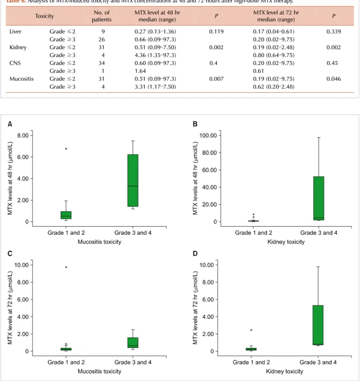
Fig. 2. Plasma methotrexate levels at 48 (A, B) and 72 hours (C, D) after high-dose methotrexate infusion were significantly associated with a development of grade 3 and 4 of mucositis and kidney toxicity.
Table 6. Analysis of MTX-induced toxicity and MTX concentrations at 48 and 72 hours after high-dose MTX therapy.
Toxicity No. of
patients MTX level at 48 hr
median (range) P MTX level at 72 hr
median (range) P
Liver Grade ≤2 9 0.27 (0.13–1.36) 0.119 0.17 (0.04–0.61) 0.339
Grade ≥3 26 0.66 (0.09–97.3) 0.20 (0.02–9.75)
Kidney Grade ≤2 31 0.51 (0.09–7.50) 0.002 0.19 (0.02–2.48) 0.002
Grade ≥3 4 4.36 (1.35–97.3) 0.80 (0.64–9.75)
CNS Grade ≤2 34 0.60 (0.09–97.3) 0.4 0.20 (0.02–9.75) 0.45
Grade ≥3 1 1.64 0.61
Mucositis Grade ≤2 31 0.51 (0.09–97.3) 0.007 0.19 (0.02–9.75) 0.046
Grade ≥3 4 3.31 (1.17–7.50) 0.62 (0.20–2.48)
according to Common Terminology Criteria for Adverse Events version (CTCAE) 3.0. At least one episode of grade 3–4 liver toxicity was experienced by 26 patients (70.3%).
In addition, grade 3 to 4 kidney toxicity and neurotoxicity were observed in 4 and 1 patients (10.8% and 2.7%), respecti- vely. Mucositis higher than grade 3 was experienced by 4 patients (10.8%). The results of the analysis on MTX-in- duced toxicity and plasma MTX concentrations at 48 and
72 hours after high-dose MTX infusion are displayed in Table 6. Severe liver, kidney, and neurologic toxicities and mucosi- tis were as defined by the presence of grade 3 or higher symptoms. Higher plasma MTX levels at 48 hours were asso- ciated with severe mucositis and kidney toxicity (P=0.007 and P=0.002), and MTX levels at 72 hours also showed associ- ations with development of severe mucositis and kidney toxicity (P=0.046 and P=0.002), (Fig. 2). Plasma MTX levels
Table 7. Analysis of SNPs and methotrexate-induced toxicity.
Gene SNPs
Liver toxicity Kidney toxicity Mucositis
≤Grade 2
N (%) ≥Grade 3
N (%) OR P 95% CI ≤Grade 2
N (%) ≥Grade 3
N (%) OR P 95% CI ≤Grade 2
N (%) ≥Grade 3
N (%) OR P 95% CI
ATIC 347
(N=27) CC 5 (28) 13 (72)
0.30
16 (89) 2 (11) 1.0 15 (83) 3 (17)
0.63
CG 4 (57) 3 (43) 6 (86) 1 (14) 7 (100) 0 (0)
GG 1 (50) 1 (50) 2 (100) 0 (0) 2 (100) 0 (0)
CG/GG 5 (56) 4 (44) 0.31 0.22 0.06–1.64 8 (89) 1 (11) 1.0 1.0 0.08–12.76 9 (100) 0 (0) 0.83 0.53 0.68–1.03 MTHFR 677
(N=35) CC 3 (33) 6 (67)
1.81
9 (100) 0 (0) 0.152 9 (100) 0 (0)
0.07
CT 7 (39) 11 (61) 14 (78) 4 (22) 17 (94) 1 (6)
TT 1 (13) 7 (88) 8 (100) 0 (0) 5 (63) 3 (38)
CT/TT 8 (31) 18 (69) 1.13 1.00 0.22–5.67 22 (85) 4 (15) 1.18 0.553 1.00–1.39 22 (85) 4 (15) 1.18 0.55 1.00–1.39 MTHFR 1298
(N=37) AA 5 (24) 16 (76)
0.31
18 (86) 3 (14) 0.67 18 (86) 3 (14)
0.67
AC 5 (33) 10 (67) 14 (93) 1 (7) 14 (93) 1 (7)
CC 1 (100) 0 (0) 1 (100) 0 (0) 1 (100) 0 (0)
AC/CC 6 (38) 10 (63) 0.52 0.48 0.13–2.17 15 (94) 1 (6) 0.4 0.62 0.04–4.26 15 (94) 1 (6) 0.40 0.62 0.04–4.26 SLC19A1
(N=37) GG 1 (13) 7 (88)
0.22
7 (88) 1 (13) 1.0 5 (63) 3 (38)
0.01
GA 7 (47) 8 (53) 13 (87) 2 (13) 15 (100) 0 (0)
AA 3 (21) 11 (79) 13 (93) 1 (7) 13 (93) 1 (7)
GA/AA 10 (34) 19 (66) 0.27 0.39 0.03–2.53 26 (90) 3 (10) 0.81 1.0 0.07–9.01 28 (97) 1 (3) 0.06 0.03 0.01–0.69
at 24 hours after the high-dose MTX infusion did not show significant associations with MTX-induced toxicities.
Associations between genetic polymorphisms and high-dose MTX-induced toxicity
Results of the analysis about the associations between the candidate gene SNPs and development of grade 3 or 4 tox- icities are described in Table 7. SLC19A1 80G>A had a lower risk of developing severe mucositis (OR, 0.06; P=0.026;
95% CI, 0.005–0.693). Any of the remaining SNPs did not show significant correlations with severe liver and kidney toxicity or mucositis.
DISCUSSION
Identifying genetic predictors of MTX-related toxicity may help to determine individual dosage adjustment and to mini- mize adverse effects. This study analyzed polymorphisms of several candidate genes involved in the folate-MTX meta- bolic pathway and investigated possible associations between these SNPs and clinical data of osteosarcoma patients after high-dose MTX therapy. In clinical practice, plasma MTX monitoring is essential after high-dose MTX infusion to de- tect those who are at risk for MTX-related toxicity and to determine the dose and duration of leucovorin rescue.
Various cutoff points based on the MTX plasma half-life have been used to determine the rate of MTX clearance and leucovorin rescue to prevent or minimize toxicity.
Studies evaluating plasma MTX concentrations at 24, 48, and 72 hours have concluded that significant effects were observed at 48 hours, and plasma MTX levels at 48 hours after high-dose MTX infusion were found to be independent of both treatment protocol and patient age [10-12].
In this study, MTX plasma levels at 48 hours after high-dose MTX therapy showed an association with the SLC19A1 80
G>A polymorphism. Patients with the GG allele at the SLC19A1 80G>A polymorphism exhibited higher plasma MTX levels. SLC19A1, a major route for the transport of folates in mammalian cells, is an anion exchanger that trans- ports both folate and methotrexate into cells. The SLC19A1 80G>A is associated with an Arg27His amino acid substitution. Some studies reported that higher MTX plasma levels were found for SLC19A1 80AA genotype [13], but others demonstrated no significant association with MTX plasma levels for SLC19A1 G80A [11, 14]. Although a func- tional difference between the 80A allele and the 80G allele has yet to be determined, low expression or changes in the molecular structure, function, and activity of SLC19A1 can affect MTX transmembrane transport capacity and plasma and intracellular MTX concentrations [15, 16].
High-dose MTX therapy can cause significant side effects, including myelosuppression, mucositis, nausea, vomiting, di- arrhea, hepatotoxicity, and reduced kidney function as well as neurotoxicity [17]. Prior to the routine monitoring of plasma MTX concentrations and dosing of leucovorin accord- ingly, the incidence of fatal toxicity after high-dose MTX reached approximately 5%, and early studies revealed that a high risk for bone marrow and gastrointestinal mucosal toxicities was related to MTX concentrations greater than 5 to10 mol/L at 24 hours, 1 mol/L at 48 hours, and 0.1
mol/L at 72 hours [18-20]. However, it has become man- datory to monitor plasma MTX during high-dose MTX ther- apy to adjust hydration, alkalization, and leucovorin rescue individually. Thus, in most cases, these toxicities can be prevented and ameliorated by the administration of leuco- vorin [21, 22]. Nevertheless, this study showed that plasma MTX concentrations at 48 and 72 hours were significantly associated with MTX-related toxicity, especially mucositis and kidney toxicity.
Toxic response to high-dose MTX displays great inter-in- dividual variability. Many studies have suggested that genetic
factors contribute to the occurrence of MTX-related toxicity and that MTX toxicity can be modified by SNPs in genes involved in the metabolism, transport, and functions of fo- lates and MTX [23-25]. This study also identified that the SLC19A1 80G>A had a protective role against developing significant mucositis. There are several proposed mechanisms for MTX-induced mucositis. MTX may be secreted in the saliva, leading to increased direct mucosal toxicity, and causes alterations in glutathione metabolism, variations in gastro- intestinal microflora, and variable inflammatory responses by TNF-, IL-2, IL-6, and C-reactive protein [26, 27].
Variations in these proinflammatory cytokines and folate metabolic pathway genes have been suggested to play an important role in MTX-induced mucositis [28, 29]. The SLC19A1 gene is located on chromosome 21, which explains the increased MTX sensitivity in children with high hyper- diploidy (which nearly includes trisomy 21) or Down syn- drome [30]. Although functional studies of the SLC19A1 80G>A SNP have led to somewhat ambiguous results, the amino acid change in the first transmembrane domain of SLC19A1 caused by the G>A substitution alters its transport capacity and the ratio of SLC19A1 affinity to MTX versus other folate substrates. Carriers of the 80A allele are assumed to have higher affinity for other folate substrates [13, 24].
This also explains our results showing that homozygous carriers of the GG allele had a significantly increased risk of severe mucositis. Regarding gastrointestinal toxicity, Shimasaki et al. reported that serious vomiting episodes, as defined by the presence of grade 2 symptoms or worse, occurred more fre- quently in individuals with an increasing number of G alleles [11]. Beyond the cause of lower MTX plasma level, with leucovorin rescue after high-dose MTX therapy, the SLC19A1 80G>A polymorphism might impart a protective role against severe MTX-induced mucositis.
In conclusion, our study demonstrated the associations between MTX plasma levels at 48 and 72 hours after MTX infusion and renal toxicity and mucositis. In addition, we identified the influence of SLC19A1 80G>A on high-dose MTX-related toxicity and plasma levels. Polymorphisms of SLC19A1 80G>A need to be further investigated as this result suggested that they may have significant roles in MTX toxicity and in increased plasma MTX level. Identification of genetic variants that contribute to serious toxicities would be the first step toward developing predictive diagnostic markers that reduce the incidence of severe toxicities and improve treatment outcomes. Individual dose adjustment based partly on genetic predisposition holds promise as a strategy to minimize the risk of serious toxicity without compromising efficacy.
AuthorsÊ Disclosures of Potential Conflicts of Interest
No potential conflicts of interest relevant to this article were reported.
REFERENCES
1. Relling MV, Fairclough D, Ayers D, et al. Patient characteristics associated with high-risk methotrexate concentrations and toxicity. J Clin Oncol 1994;12:1667-72.
2. Rask C, Albertioni F, Bentzen SM, Schroeder H, Peterson C. Clinical and pharmacokinetic risk factors for high-dose methotrexate- induced toxicity in children with acute lymphoblastic leukemia-a logistic regression analysis. Acta Oncol 1998;37:277-84.
3. Johansson ÅM, Hill N, Perisoglou M, Whelan J, Karlsson MO, Standing JF. A population pharmacokinetic/pharmacodynamic model of methotrexate and mucositis scores in osteosarcoma. Ther Drug Monit 2011;33:711-8.
4. de Jonge R, Tissing WJ, Hooijberg JH, et al. Polymorphisms in folate-related genes and risk of pediatric acute lymphoblastic leukemia. Blood 2009;113:2284-9.
5. Gorlick R, Goker E, Trippett T, Waltham M, Banerjee D, Bertino JR. Intrinsic and acquired resistance to methotrexate in acute leukemia. N Engl J Med 1996;335:1041-8.
6. Stamp LK, Roberts RL. Effect of genetic polymorphisms in the folate pathway on methotrexate therapy in rheumatic diseases.
Pharmacogenomics 2011;12:1449-63.
7. Meyers PA, Schwartz CL, Krailo M, et al. Osteosarcoma: a randomized, prospective trial of the addition of ifosfamide and/or muramyl tripeptide to cisplatin, doxorubicin, and high-dose methotrexate. J Clin Oncol 2005;23:2004-11.
8. Marina N, Bielack S, Whelan J, et al. International collaboration is feasible in trials for rare conditions: the EURAMOS experience.
Cancer Treat Res 2009;152:339-53.
9. Trotti A, Colevas AD, Setser A, et al. CTCAE v3.0: development of a comprehensive grading system for the adverse effects of cancer treatment. Semin Radiat Oncol 2003;13:176-81.
10. Perez C, Wang YM, Sutow WW, Herson J. Significance of the 48-hour plasma level in high-dose methotrexate regimens.
Cancer Clin Trials 1978;1:107-11.
11. Shimasaki N, Mori T, Samejima H, et al. Effects of methyle- netetrahydrofolate reductase and reduced folate carrier 1 polymorphisms on high-dose methotrexate-induced toxicities in children with acute lymphoblastic leukemia or lymphoma. J Pediatr Hematol Oncol 2006;28:64-8.
12. Imanishi H, Okamura N, Yagi M, et al. Genetic polymorphisms associated with adverse events and elimination of methotrexate in childhood acute lymphoblastic leukemia and malignant lymphoma. J Hum Genet 2007;52:166-71.
13. Laverdière C, Chiasson S, Costea I, Moghrabi A, Krajinovic M.
Polymorphism G80A in the reduced folate carrier gene and its relationship to methotrexate plasma levels and outcome of childhood acute lymphoblastic leukemia. Blood 2002;100:3832-4.
14. Huang L, Tissing WJ, de Jonge R, van Zelst BD, Pieters R.
Polymorphisms in folate-related genes: association with side effects of high-dose methotrexate in childhood acute lympho- blastic leukemia. Leukemia 2008;22:1798-800.
15. Zhao R, Seither R, Brigle KE, Sharina IG, Wang PJ, Goldman ID.
Impact of overexpression of the reduced folate carrier (RFC1), an anion exchanger, on concentrative transport in murine L1210 leukemia cells. J Biol Chem 1997;272:21207-12.
16. Matherly LH, Goldman DI. Membrane transport of folates. Vitam Horm 2003;66:403-56.
17. Holmboe L, Andersen AM, Mørkrid L, Slørdal L, Hall KS. High dose methotrexate chemotherapy: pharmacokinetics, folate and toxicity in osteosarcoma patients. Br J Clin Pharmacol 2012;73:
106-14.
18. Widemann BC, Adamson PC. Understanding and managing methotrexate nephrotoxicity. Oncologist 2006;11:694-703.
19. Evans WE, Pratt CB, Taylor RH, Barker LF, Crom WR.
Pharmacokinetic monitoring of high-dose methotrexate. Early recognition of high-risk patients. Cancer Chemother Pharmacol 1979;3:161-6.
20. Stoller RG, Hande KR, Jacobs SA, Rosenberg SA, Chabner BA. Use of plasma pharmacokinetics to predict and prevent methotrexate toxicity. N Engl J Med 1977;297:630-4.
21. Bernard S, Etienne MC, Fischel JL, Formento P, Milano G. Critical factors for the reversal of methotrexate cytotoxicity by folinic acid. Br J Cancer 1991;63:303-7.
22. Fabre I, Fabre G, Goldman ID. Polyglutamylation, an important element in methotrexate cytotoxicity and selectivity in tumor versus murine granulocytic progenitor cells in vitro. Cancer Res 1984;44:3190-5.
23. Gervasini G. Polymorphisms in methotrexate pathways: what is
clinically relevant, what is not, and what is promising. Curr Drug Metab 2009;10:547-66.
24. Schmiegelow K. Advances in individual prediction of metho- trexate toxicity: a review. Br J Haematol 2009;146:489-503.
25. Ross CJ, Visscher H, Rassekh SR, et al. Pharmacogenomics of serious adverse drug reactions in pediatric oncology. J Popul Ther Clin Pharmacol 2011;18:e134-51.
26. Stringer AM, Gibson RJ, Bowen JM, Keefe DM. Chemotherapy- induced modifications to gastrointestinal microflora: evidence and implications of change. Curr Drug Metab 2009;10:79-83.
27. Pico JL, Avila-Garavito A, Naccache P. Mucositis: Its occurrence, consequences, and treatment in the oncology setting. Oncologist 1998;3:446-51.
28. Epstein JB. Mucositis in the cancer patient and immunosuppressed host. Infect Dis Clin North Am 2007;21:503-22, vii.
29. de Koning BA, van Dieren JM, Lindenbergh-Kortleve DJ, et al.
Contributions of mucosal immune cells to methotrexate-induced mucositis. Int Immunol 2006;18:941-9.
30. Gregers J, Christensen IJ, Dalhoff K, et al. The association of reduced folate carrier 80G>A polymorphism to outcome in childhood acute lymphoblastic leukemia interacts with chromo- some 21 copy number. Blood 2010;115:4671-7.