ABSTRACT
Allergic bronchopulmonary mycosis (ABPM) develops mainly in patients with asthma or cystic fibrosis via types I and III hypersensitivity reactions to filamentous fungi. Aspergillus spp., especially Aspergillus fumigatus, is the major causative fungus because of its small conidia, thermophilic hyphae, and ability to secrete serine proteases. The cardinal histological feature of ABPM is allergic (eosinophilic) mucin-harboring hyphae in the bronchi, for which the formation of extracellular DNA trap cell death (ETosis) of eosinophils induced by viable fungi is essential. Clinically, ABPM is characterized by peripheral blood eosinophilia, increased IgE levels in the serum, IgE and IgG antibodies specific for fungi, and characteristic radiographic findings; however, there are substantial differences in the clinical features of this disease between East and South Asian populations. Systemic corticosteroids and/or antifungal drugs effectively control acute diseases, but recurrences are quite common, and development of novel treatments are warranted to avoid adverse effects and emergence of drug-resistance due to prolonged treatment with corticosteroids and/or antifungal drugs.
Keywords: Fungus; Aspergillus fumigatus; Eosinophils; Allergy
INTRODUCTION
Allergic bronchopulmonary mycosis (ABPM) is a disease first recognized by Hinson et al. [1]
as bronchopulmonary aspergillosis in 1952. Four cases have been reported, demonstrating the clinical characteristics typical for allergic bronchopulmonary aspergillosis (ABPA), including bronchitis (possible asthma), eosinophilia, bronchiectasis and/or mucus plugs, and isolation of Aspergillus fumigatus in lung tissue.
ABPM develops mainly in adult patients with asthma or in adult/pediatric patients with cystic fibrosis. Reports from South Africa, Ireland, New Zealand, Saudi Arabia, and China suggest that 2.5% (range, 0.7%–3.5%) of adult patients with asthma suffer from ABPA, whereas the prevalence is higher (>5%) in South Asia, especially in India [2]. Pathophysiologically, the disease is closely associated with types I and III hypersensitivity reactions to filamentous fungi inhaled as conidia (spores) and germinated in the respiratory tract. Clinically, ABPM is characterized by peripheral blood eosinophilia, high IgE levels in serum, positive fungus-
Current Review
Received: Jun 9, 2018 Accepted: Jul 9, 2018
*Correspondence to Koichiro Asano
Division of Pulmonary Medicine, Department of Medicine, Tokai University School of Medicine, 143 Shimokasuya, Isehara, Kanagawa 259-1193, Japan.
Tel: +81-463-93-1121 Fax: +81-463-93-0381 E-mail: ko-asano@tokai-u.jp
Copyright © 2018. Asia Pacific Association of Allergy, Asthma and Clinical Immunology.
This is an Open Access article distributed under the terms of the Creative Commons Attribution Non-Commercial License (https://
creativecommons.org/licenses/by-nc/4.0/) which permits unrestricted non-commercial use, distribution, and reproduction in any medium, provided the original work is properly cited.
ORCID iDs Koichiro Asano
https://orcid.org/0000-0002-9044-3061 Author Contributions
Conceptualization: Koichiro Asano, Katsuhiko Kamei, Akira Hebisawa. Data curation: Koichiro Asano, Katsuhiko Kamei, Akira Hebisawa.
Funding acquisition: Koichiro Asano, Katsuhiko Kamei, Akira Hebisawa. Investigation: Koichiro Asano, Katsuhiko Kamei, Akira Hebisawa.
Supervision: Koichiro Asano. Validation:
Koichiro Asano, Katsuhiko Kamei, Akira Hebisawa. Writing - original draft: Koichiro Asano, Katsuhiko Kamei, Akira Hebisawa.
Writing - review & editing: Koichiro Asano, Katsuhiko Kamei, Akira Hebisawa.
Koichiro Asano 1,*, Katsuhiko Kamei2, and Akira Hebisawa3
1 Division of Pulmonary Medicine, Department of Medicine, Tokai University School of Medicine, Kanagawa 259-1193, Japan
2Medical Mycology Research Center, Chiba University, Chiba 263-8522, Japan
3Clinical Research Center, National Hospital Organization Tokyo National Hospital, Tokyo 152-8902, Japan
Allergic bronchopulmonary mycosis –
pathophysiology, histology, diagnosis,
and treatment
specific IgE and/or IgG antibodies, immediate and/or delayed skin reactions to fungal antigen, and radiographic findings including pulmonary opacities, central bronchiectasis, and mucus plugs [3-5]. Although treatment with corticosteroids and/or antifungal drugs are effective in most cases, recurrence is common and occurs repeatedly [6, 7]. Patients that are untreated or inadequately treated may develop cystic and fibrotic changes in the lungs, resulting in respiratory failure [8].
PATHOPHYSIOLOGY OF ABPM
In contrast to conidia that are immunologically silent, fungal hyphae express and release various proteases and pathogen-associated molecular patterns [9-11]. For example, β-glucan, a major fungus-specific pathogen-associated molecular pattern, is not accessible in resting conidia but expressed on the surface of swollen conidia and hyphae, and activates dendritic cells via the type-C lectin receptor Dectin-1 [12]. Therefore, repeated exposures to airborne fungal fragments, either dead or alive, can induce various immune responses related with various respiratory diseases such as asthma and hypersensitivity pneumonitis.
In contrast, for the development of ABPM, it is essential that viable fungi, but not their fragments, are inhaled into the lower airways. There are several steps for the inhaled fungi to induce ABPM pathology; the first step is that conidia, but not hyphae, are inhaled into the lower airways because of their small size. In the second step, the conidia germinate and form hyphae, which activates the immune system of the host. Unlike fungal infections, hyphae do not penetrate the lung tissues but stay in the mucus plugs of the bronchi. Then, eosinophilic mucus plugs containing fungal hyphae are formed in the respiratory tracts, which plays a major role in the pathophysiology of ABPM.
CAUSATIVE FUNGI OF ABPM
A. fumigatus is the major causative fungus, but other Aspergillus spp. such as A. flavus, A. niger, and A. oryzae can result in ABPA, although less frequently. In contrast, there have been few studies on the causative fungal species other than Aspergillus spp. Chowdhary et al. [13]
reviewed 143 cases of non-Aspergillus ABPM published worldwide, and reported that the top three species, Candida albicans, Bipolaris spp., and Shizophyllum commune accounts for 84% of the reported cases of ABPM. However, care must be taken in the interpretation of these data. Firstly, the diagnostic criteria currently used are specific for ABPA, and no criteria are available to diagnose ABPM caused by other fungi. Secondly, C. albicans, reported as the most common pathogen to cause non-Aspergillus ABPM, is a yeast-like fungus that is unlikely to induce an ABPM-like pathology. C. albicans is often colonized in the upper airway and is expectorated frequently in the sputum, whereas C. albicans-specific IgE, IgG, and precipitins can be positive even in healthy individuals. Therefore, it can be easily misdiagnosed as a causative fungus when the true causative fungi could not be identified serologically or microbiologically. If the cases associated with C. albicans are excluded, Bipolaris spp. (31%), S. commune (28%), and Curvularia spp. (9%) might be the major causative fungi of non- Aspergillus ABPM. Penicillium spp. are also found in a substantial number of cases [14], whereas Alternaria spp. and Cladosporium spp., although common as airborne fungi, rarely causes ABPM.
CHARACTERISTICS OF CAUSATIVE FUNGI
Atopic sensitization to fungi occurs proportionally to the amount of airborne fungi; for example, in patients with severe asthma, the sensitization rates to Aspergillus spp. (36%) were the highest, followed by Penicillium spp. (21%), Alternalia spp. (19%), and Cladosporium spp.
(15%) [15]. In sharp contrast, there was significant disproportionality in the frequency of fungi causing ABPM as previously mentioned [13].
This can be explained to some extent by the difference in the size of fungal conidia. Conidia of Alternalia are as large as 25–60 × 3–3.5 μm, and conidia of Cladosporium are generally 15–25
× 7–10 μm. However, conidia of Aspergillus and Penicillium are spherical and smaller in size, approximately 3–6 μm, and therefore can easily reach the lower airways. The conidial size of S. commune is as small as 3–4 × 1–1.5 μm.
Another important factor for the causative fungus of ABPM is thermophilicity, which can allow the fungi to germinate even at human body temperatures. The optimum germination temperature for common fungi is 18°C–22°C, which is close to room temperature, and the optimum germination temperature for A. niger is approximately 30°C. On the other hand, the optimum germination temperature of A. fumigatus is 37°C–42°C [16], which is approximately the same as that of the human body; therefore, it can easily germinate in the lower airway and form hyphae. The optimum germination temperature of S. commune is 30°C–35°C as well [17].
There are also important differences in the fungus-host interaction according to the type of fungi. When gene expression profiles were examined in the bronchial epithelial cells stimulated with extracts of A. fumigatus, Penicillium notatum, or Alternalia alternata, the mucin gene Muc5ac was specifically induced by A. fumigatus [18]. Induction of Muc5ac and mucus production were dependent on the serine protease activity of A. fumigatus, which activates a tumor necrosis factor α-converting enzyme on the epithelial cell membrane, cleaves membrane-bound transforming growth factor-α, and activates epidermal growth factor receptors. Compared to A. fumigatus, other fungi such as P. notatum and A. alternata have lower serine protease activity [18]. Among Aspergillus spp., serine protease activity was highest in A. fumigatus, followed by A. flavus, and the lowest in A. niger [19]. Mucus production promoted by A. fumigatus may be helpful to create a suitable environment for fungal colonization and germination.
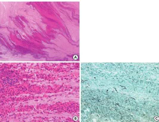
HISTOPATHOLOGY OF ABPM (Fig. 1)
Morphologically, ABPM is characterized by (1) allergic (eosinophilic) mucin in the bronchi (mucoid impaction of bronchi), (2) expansion of the bronchi with mucus plug (central bronchiectasis), and (3) bronchocentric granulomatosis with tissue eosinophilia, eosinophilic pneumonia, and organized pneumonia in the peripheral lungs [20]. In 1988, Bosken et al. [21] proposed pathological diagnostic criteria for ABPM based on observations in surgically-resected lung specimen. In their criteria, the presence of (1) eosinophilic mucus plugs or bronchocentric granulomatosis with advanced eosinophil infiltration, and (2) fungal hyphae in these lesions without tissue invasion suggest the diagnosis of ABPM.
Hebisawa et al. [22] examined the pathology of ABPM in the specimens obtained by lobectomy including central bronchi, and found that (1) the oldest lesions were eosinophilic mucus plugs in the bronchi, (2) central bronchiectasis was formed secondarily because of the expansion of
mucus plugs stretching the airway wall, which has already been weakened by severe eosinophilic inflammation, and (3) the peripheral lung lesions were caused by the transluminal spreading of fungal components from the eosinophilic mucus plugs in the central bronchi. Based on these observations, they concluded that the primary lesions of ABPM were eosinophilic plasmacytoid/
lymphocytic mucus plugs but not central bronchiectasis or pulmonary infiltration as often cited in the diagnostic criteria [22, 23]. Another example of eosinophilic mucus plugs deviating anatomical structures has been reported in cases of allergic fungal rhinosinusitis, in which eosinophilic mucus plugs deflect the paranasal sinus bony structures [24, 25].
Mucus plugs in ABPM contain many eosinophils, Charcot-Leyden crystals, and fibrin exudates, and are packed in the bronchi, forming a fir tree-like structure [23], suggesting a dehydration process. Filamentous fungi are found in mucus plugs without gathering densely as seen in chronic pulmonary mycoses. Neutrophils are sometimes observed on the periphery of eosinophilic mucus plugs, but the number is much smaller compared to eosinophils. In comparison to the mucus plugs in resected lungs, mucus plugs in sputum or collected by bronchoscopy were softer, and less often accompanied by fir tree-like structures [22].
CHARACTERISTICS OF EOSINOPHIL MUCUS PLUGS IN ABPM
Extracellular DNA trap cell death (ETosis) of eosinophils has been suggested as a mechanism for the formation of eosinophilic mucus plugs in various diseases [26, 27]. ETosis was originally reported in neutrophils, which release granular proteins together with nuclear
A
B C
Fig. 1. Histology of allergic (eosinophilic) mucin in the bronchi. Mucus plugs packed in the bronchi of surgically- resected lungs from a patient with allergic bronchopulmonary mycosis demonstrate a fir tree-like structure (A:
H&E, x5), which contain many eosinophils, Charcot-Leyden crystals, and fibrin exudates (B: H&E, x40). (C) Hyphae of fungi are sparsely found in mucus plugs (Grocott staining, x40).
chromatin DNA (neutrophil extracellular traps [NETs]) to exert bactericidal activity [28].
Fungi cause ETosis of neutrophils, but recent studies have revealed that NETs have no fungicidal action [29].
Cells other than neutrophils can also cause ETosis and release chromatin DNA and eosinophils as well. Ueki et al. [30] investigated eosinophilic ETosis and demonstrated that the extracellular DNA trap released by eosinophils was more aggregated in structure and more stable than the extracellular DNA trap produced by neutrophils. Although NETs are viscous owing to the physical properties of DNA, the extracellular DNA trap derived from eosinophils exhibits greater viscosity. We and other researchers have demonstrated that eosinophilic mucus plugs of ABPA, eosinophilic sinusitis, and eosinophilic otitis media have a net-like structure with DNA and citrullinated histones suggesting the extracellular DNA traps [30-32].
CLINICAL CHARACTERISTICS OF ABPA IN EAST AND SOUTH ASIA
Table 1 summarizes the clinical characteristics of ABPA/ABPM in cohort studies reported from Japan, Korea, China, and India [6, 7, 33-41]. The inclusion criteria for each study were different; the presence of asthma was indispensable in the studies by Agarwal et al. [6, 33], and central bronchodilation or mucous plugs were required for the study by Oguma et al.
[7]. However, there were obvious differences in the clinical characteristics of ABPA/ABPM between East Asian countries such as Japan, Korea, and China versus India.
Age and age at onset
There was a significant difference in the age of the recruited patients with ABPA between the studies in Japan and Korea versus those in India. It has been suggested that ABPA mostly develops in patients aged 30–40 years old, which is in agreement with the cases in India [6, 33-35, 38]. Meanwhile, the median age of patients with ABPA in Japan and Korea was 50–70 years [7, 36, 37, 39], and that of Chinese patients was 40–50 years [40, 41]. Two studies in Japan and Korea that examined the age of onset for ABPA demonstrated that the median age at disease onset was 50–60 years [7, 37]. Two-thirds of cases developed ABPA at 50 years or older as reported in a Japanese nation-wide survey of this disease [7].
There are 3 possible reasons that might explain the difference in age distribution. The first possibility involves genetic predisposition, but little is known about this except for the cases with CFTR mutation in patients with cystic fibrosis. The second possibility is the difference in the age structure of populations in East Asian countries versus India. The elderly population aged 55 years or older makes up less than 15% of the total population in India, whereas it accounts for 22%, 30%, and 40% of the population in China, Korea and Japan, respectively (https://www.cia.gov/library/publications/the-world-factbook/fields/2010.
html). These differences in the age structure may have skewed the age distribution of the patients to younger generation in India, and to older population in Japan and South Korea.
We performed a cluster analysis using a Japanese nationwide survey data and identified three clusters with different ages of onset, with the median age of onset being 30–40, 50–60, and 60–70 years, respectively (Oguma T and Asano K, unpublished data), suggesting that there are multiple phenotypes of ABPA with different ages of onset. One phenotype of Japanese ABPA had a median age at onset of 37 years, compatible with that of Indian ABPA.
Table 1. Clinical characteristics of allergic bronchopulmonary aspergillosis (ABPA) in East and South Asia CharacteristicEast AsiaSouth Asia JapanKoreaChinaIndia Study Author [reference number]Tanimoto [39]Ishiguro [36]Oguma [7]Kim [37]Ye [40]Zhang [41]Behera [34]Chakrabarti [35]Prasad [38]Agarwal [6]Agarwal [33] Publication year20152016201820122009201719942002200920102018 No. of cases5342358105777358942234131 Age (yr)5757646341423436313437 Women42%-57%20%42%51%60%39%36%47%47% Asthma79%67%81%100%-96%94%80%100%100%100% Duration between onset of asthma and ABPA (yr)31-1415--111212612 Peripheral blood eosinophil counts (/µL)580-1,0751,198-----8471,271 Serum IgE levels (IU/mL)1,170-1,913927-----5,01510,540 Serum IgE levels > 1,000 IU/mL-81%68%50%-71%----100% Specific IgE or immediate skin reaction to Aspergillus antigen100%96%100%100%-89%51%82%98%100%95% Precipitins for Aspergillus antigen-77%73%--57%77%72%100%83%50% Culture-positive for Aspergillus spp.-60%59%--38%-63%69%-- Radiographic findings----------- Pulmonary opacities-100%88%20%56%79%-43%95%-66% Central bronchiectasis-98%89%80%44%81%71%69%100%77%93% High attenuation mucus--41%------21%41%
Therefore, a classic, early onset phenotype of ABPA was also present in Japan as in India, but later-onset phenotype(s) may dominate in these populations. Importantly, there are no substantial differences in the duration between the onset of asthma and ABPA in Indians and East Asians, suggesting that the age of onset for asthma may be higher in the latter population. The third possibility is that greater exposure to environmental fungi in India, possibly because of higher burden of airborne fungi in the hot and humid environments, accelerates the sensitization and development of ABPA in younger age. This hypothesis is supported by the remarkably high incidence of ABPA In India compared to other areas of the world [2, 42].
ABPA rarely develops during childhood in patients with asthma, with some exception in India [43]. This trend matches well with the findings among other eosinophilic diseases in the upper and lower airways, such as rhinosinusitis with nasal polyps, eosinophilic otitis media, aspirin-exacerbated respiratory diseases, and chronic eosinophilic pneumonia, suggesting a common pathology underlying these adult-onset eosinophilic airway diseases [44].
Predisposing condition
Cystic fibrosis, a major predisposing condition of ABPM in Europe and the United States, is rare in Asians, for whom the major predisposing disease of ABPM is asthma. Asthma was included as one of the essential components in the current diagnostic criteria for ABPA.
However, there have been reports of cases with ABPA that were not accompanied by asthma [45]. In a study of pathologically diagnosed cases of ABPM, 7 of 17 patients (41%) reported that they did not have asthma nor cystic fibrosis [46]. Among 453 Japanese patients with ABPM, approximately 20% (93 patients) did not present with asthma [7,36,39]. In contrast, only 5% (20 patients) of 400 Indian patients with ABPA did not have asthma [6, 34, 35, 38].
This difference in the incidence of asthma among patients with ABPA may be due to the difference in the diagnostic criteria used in these studies, or may reflect the presence of a specific phenotype without asthma among Japanese ABPA.
Serum levels of total IgE
Increased levels of total IgE in serum are characteristic of ABPA, and are useful not only for its diagnosis but also for monitoring disease activity. However, different cutoff values, such as >417 IU/mL or >1,000 IU/mL, have been used depending on the diagnostic criteria [3, 47, 48]. A survey of the members of the American Academy of Allergy Asthma Immunology reported that 44.9% used the cutoff value of 417 IU/mL whereas 42% used 1,000 IU/mL [49].
The diagnostic criteria proposed by the International Society for Human Animal Mycology (ISHAM) in 2013 emphasize the usefulness of higher cutoff values, such as 1,000 IU/mL, to distinguish ABPA from severe asthma with fungal sensitization [3]. A recent Indian study recommends even higher cutoff value, 2,347 IU/mL [50]. However, we should be cautious not to exclude patients with relatively low levels of total IgE in serum, because its levels in East Asian patients were lower than in Indian patients, and 20%–50% of the patients showed IgE
< 1,000 IU/mL [7, 36, 37, 41].
Fungal culture
Although the efficacy to detect filamentous fungi with culture methods may vary depending on culture conditions such as media, duration, and temperature [51], most of the previous studies demonstrate the detection rate of Aspergillus spp. between 60%–70% [7, 35, 36, 38]. What was unique in Japanese studies was the presence of S. commune in 6%–14% [7, 36]. S. commune was the most common filamentous basidiomycetes of mushroom that causes human airway diseases
[52], commonly found on the rotten wood of trees worldwide. The difference in the incidence of S. commune-induced ABPM between Japan and other countries may be because of the difficulty in identifying this fungus in the laboratory, which grows as a nonsporulating white mold.
DIAGNOSTIC CRITERIA FOR ABPA/ABPM
ABPM should be suspected, and more detailed diagnostic tests should be performed when peripheral blood eosinophil counts or serum IgE levels were increased in patients with asthma sensitized to fungal antigens, or when pulmonary opacities, central bronchiectasis, or mucus plugs were accompanied by peripheral blood eosinophilia even in the absence of asthma.
The classic diagnostic criteria were proposed in 1977 by Rosenberg, Patterson, and their colleagues [48] based on the clinical characteristics of 20 cases suspected of ABPA; which included 7 primary components: asthma, peripheral blood eosinophilia, positive immediate skin reaction to Aspergillus antigen, presence of precipitating antibody to Aspergillus antigen, increased serum total IgE levels, transient or fixed pulmonary opacities, and central bronchiectasis. Agarwal et al. [53] examined these criteria with an additional component, the presence of A. fumigatus-specific IgE, and demonstrated that the specificity and specificity were high when 6 components were satisfied, but its sensitivity decreased to 39% when 7 components were needed to be satisfied as originally proposed.
In 1988, Greenberger and Patterson [47] added a new disease concept, ABPA-seropositive, which refers to cases that lack central bronchiectasis, but satisfy other criteria including serological tests. In 2013, ISHAM proposed new diagnostic criteria [3], which defines asthma or cystic fibrosis as predisposing conditions and proposes 2 obligatory criteria that must be met, immediate cutaneous hypersensitivity to Aspergillus antigen or elevated IgE levels against A. fumigatus, and elevated total IgE levels > 1,000 IU/mL. In addition, at least two of the following three criteria should be satisfied for the diagnosis of ABPA, which include the presence of precipitating or IgG antibodies against A. fumigatus in serum, radiographic features in the lungs consistent with ABPA, and total eosinophil count > 500 cells/μL. A comparison of these diagnostic criteria is summarized in Table 2.
However, these diagnostic criteria are specific to ABPA caused by A. fumigatus. The ABPM research program supported by the Japan Medical Research and Development Organization Table 2. Comparison of diagnostic criteria
Diagnostic criteria Rosenberg-Patterson 1977 Greenberger-Patterson 1986 ISHAM 2013
Asthma or cystic fibrosis ● ● ●
Peripheral blood eosinophilia ● ○
Increased levels of IgE in serum ● ● ●
Immediate skin reaction to Aspergillus antigen ● ● ●
Specific IgE to Aspergillus fumigatus ● ●
Specific IgG to Aspergillus fumigatus ● ○
Precipitin to Aspergillus fumigatus ● ○
Pulmonary opacities ● ○
Central bronchiectasis ● ● ○
Positive sputum culture of Aspergillus fumigatus △
Expectoration of brownish mucous plugs △
Arthus-type skin reaction to Aspergillus antigen △
Closed circles are mandatory components. In International Society for Human Animal Mycology (ISHAM) criteria, 2 out of 3 components (open circles) should also be fulfilled. Triangles are secondary components, further supporting the diagnosis of allergic bronchopulmonary aspergillosis.
are developing new diagnostic criteria for ABPM. Based on the preliminary results, the new diagnostic criteria may improve sensitivity and specificity for ABPA/ABPM.
TREATMENT
Corticosteroids
Systemic corticosteroids improve symptoms such as wheezing, as well as laboratory and radiographic abnormalities such as high IgE levels in the serum and pulmonary opacities during the acute phase of ABPA. There are 2 standard protocols for systemic corticosteroid treatment: the administration of high-dose prednisolone (0.75 mg/kg/day) for 6 weeks, followed by 0.5 mg/kg/day for 6 weeks, and then tapered by 5 mg per week for 6–12 months;
and 2 weeks of moderate-dose prednisolone (0.5 mg/kg/day), followed by the same dose administered every other day for 8 weeks, and then tapered by 5 to 10 mg per every 2 weeks.
Randomized controlled trials comparing these 2 protocols were performed in 92 patients with ABPA that were previously untreated [54]. There was no significant difference between the 2 protocols in the relapse rate after treatment, which was 46% for each group. The rate of clinical improvement based on symptoms, serum IgE levels, and radiographic findings after the 6-week-treatment was 100% in the high-dose corticosteroid group and 88% in the middle-dose group, but the number of patients that experienced side effects due to corticosteroid treatment was 80% in the high-dose protocol compared to 29% in the middle- dose protocol [54].
Treatment efficacy can be evaluated by a radiographic improvement, with serum IgE levels decreased by 20%–25% or more from the baseline [3, 55]. The recurrence rate was high after decreasing/stopping corticosteroids regardless of the initial dose or treatment period, and often continuous administration of low dose corticosteroids is required [7, 54]. Long-term corticosteroid administration in the presence of bronchiectasis may be complicated with chronic lower respiratory tract infections with Pseudomonas aeruginosa or nontuberculous mycobacteria [56].
Inhaled corticosteroids may be useful to control asthmatic symptoms, but there has been no large-scale trial showing its effectiveness as a therapeutic agent for ABPA. A small, single-arm study demonstrated that a combination of inhaled high-dose corticosteroids with the long- acting β-agonist formoterol/budesonide (24/1,600 μg/day) for 6 months resulted in subjective improvements, but did not decrease the levels of IgE [57], suggesting a failure to suppress the disease activity of ABPA.
Antifungal drugs
The latest systematic review on the treatment of ABPA with antifungal drugs, which summarizes 38 observational studies and 4 randomized controlled trials, confirms that antifungal drugs might improve symptoms, pulmonary function, and radiographic findings, and reduce the exacerbation rate and dose of corticosteroids, but the evidence is not robust because of the lack of controlled studies and heterogeneity of study design [58]. Therefore, there are some differences in the positioning of antifungal drugs among the guidelines. In the clinical practice guidelines from the Infectious Diseases Society of America [59], oral itraconazole therapy is recommended when asthmatic patients with bronchiectasis or mucoid impaction are symptomatic despite oral or inhaled corticosteroid therapy. Meanwhile, the Japanese guidelines for management of deep-seated mycosis [60]
recommend a more cautious approach for using antifungal drugs; the identification of the causative fungi in culture and short-term treatment (16 weeks) is recommended to avoid the induction of azol-resistance. However, in more than half of the cases with ABPM in Japan, antifungal drugs were used for more than a year [7].
In an open-labeled study, 131 patients with ABPA complicating asthma were randomly assigned to the treatment groups with either itraconazole or systemic corticosteroids [33]. In 6 weeks, 88% and 100% of the subjects receiving itraconazole and systemic corticosteroids, respectively, showed partial/total improvement in symptoms and radiographic findings, with serum IgE levels having decreased by 25% or more. Although the corticosteroid group showed quicker responses, both treatment groups show 100% treatment response within 3 months [33].
Anti-IgE antibody
An anti-IgE antibody (omalizumab) was effective in patients with severe asthma in improving symptoms and reducing the frequency of exacerbation. There is an increasing number of reported cases demonstrating the effectiveness of omalizumab in patients with ABPM, but only a few studies have examined the effect of omalizumab prospectively. Tillie- Leblond et al. [61] performed a single-arm study in 16 cases with ABPA, and omalizumab significantly reduced the exacerbation rate and dose of systemic corticosteroids.
Additionally, Voskamp et al. [62] performed a randomized, placebo-controlled study in cases, revealing a decrease in the exacerbation rate and fraction of exhaled nitric oxide, but not in pulmonary functions.
CONCLUSIONS
It has been more than 65 years since ABPA was first reported; however, there are still several unsolved problems for ABPA/ABPM. First, the current diagnostic criteria do not necessarily cover the whole spectrum of this complex disease, and there is a substantial number of patients who are misdiagnosed. Secondly, systemic corticosteroids and/or antifungal drugs were effective for improving the acute-phase symptoms, but recurrences are quite common, and side effects of the drugs and emergence of azol-resistant fungi are problematic.
Histopathological and biological approaches are now being used to elucidate the pathophysiology of this condition for subsequent development of new therapies.
ACKNOWLEDGEMENTS
This study was funded in part by a Research Grant on Allergic Disease and Immunology from the Japan Agency for Medical Research and Development under Grant Number JP18ek0410026 and JSPS KAKENHI 16K09925.
We would like to thank Masami Taniguchi, Trufumi Shimoda, Noboru Takayanagi, Hiroto Matsuse, Tsuyoshi Oguma, Satoshi Konno, Koichi Fukunaga, Shigeharu Ueki, Yuma Fukutomi, and other members in the research program of ABPM in Japan for the useful discussion. All the authors contributed the writing of the manuscript and the decision to submit the manuscript for publication. The authors declare no competing financial interests.
REFERENCES
1. Hinson KF, Moon AJ, Plummer NS. Broncho-pulmonary aspergillosis; a review and a report of eight new cases. Thorax 1952;7:317-33.
PUBMED | CROSSREF
2. Denning DW, Pleuvry A, Cole DC. Global burden of allergic bronchopulmonary aspergillosis with asthma and its complication chronic pulmonary aspergillosis in adults. Med Mycol 2013;51:361-70.
PUBMED | CROSSREF
3. Agarwal R, Chakrabarti A, Shah A, Gupta D, Meis JF, Guleria R, Moss R, Denning DWABPA complicating asthma ISHAM working group. Allergic bronchopulmonary aspergillosis: review of literature and proposal of new diagnostic and classification criteria. Clin Exp Allergy 2013;43:850-73.
PUBMED | CROSSREF
4. Knutsen AP, Bush RK, Demain JG, Denning DW, Dixit A, Fairs A, Greenberger PA, Kariuki B, Kita H, Kurup VP, Moss RB, Niven RM, Pashley CH, Slavin RG, Vijay HM, Wardlaw AJ. Fungi and allergic lower respiratory tract diseases. J Allergy Clin Immunol 2012;129:280-91.
PUBMED | CROSSREF
5. Kosmidis C, Denning DW. The clinical spectrum of pulmonary aspergillosis. Thorax 2015;70:270-7.
PUBMED | CROSSREF
6. Agarwal R, Khan A, Gupta D, Aggarwal AN, Saxena AK, Chakrabarti A. An alternate method of classifying allergic bronchopulmonary aspergillosis based on high-attenuation mucus. PLoS One 2010;5:e15346.
PUBMED | CROSSREF
7. Oguma T, Taniguchi M, Shimoda T, Kamei K, Matsuse H, Hebisawa A, Takayanagi N, Konno S, Fukunaga K, Harada K, Tanaka J, Tomomatsu K, Asano K. Allergic bronchopulmonary aspergillosis in Japan: a nationwide survey. Allergol Int 2018;67:79-84.
PUBMED | CROSSREF
8. Chetty A. Pathology of allergic bronchopulmonary aspergillosis. Front Biosci 2003;8:e110-4.
PUBMED | CROSSREF
9. Fukutomi Y, Taniguchi M. Sensitization to fungal allergens: resolved and unresolved issues. Allergol Int 2015;64:321-31.
PUBMED | CROSSREF
10. Portnoy JM, Williams PB, Barnes CS. Innate immune responses to fungal allergens. Curr Allergy Asthma Rep 2016;16:62.
PUBMED | CROSSREF
11. Romani L. Immunity to fungal infections. Nat Rev Immunol 2011;11:275-88.
PUBMED | CROSSREF
12. Chamilos G, Ganguly D, Lande R, Gregorio J, Meller S, Goldman WE, Gilliet M, Kontoyiannis DP.
Generation of IL-23 producing dendritic cells (DCs) by airborne fungi regulates fungal pathogenicity via the induction of T(H)-17 responses. PLoS One 2010;5:e12955.
PUBMED | CROSSREF
13. Chowdhary A, Agarwal K, Kathuria S, Gaur SN, Randhawa HS, Meis JF. Allergic bronchopulmonary mycosis due to fungi other than Aspergillus: a global overview. Crit Rev Microbiol 2014;40:30-48.
PUBMED | CROSSREF
14. Oshikata C, Watanabe M, Saito A, Yasueda H, Akiyama K, Kamata Y, Tsurikisawa N. Allergic bronchopulmonary mycosis caused by Penicillium luteum. Med Mycol Case Rep 2016;15:9-11.
PUBMED | CROSSREF
15. O'Driscoll BR, Hopkinson LC, Denning DW. Mold sensitization is common amongst patients with severe asthma requiring multiple hospital admissions. BMC Pulm Med 2005;5:4.
PUBMED | CROSSREF
16. Araujo R, Rodrigues AG. Variability of germinative potential among pathogenic species of Aspergillus. J Clin Microbiol 2004;42:4335-7.
PUBMED | CROSSREF
17. Imtiaj A, Jayasinghe C, Lee GW, Kim HY, Shim MJ, Rho HS, Lee HS, Hur H, Lee MW, Lee UY, Lee TS.
Physicochemical requirement for the vegetative growth of schizophyllum commune collected from different ecological origins. Mycobiology 2008;36:34-9.
PUBMED | CROSSREF
18. Oguma T, Asano K, Tomomatsu K, Kodama M, Fukunaga K, Shiomi T, Ohmori N, Ueda S, Takihara T, Shiraishi Y, Sayama K, Kagawa S, Natori Y, Lilly CM, Satoh K, Makimura K, Ishizaka A. Induction of mucin and MUC5AC expression by the protease activity of Aspergillus fumigatus in airway epithelial cells.
J Immunol 2011;187:999-1005.
PUBMED | CROSSREF
19. Alp S, Arikan S. Investigation of extracellular elastase, acid proteinase and phospholipase activities as putative virulence factors in clinical isolates of Aspergillus species. J Basic Microbiol 2008;48:331-7.
PUBMED | CROSSREF
20. Katzenstein AL, Liebow AA, Friedman PJ. Bronchocentric granulomatosis, mucoid impaction, and hypersensitivity reactions to fungi. Am Rev Respir Dis 1975;111:497-537.
PUBMED
21. Bosken CH, Myers JL, Greenberger PA, Katzenstein AL. Pathologic features of allergic bronchopulmonary aspergillosis. Am J Surg Pathol 1988;12:216-22.
PUBMED | CROSSREF
22. Hebisawa A, Tamura A, Kurashima A, Oobayashi C, Kawamata M, Maeda M, Saiki S, Komatsu H, Yoneda R. Pathologic reconsideration on allergic bronchopulmonary aspergillosis and mycosis. Nihon Kokyuki Gakkai Zasshi 1998;36:330-7.
PUBMED
23. Jelihovsky T. The structure of bronchial plugs in mucoid impaction, bronchocentric granulomatosis and asthma. Histopathology 1983;7:153-67.
PUBMED | CROSSREF
24. deShazo RD, Swain RE. Diagnostic criteria for allergic fungal sinusitis. J Allergy Clin Immunol 1995;96:24-35.
PUBMED | CROSSREF
25. Mukherji SK, Figueroa RE, Ginsberg LE, Zeifer BA, Marple BF, Alley JG, Cooper LL, Nemzek WR, Yousem DM, Jones KR, Kupferberg SB, Castillo M. Allergic fungal sinusitis: CT findings. Radiology 1998;207:417-22.
PUBMED | CROSSREF
26. Ueki S, Melo RC, Ghiran I, Spencer LA, Dvorak AM, Weller PF. Eosinophil extracellular DNA trap cell death mediates lytic release of free secretion-competent eosinophil granules in humans. Blood 2013;121:2074-83.
PUBMED | CROSSREF
27. Ueki S, Tokunaga T, Fujieda S, Honda K, Hirokawa M, Spencer LA, Weller PF. Eosinophil ETosis and DNA traps: a new look at eosinophilic inflammation. Curr Allergy Asthma Rep 2016;16:54.
PUBMED | CROSSREF
28. Brinkmann V, Reichard U, Goosmann C, Fauler B, Uhlemann Y, Weiss DS, Weinrauch Y, Zychlinsky A.
Neutrophil extracellular traps kill bacteria. Science 2004;303:1532-5.
PUBMED | CROSSREF
29. Gazendam RP, van Hamme JL, Tool AT, Hoogenboezem M, van den Berg JM, Prins JM, Vitkov L, van de Veerdonk FL, van den Berg TK, Roos D, Kuijpers TW. Human neutrophils use different mechanisms to kill Aspergillus fumigatus conidia and hyphae: evidence from phagocyte defects. J Immunol 2016;196:1272-83.
PUBMED | CROSSREF
30. Ueki S, Konno Y, Takeda M, Moritoki Y, Hirokawa M, Matsuwaki Y, Honda K, Ohta N, Yamamoto S, Takagi Y, Wada A, Weller PF. Eosinophil extracellular trap cell death-derived DNA traps: their presence in secretions and functional attributes. J Allergy Clin Immunol 2016;137:258-67.
PUBMED | CROSSREF
31. Muniz VS, Silva JC, Braga YAV, Melo RCN, Ueki S, Takeda M, Hebisawa A, Asano K, Figueiredo RT, Neves JS. Eosinophils release extracellular DNA traps in response to Aspergillus fumigatus. J Allergy Clin Immunol 2018;141:571-585.e7.
PUBMED | CROSSREF
32. Ueki S, Ohta N, Takeda M, Konno Y, Hirokawa M. Eosinophilic otitis media: the aftermath of eosinophil extracellular trap cell death. Curr Allergy Asthma Rep 2017;17:33.
PUBMED | CROSSREF
33. Agarwal R, Dhooria S, Singh Sehgal I, Aggarwal AN, Garg M, Saikia B, Behera D, Chakrabarti A. A randomized trial of itraconazole vs prednisolone in acute-stage allergic bronchopulmonary aspergillosis complicating asthma. Chest 2018;153:656-64.
PUBMED | CROSSREF
34. Behera D, Guleria R, Jindal SK, Chakrabarti A, Panigrahi D. Allergic bronchopulmonary aspergillosis: a retrospective study of 35 cases. Indian J Chest Dis Allied Sci 1994;36:173-9.
PUBMED
35. Chakrabarti A, Sethi S, Raman DS, Behera D. Eight-year study of allergic bronchopulmonary aspergillosis in an Indian teaching hospital. Mycoses 2002;45:295-9.
PUBMED | CROSSREF
36. Ishiguro T, Takayanagi N, Uozumi R, Baba Y, Kawate E, Kobayashi Y, Takaku Y, Kagiyama N, Shimizu Y, Morita S, Sugita Y. Diagnostic criteria that can most accurately differentiate allergic bronchopulmonary mycosis from other eosinophilic lung diseases: a retrospective, single-center study. Respir Investig 2016;54:264-71.
PUBMED | CROSSREF
37. Kim JH, Jin HJ, Nam YH, Hwang EK, Ye YM, Park HS. Clinical features of allergic bronchopulmonary aspergillosis in Korea. Allergy Asthma Immunol Res 2012;4:305-8.
PUBMED | CROSSREF
38. Prasad R, Garg R, Sanjay , Shukla AD. Allergic bronchopulmonary aspergillosis: a review of 42 patients from a tertiary care center in India. Lung India 2009;26:38-40.
PUBMED | CROSSREF
39. Tanimoto H, Fukutomi Y, Yasueda H, Takeuchi Y, Saito A, Watai K, Sekiya K, Tsuburai T, Asano K, Taniguchi M, Akiyama K. Molecular-based allergy diagnosis of allergic bronchopulmonary aspergillosis in Aspergillus fumigatus-sensitized Japanese patients. Clin Exp Allergy 2015;45:1790-800.
PUBMED | CROSSREF
40. Ye F, Zhang NF, Zhong NS. Clinical and pathological analysis of allergic bronchopulmonary aspergillosis in China. Zhonghua Jie He He Hu Xi Za Zhi 2009;32:434-8.
PUBMED
41. Zhang M, Gao J. Clinical analysis of 77 patients with allergic bronchopulmonary Aspergillosis in Peking Union Medical College Hospital. Zhongguo Yi Xue Ke Xue Yuan Xue Bao 2017;39:352-7.
PUBMED
42. Agarwal R, Aggarwal AN, Gupta D, Jindal SK. Aspergillus hypersensitivity and allergic
bronchopulmonary aspergillosis in patients with bronchial asthma: systematic review and meta-analysis.
Int J Tuberc Lung Dis 2009;13:936-44.
PUBMED
43. Shah A, Kunal S. A review of 42 asthmatic children with allergic bronchopulmonary aspergillosis. Asia Pac Allergy 2017;7:148-55.
PUBMED | CROSSREF
44. Asano K. Innate immune systems: a clue in the pathogenesis of adult-onset, eosinophilic lung diseases.
Respir Investig 2017;55:93.
PUBMED | CROSSREF
45. Glancy JJ, Elder JL, McAleer R. Allergic bronchopulmonary fungal disease without clinical asthma.
Thorax 1981;36:345-9.
PUBMED | CROSSREF
46. Ishiguro T, Takayanagi N, Kagiyama N, Shimizu Y, Yanagisawa T, Sugita Y. Clinical characteristics of biopsy-proven allergic bronchopulmonary mycosis: variety in causative fungi and laboratory findings.
Intern Med 2014;53:1407-11.
PUBMED | CROSSREF
47. Greenberger PA, Patterson R. Allergic bronchopulmonary aspergillosis and the evaluation of the patient with asthma. J Allergy Clin Immunol 1988;81:646-50.
PUBMED | CROSSREF
48. Rosenberg M, Patterson R, Mintzer R, Cooper BJ, Roberts M, Harris KE. Clinical and immunologic criteria for the diagnosis of allergic bronchopulmonary aspergillosis. Ann Intern Med 1977;86:405-14.
PUBMED | CROSSREF
49. Greenberger PA, Bush RK, Demain JG, Luong A, Slavin RG, Knutsen AP. Allergic bronchopulmonary aspergillosis. J Allergy Clin Immunol Pract 2014;2:703-8.
PUBMED | CROSSREF
50. Agarwal R, Aggarwal AN, Garg M, Saikia B, Chakrabarti A. Cut-off values of serum IgE (total and A.
fumigatus -specific) and eosinophil count in differentiating allergic bronchopulmonary aspergillosis from asthma. Mycoses 2014;57:659-63.
PUBMED | CROSSREF
51. Pashley CH, Fairs A, Morley JP, Tailor S, Agbetile J, Bafadhel M, Brightling CE, Wardlaw AJ. Routine processing procedures for isolating filamentous fungi from respiratory sputum samples may underestimate fungal prevalence. Med Mycol 2012;50:433-8.
PUBMED | CROSSREF
52. Chowdhary A, Kathuria S, Agarwal K, Meis JF. Recognizing filamentous basidiomycetes as agents of human disease: a review. Med Mycol 2014;52:782-97.
PUBMED | CROSSREF
53. Agarwal R, Maskey D, Aggarwal AN, Saikia B, Garg M, Gupta D, Chakrabarti A. Diagnostic performance of various tests and criteria employed in allergic bronchopulmonary aspergillosis: a latent class analysis.
PLoS One 2013;8:e61105.
PUBMED | CROSSREF
54. Agarwal R, Aggarwal AN, Dhooria S, Singh Sehgal I, Garg M, Saikia B, Behera D, Chakrabarti A. A randomised trial of glucocorticoids in acute-stage allergic bronchopulmonary aspergillosis complicating asthma. Eur Respir J 2016;47:490-8.
PUBMED | CROSSREF
55. Agarwal R, Aggarwal AN, Sehgal IS, Dhooria S, Behera D, Chakrabarti A. Utility of IgE (total and Aspergillus fumigatus specific) in monitoring for response and exacerbations in allergic bronchopulmonary aspergillosis. Mycoses 2016;59:1-6.
PUBMED | CROSSREF
56. Ishiguro T, Takayanagi N, Takaku Y, Kagiyama N, Shimizu Y, Yanagisawa T, Kawabata Y, Sugita Y. Allergic bronchopulmonary aspergillosis with repeated isolation of nontuberculous mycobacteria. Intern Med 2013;52:1721-6.
PUBMED | CROSSREF
57. Agarwal R, Khan A, Aggarwal AN, Saikia B, Gupta D, Chakrabarti A. Role of inhaled corticosteroids in the management of serological allergic bronchopulmonary aspergillosis (ABPA). Intern Med 2011;50:855-60.
PUBMED | CROSSREF
58. Moreira AS, Silva D, Ferreira AR, Delgado L. Antifungal treatment in allergic bronchopulmonary aspergillosis with and without cystic fibrosis: a systematic review. Clin Exp Allergy 2014;44:1210-27.
PUBMED | CROSSREF
59. Patterson TF, Thompson GR 3rd, Denning DW, Fishman JA, Hadley S, Herbrecht R, Kontoyiannis DP, Marr KA, Morrison VA, Nguyen MH, Segal BH, Steinbach WJ, Stevens DA, Walsh TJ, Wingard JR, Young JA, Bennett JE. Practice guidelines for the diagnosis and management of Aspergillosis: 2016 update by the Infectious Diseases Society of America. Clin Infect Dis 2016;63:e1-60.
PUBMED | CROSSREF
60. Kohno S, Tamura K, Niki Y, Izumikawa K, Oka S, Ogawa K, Kadota J, Kamei K, Kanda Y, Kiuchi T, Shibuya K, Takakura S, Takata T, Takesue Y, Teruya K, Tokimatsu I, Fukuda T, Maesaki S, Makimura K, Mikamo H, Mitsutake K, Miyazaki Y, Mori M, Yasuoka A, Yano K, Yamanaka N, Yoshida M. Executive summary of Japanese domestic guidelines for management of deep-seated mycosis 2014. Med Mycol J 2016;57:E117-63.
PUBMED | CROSSREF
61. Tillie-Leblond I, Germaud P, Leroyer C, Tétu L, Girard F, Devouassoux G, Grignet JP, Prudhomme A, Dusser D, Wallaert B. Allergic bronchopulmonary aspergillosis and omalizumab. Allergy 2011;66:1254-6.
PUBMED | CROSSREF
62. Voskamp AL, Gillman A, Symons K, Sandrini A, Rolland JM, O'Hehir RE, Douglass JA. Clinical efficacy and immunologic effects of omalizumab in allergic bronchopulmonary aspergillosis. J Allergy Clin Immunol Pract 2015;3:192-9.
PUBMED | CROSSREF
![Table 1. Clinical characteristics of allergic bronchopulmonary aspergillosis (ABPA) in East and South Asia CharacteristicEast AsiaSouth Asia JapanKoreaChinaIndia Study Author [reference number]Tanimoto [39]Ishiguro [36]Oguma [7]Kim [37]Ye [40]Zhang [41]Beh](https://thumb-ap.123doks.com/thumbv2/123dokinfo/5206312.119240/6.892.278.613.129.1090/characteristics-bronchopulmonary-aspergillosis-characteristiceast-asiasouth-japankoreachinaindia-reference-ishiguro.webp)
