196
This is an Open Access article distributed under the terms of the Creative Commons Attribution Non-Commercial License (http://creativecommons.org/licenses/by-nc/3.0) which permits unrestricted non-commercial use, distribution, and reproduction in any medium, provided the original work is properly cited.
BLOOD RESEARCH Volume 49ㆍNumber 3ㆍSeptember 2014
Letters to the Editor
Mixed-phenotypic acute leukemia:
cytochemically myeloid and
phenotypically early T-cell precursor acute lymphoblastic leukemia
TO THE EDITOR: Early T-cell precursor acute lymphoblastic leukemia (ETP-ALL) is characterized by the specific im- munophenotype of CD1a-, CD8-, and CD5weak/- with stem cell or myeloid marker expression. This leukemia presents a particular flow cytometric diagnostic challenge, as it fre- quently exhibits sufficient myeloid-lineage differentiation to be classified as mixed-phenotype acute leukemia (MPAL).
The 2008 World Health Organization classification has es- tablished new strict criteria for the diagnosis of MPAL;
however, the exact MPO positivity cut-off percentage re- quired for establishing a myeloid lineage was not mentioned.
Herein, we report a case of ETP-ALL with trisomy 4 and t(6;14)(q27:q22) in a 25-year-old man, the final diagnosis of which was MPAL (T/Myeloid) due to the morphologic presence of Auer rods and myeloperoxidase staining, despite exhibiting a flow cytometric ETP-ALL phenotype. To the best of our knowledge, this is the first report on a case of ETP-ALL with Auer rods.
INTRODUCTION
Early T-cell precursor acute lymphoblastic leukemia (ETP-ALL) is characterized by the specific immunopheno- type of CD1a-, CD8-, and CD5weak/- with stem cell or myeloid marker expression [1]. This leukemia is associated with in- creased genomic instability, a high remission failure fre- quency, and hematological relapse. ETP-ALL patients also represent a distinct molecular T-ALL subgroup with a low frequency of NOTCH1 mutations and a high frequency of FLT3 mutations. ETP-ALL with FLT3 mutation has also been described as a new ETP-ALL subgroup with unique immunophenotypic features and high levels of CD2, myeloid antigen CD13, and CD117 expression, indicating a stem cell phenotype [2]. Mixed-phenotypic acute leukemia (MPAL), T/Myeloid subtype is rarely suspected from mor-
phology, except in a small subset of cases, which have a distinct dual-blast population with both lymphoid and mye- loid features. Therefore, final diagnoses of MPAL are based on flow cytometric immunophenotyping. Common cytoge- netic abnormalities associated with MPAL include t(9;22) and MLL rearrangements, which may be restricted to B-Myeloid cases, whereas T-myeloid cases exhibit generally non-recurring chromosomal abnormalities [3]. Herein, we report a case of MPAL (T/Myeloid) with trisomy 4 and t(6;14)(q27:q22) in a 25-year-old man, the diagnosis of which was directed by the morphological presence of Auer rods and myeloperoxidase (MPO) staining, despite exhibiting a flow cytometric ETP-ALL phenotype.
CASE
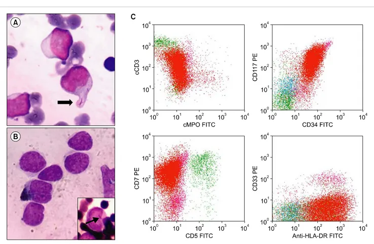
A 25-year-old man presented with multiple bilateral neck swelling along with a nasal vocal intonation, weight loss, and low-grade fever since 1 month. He was externally eval- uated via neck ultrasonography and nasal endoscopy, which revealed bilateral cervical lymphadenopathy and nasophar- yngeal lymphoid hyperplasia, respectively. The results of fine needle aspiration of a cervical lymph node were sugges- tive of a hemato-lymphoid malignancy, and the patient was referred to our hospital for further evaluation. Upon examination, he presented with bilateral cervical lympha- denopathy and splenomegaly. His lactate dehydrogenase level was elevated (1,703 U/L; reference range, 313–618 U/L) and liver and renal functions were normal. Hematological parameters revealed a hemoglobin level of 55 g/L, total leukocyte count of 193.8×10⁹ cells/L, and platelet count of 40×109 cells/L. A peripheral blood smear revealed a blast frequency of 88%; these were large with scant light blue agranular cytoplasm, open nuclear chromatin, and 1–2 indis- tinct nucleoli. The bone marrow showed near total replace- ment by blasts. Most blasts exhibited the previously de- scribed morphology, but few had moderate amounts of gran- ular cytoplasm, fine nuclear chromatin, and 2–3 prominent nucleoli, and occasional blasts contained Auer rods (Fig.
1A). Myeloperoxidase (MPO) cytochemistry revealed ap- proximately 3% MPO-positive blasts, including an occa- sional Auer rod (Fig. 1B). Flow cytometric immuno- phenotyping revealed that the blasts were positive for CD45;
bloodresearch.or.kr Blood Res 2014;49:196-207.
Letters to the Editor 197
Fig. 1. (A) Bone marrow aspirate showing blasts with a single Auer rod (thick arrow; Giemsa, ×100). (B) Few myeloperoxidase (MPO)-positive blasts; inset shows an MPO-positive Auer rod (thin arrow; MPO, ×100). (C) Blasts are cCD3, CD34, CD117, and CD7 positive and cMPO, CD5, and CD33 negative.
the T cell markers cCD3, CD2, and CD7; and the mye- loid/stem cell markers CD34, CD117, CD13, HLA-DR, CD123, and CD99, but were negative for CD1a, CD8, and CD5 as well as CD19, CD10, CD4, sCD3, CD64, CD14, CD20, CD33, CD56, CD16, CD66b, CD15, CD11b, and MPO, suggesting a diagnosis of ETP-ALL (Fig. 1C). Given the morphological presence of MPO-positive Auer rods and the flow cytometric ETP-ALL phenotype, a final diagnosis of MPAL (T/Myeloid) was made. Conventional karyotyping revealed 47, XY, +4, and t(6;14)(q27:q22). The real-time polymerase chain reaction analysis was positive for FLT3-ITD mutation and negative for NPM1 mutation.
DISCUSSION
The 2008 World Health Organization (WHO) classi- fication established new and strict criteria for the diagnosis of MPAL [4]. The WHO definition of MPAL, T/Myeloid was based on the expression of specific T-cell (cytoplas- mic/surface CD3) and myeloid (MPO) antigens; the latter is determined using flow cytometry, cytochemistry, or im- munohistochemistry and/or from clear evidence of mono- cytic differentiation. The newly identified entity ETP-ALL, which is not yet included in the WHO classification, presents a particular flow cytometric diagnostic challenge because it frequently presents with sufficient myeloid-lineage differ-
entiation to be classified as MPAL, T/Myeloid [5]. Recently, Borowitz [6] suggested that these 2 types of leukemia have more similarities than differences; however, they are treated differently because of the central importance of MPO in labeling a disease as myeloid and the manner, in which leukemia treatment protocols are structured. However, the exact MPO positivity cut-off percentage required to establish a myeloid lineage was not mentioned in the WHO classification. Our case fulfilled all the flow cytometric cri- teria for ETP-ALL, including MPO negativity [1]. However, an extensive light microscopic evaluation of the bone mar- row revealed approximately 3% blasts with cytochemical MPO positivity and Auer rods (also positive for MPO); thus, this case was classified as MPAL, T/myeloid phenotype.
To the best of our knowledge, this is the first case of ETP-ALL with Auer rods reported in the literature.
ACKNOWLEDGEMENTS
We would like to thank our director, Dr. Raina, chairman, Dr. Kher, and Dr. Vaid and above all, our mentor and inspira- tion, Dr. Naresh Trehan.
Smeeta Gajendra, Ritesh Sachdev, Pranav Dorwal, Shalini Goel, Bhawna Jha, Tushar Sahni
Blood Res2014;49:196-207. bloodresearch.or.kr
198 Letters to the Editor
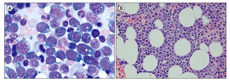
Fig. 1. Bone marrow (BM) aspiration and biopsy findings in the first case. BM aspiration showed the presence of neoplastic cells (27.8%) with oval to round nuclei, prominent nucleoli, and abundant amount of bluish cytoplasm (A, Wright staining, ×1,000). The BM clot section also showed interstitial infiltration of neoplastic cells (B, hematoxylin and eosin staining, ×400) positive for CD4, CD56, and CD123 on immunohistochemical staining.
Department of Pathology and Laboratory Medicine, Medanta-The Medicity, Gurgaon, India
Correspondence to: Ritesh Sachdev Department of Pathology and Laboratory Medicine,
Medanta-The Medicity, Sector - 38, Gurgaon, Haryana 122 001, India E-mail: sachdev05@gmail.com
Received on Mar. 26, 2014; Revised on Mar. 29, 2014; Accepted on Jun. 11, 2014 http://dx.doi.org/10.5045/br.2014.49.3.196
AuthorsÊ Disclosures of Potential Conflicts of Interest No potential conflicts of interest relevant to this article were reported.
REFERENCES
1. Coustan-Smith E, Mullighan CG, Onciu M, et al. Early T-cell pre- cursor leukaemia: a subtype of very high-risk acute lympho- blastic leukaemia. Lancet Oncol 2009;10:147-56.
2. Neumann M, Coskun E, Fransecky L, et al. FLT3 mutations in ear- ly T-cell precursor ALL characterize a stem cell like leukemia and imply the clinical use of tyrosine kinase inhibitors. PLoS One 2013;8:e53190.
3. Weir EG, Ali Ansari-Lari M, Batista DA, et al. Acute bilineal leu- kemia: a rare disease with poor outcome. Leukemia 2007;21:
2264-70.
4. Borowitz MJ, Bene MC, Harris NL, Porwit A, Matutes E. Acute leukaemias of ambiguous lineage. In: Swerdlow SH, Campo E, Harris NL, et al, eds. WHO classification of tumours of haemato- poietic and lymphoid tissues, 4th ed. Lyon, France: IARC Press, 2008:150-5.
5. Chen W. Case study interpretation-New Orleans: case 2. Mixed phenotype acute leukemia, T/myeloid. Cytometry B Clin Cytom 2013;84:342-5.
6. Borowitz MJ. Mixed phenotype acute leukemia. Cytometry B
Clin Cytom 2014;86:152-3.
Leukemic manifestation of blastic plasmacytoid dendritic cell neoplasm:
laboratory approaches in 2 cases
TO THE EDITOR: The blastic plasmacytoid dendritic cell neoplasm (BPDCN) is a rare subtype of the myeloid neo- plasm, with an incidence of 0.44% among all hematologic malignancies and characterized by clonal plasmacytoid den- dritic cell presence [1, 2]. Patients with BPDCN typically display skin lesions and aggressive clinical features with rapid systemic dissemination, exhibiting bone marrow (BM) involvement in 60–90% of cases [3, 4]. However, leukemic presentation of BPDCN is rare, with a reported incidence of less than 1% among those with acute leukemia [5].
Moreover, BPDCN with leptomeningeal involvement has been reported to be more infrequent compared with lymph node or liver/spleen involvement, as only 4 of 43 BPDCN patients experienced cerebrospinal fluid (CSF) involvement in a large-scale multicenter study [1]. Herein, we report 2 patients with BPDCN who exhibited leukemic manifes- tation (1 with simultaneous leptomeningeal involvement) diagnosed by using immunohistochemical staining and flow cytometry analysis for CD4, CD56, and CD123.
CASES
The first case of BPDCN was observed in a 36-year-old woman who was admitted to the authors’ institution for workup of a left cheek skin lesion that had developed 6 months previously. An incisional biopsy of the skin lesion