Patients with rectal cancer in the lower third of the rectum have three main options for surgical treatment
including transanal resection, abdominoperineal resection (APR), total mesorectal excision (TME) and preoperative concurrent chemoradiotherapy (CCRT) with surgical management. However, APR can lead to a decreased quality of life, viewed in colostomy and compared to other surgical treatments. To achieve an acceptable quality of life, sphincter-saving procedures have become increasingly popular (1, 2). In recent years, high-resolution pelvic magnetic resonance imaging (MRI) has achieved an essential role in the determination of circumferential resection margin (CRM) and the evaluation of sphincter invasion. The prediction of sphincter involvement and measurement of the distance from the anal verge are important for treatment planning in patients with lower rectal cancer INTRODUCTION
�Received; October 4, 2014�Revised; November 12, 2014
�Accepted; November 18, 2014 Corresponding author : Joo Hee Kim, M.D.
Department of Radiology, Gangnam Severance Hospital, Yonsei University College of Medicine, 211 Eonjuro, Gangnam-gu, Seoul 135- 720, Korea.
Tel. 82-2-2019-3510, Fax. 82-2-3462-5472 E-mail : [email protected]
This is an Open Access article distributed under the terms of the Creative Commons Attribution Non-Commercial License (http://creativecommons.org/licenses/by- nc/3.0/) which permits unrestricted non-commercial use, distribution, and reproduction in any medium, provided the original work is properly cited.
Preoperative Evaluation of Lower Rectal Cancer by Pelvic MR with and without Gel Filling
Dae Jung Kim1, 2, Joo Hee Kim1, Joon Seok Lim3, Jae-Joon Chung1, Jeong-Sik Yu1, Myeong-Jin Kim3, Ki Whang Kim3
1Department of Radiology, Gangnam Severance Hospital, Yonsei University College of Medicine, Seoul, Korea
2Department of Radiology, CHA Bundang Medical Center, CHA University, Gyeonggi-do, Korea
3Department of Radiology, Severance Hospital, Yonsei University College of Medicine, Seoul, Korea
Purpose : To assess the usefulness of rectal filling using ultrasonographic gel in patients with lower rectal cancer.
Materials and Methods: Twenty five patients with lower rectal cancer were enrolled. High resolution pelvic MR was per- formed twice before and after gel filling. Independently and retrospectively, two radiologists reviewed each set of MR images using five-grade scales for sphincter involvement, CRM (circumferential resection margin) involvement and depic- tion of the tumor. Same two radiologists retrospectively performed consensus review of each set of MR images for tumor distance from the anal verge and T&N staging.
Results: Tumor depiction scores from MR with gel filling were significantly higher than those of MR without distention (p<0.001). Compared to MR without distension, MR with gel filling had no significant differences in prediction of CRM or sphincter involvement (p>0.05). Distance from the anal verge was significantly different between MR with gel filling and rigid endoscopy (6.8 1.6 cm vs. 5.8 1.6 cm, p=0.001). There were no significant differences between pathological staging and MR staging with or without gel filling.
Conclusion: MR with gel filling improved tumor depiction. And also MR with gel filling revealed same ability for the pre- dictions of CRM or sphincter invasion in patients with lower rectal cancer, comparing with MR without gel filling.
Index words : Rectal cancer∙Magnetic resonance imaging (MRI)∙Rectum∙Ultrasonographic transmission gel Preoperative staging
Original Article
(2, 3).
Several studies have reported that pelvic MRI with rectal contrast filling including diluted barium suspen- sions, methylcellulose, superparamagnetic iron oxide solutions and water improve tumor depiction and estimation of tumor extension in the rectum (4-8).
The MRI jelly method, in which ultrasonographic transmission gel is injected into the vagina and rectum, has been widely used for detection of deep pelvic endometriosis for many years (9, 10). Recently, Kim, et al. (11) reported that ultrasonographic transmission gel is an effective and safe contrast material for use in the rectum on pelvic MRI. And also ultrasonographic transmission gel improves the tumor depiction and estimation of tumor extension in the rectum, it may be helpful for planning of endoscopic mucosa resection or dissection at T1 cancer, but the influence of gel insertion in the rectum is not disclosed for tumor staging, CRM involvement prediction and sphincter involvement prediction at lower rectal cancer. Our study was aimed to assess the usefulness of pelvic MR with and without gel filling in patients with lower rectal cancer with regard to tumor depiction, sphincter involvement, CRM involvement, T staging, N staging and tumor distance measurement (from the anal verge).
Patient populations
This retrospective study was designed from October 2008 to February 2009 after approval by the institu- tional review board. During this period, thirty-three patients with histologically proven lower rectal cancer (lower rectal cancer was defined as, clinically < 8 cm from the anal verge to the distal tumor margin, as determined with sigmoiodoscpy or rectal exam) were enrolled. These patients underwent 1.5 T or 3 T high resolution pelvic MRI, before and after rectal disten- tion, for treatment planning. Among the 33 patients, eight patients were excluded because they did not receive any curative treatment. Our study required pathologic or surgical results for analysis but surgical
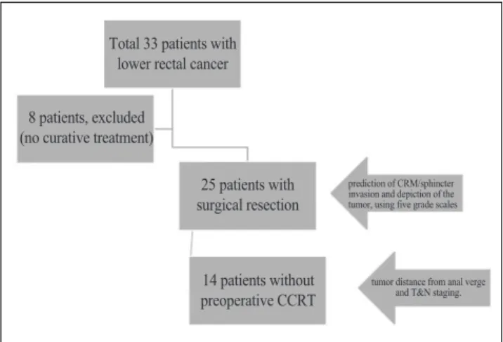
resection (ultra-lower anterior resection and lower anterior resection), and 11 of them had a preoperative CCRT due to sphincter invasion and/or CRM involve- ment (Fig. 1). Fourteen patients without CCRT underwent radical surgery within one month, after MR exam. Eleven patients with CCRT underwent radical surgery within two months, after CCRT (Fig. 1).
MRI Technique
MRIs were performed with a 1.5T scanner (MagneAvanto; Siemens Medical Systems, Erlangen, Germany) and a 3T scanner (Achieva; Philips, Eindhoven, Netherlands). The patients did not undergo bowel preparation; however, before the exam, if not contraindicated, 20 mg of intravenous scopolamine butyl bromide (Buscopan, Boehringer Ingelheim) was administered to minimize peristalsis and prevent rectal spasm and all patients in this study were administered only one time before the examina- tion. The following imaging sequences were performed before and after rectal distension on a 1.5 T scanner:
T1-weighted turbo spin-echo MRI (TR/TE 530/10 ms, matrix size 448×358, 3-mm slice thickness, 0.3-mm intersection gap, and 20 cm × 20 cm field of view) in the axial plane and T2-weighted turbo spin-echo MRI (TR/TE 3400/100 ms, matrix size 512×180, 3-mm slice thickness, 0.3-mm intersection gap, and 20 cm × 20 cm field of view) in the axial, oblique coronal, and sagittal planes. Also the following imaging sequences were performed before and after rectal distension on a 3T scanner: T1-weighted turbo spin-echo MRI (TR/TE 550/10 ms, matrix size 500× 500, 3-mm slice thickness, 0.3-mm intersection gap, and 25 cm × 25 cm field of view) in the axial plane and T2-weighted turbo spin-echo MRI (TR/TE 3500/90 ms, matrix size 500×500, 3-mm slice thickness, 0.3-mm intersection gap, and 25 cm × 25 cm field of view) in the axial, oblique coronal, and sagittal planes. In tumor level, all axial images were obtained at angled to the axis of the rectal tumor.
Approximately 50 ml to 100 ml of warm ultrasono- graphic transmission gel was injected using a balloon- tipped rectal tube. If patients were uncomfortable, the injection was stopped. The rectal tube was carefully MATERIALS AND METHODS
time for rectal gel administration, consumed approxi- mately fifty to sixty minutes in this study.
Image Analysis
Two radiologists with five years and more than ten years of experience in pelvic MRI interpretation, respectively, independently and separately analyzed each set of axial, oblique coronal and sagittal T2- weighted images from 25 patients (MR exams before and after gel filling) using five-grade scales [1-5]
without clinical information. Five grade scales were used to determine sphincter involvement (we defined that tumor infiltration/invasion is seen at the external anal sphincter and/or levator ani muscle; 1, definitely absent; 2, probably absent; 3, possibly present; 4, probably present; and 5, definitely present), CRM involvement (we defined that tumor is abutting to or out of mesorectal fascia; 1, definitely absent; 2, probably absent; 3, possibly present; 4, probably present; and 5, definitely present) and depiction of the tumor (1, could not indentify tumor ; 2, partially indentified tumor; 3, indentified tumor but could not demarcate margins; 4, presence of tumor and partially demarcated margin; and 5, well configured tumor and well demarcated tumor margin; with a focus on the
intraluminal part of the tumor).
The same two radiologists reviewed with consensus each set of MR images (before and after gel filling) of 14 patients who underwent surgery without preopera- tive CCRT to determine the tumor distance from the anal verge, tumor staging (T) and nodal staging (N, Rather than using size, we used the criteria for lymph node metastasis as a node of any size with an indistinct border, irregular margin, or mixed signal intensity) (3). We excluded preoperative CCRT cases,
Fig. 1. Flow chart of patient selection and overall study design.
a b
Fig. 2. A 40-year-old man with lower rectal cancer whose tumor distance from the anal verge was approximately 4 cm according to rigid sigmoidoscopy.
The distance to the mass was measured from the anal verge to the distal tumor margin on MRI (lines). He underwent ultra-lower anterior resection surgery.
a. Sagittal T2 weighted image obtained before rectal gel distension shows that tumor distance from the anal verge measured approximately 4.4 cm.
b. Sagittal T2 weighted image obtained after rectal gel distension shows that tumor distance from the anal verge was approximately 6.2 cm, and the distance from anal verge (distal tumor margin) was exaggerated after gel filling.
Total 33 patients with lower rectal cancer
8 patients, excluded (no curative treatment)
25 patients with surgical resection
prediction of CRM/sphincter invasion and depiction of the tumor, using five grade scales
tumor distance from anal verge and T&N staging.
14 patients without preoperative CCRT
because pathological results and tumor distances after CCRT were changed compared to those of preopera- tive MRI. The tumor distance was measured from the anal verge to the distal tumor margin on sagittal T2- weighted images (Figs. 1, 2). All images were reviewed using a full picture archiving and communication system workstation (General Electric Medical system).
Data Analysis
The scores for sphincter involvement, CRM involve- ment and tumor depiction, as rated by two radiolo- gists, were compared using Wilcoxon’s signed rank test to differentiate the methods. The weighted kappa test was applied for interobserver variability, in which a kappa value less than 0.20 indicated poor agreement;
0.21-0.40 was considered fair agreement; 0.41-0.60 represented moderate agreement; 0.61-0.80 indicated substantial agreement; and 0.81 or greater implied excellent agreement.
Measurement of tumor distance using rigid endoscopy during surgery was the defined reference method. A paired t-test was used to analyze differ- ences in tumor distance between each MRI set and rigid endoscopy during surgery. Pathological T&N staging was defined as the gold standard, and a paired t-test was used to analyze differences in T&N staging between predictions of each MRI set and pathological
results. We also calculated the accuracy of each MRI T- staging and N-staging, based on the pathological results.
Analyses were performed using SPSS version 15.0 (SPSS, Chicago, Ill). In all statistical analyses, differ- ences were considered significant when the P value was less than 0.05.
Tumor depiction scores from MRI with gel filling were significantly higher than those in MRI without gel filling (p<0.001). Compared to MRI without gel filling, MRI with gel filling revealed no significant difference in CRM or sphincter involvement (p>0.05) (Table 1, Figs. 3-5). Interobserver agreement (kappa=0.60) was moderate.
Distance from the anal verge was significantly differ- ent between MRI with gel filling and rigid endoscopy (p=0.001, rigid sigmoidoscopy vs. MR with gel filling:
5.8 1.6 cm vs. 6.8 1.6 cm) (Fig. 2), The distance from the anal verge was not significantly different between MRI without gel filling and rigid endoscopy (p=0.786, rigid sigmoidoscopy vs. MR without gel filling: 5.8 1.6 cm vs. 5.6 1.4 cm).
There were no statistical differences between RESULTS
Table 1. Ranks of tumor depiction and CRM/sphincter invasion prediction scores for each reviewer (Wilcoxon’s signed rank test)
Reviewer 1
MRI with gel filling vs. MRI without gel filling Positive Ranks * (N) Negative Ranks ** (N) Ties *** (N) P Values
Tumor depiction 22 0 3 0.000
CRM invasion prediction 3 3 19 0.739
Sphincter invasion prediction 6 4 15 0.405
Reviewer 2
MRI with gel filling vs. MRI without gel filling Positive Ranks * (N) Negative Ranks ** (N) Ties *** (N) P Values
Tumor depiction 18 1 6 0.000
CRM invasion prediction 7 6 12 0.942
Sphincter invasion prediction 4 10 11 0.071
a b
c d
Fig. 3. A 66-year-old woman with lower rectal cancer (pT1).
a, b. Oblique coronal & axial T2 weighted images obtained before rectal gel distension show that the tumor was not well defined on MRI. T3 rectal cancer was diagnosed.
c, d. Oblique coronal & axial T2 weighted image obtained after rectal gel distension shows that the tumor was well defined on MRI. T1 rectal cancer (arrows) was diagnosed.
a b
Fig. 4. A 70-year-old man with lower rectal cancer and sphincter invasion.
a. Oblique coronal T2 weighted image obtained before rectal gel distension shows that the tumor infiltrated the right external sphincter (arrow).
b. Oblique coronal T2 weighted image obtained after rectal gel distension shows more clearly that the tumor infiltrated the right external sphincter (arrow). However, there were no significantly different assessment scores between MRI with gel filling and MRI without gel filling.
pathological staging and MRI staging with and without gel filling (P>0.05) - accuracies of T staging of MRI with gel filling vs. MRI without gel filling: 71% vs.
64%; accuracies of N staging of MRI with gel filling vs. MRI without gel filling: 57% vs. 57% (Table 2).
In patient with rectal cancer within the lower third of the rectum, surgeons struggle to balance the risk of loco-regional recurrence and the patient’s quality of life. Recently, high-resolution pelvic MRI provides useful information for the determination of sphincter- preserving surgery. And pre-operative CCRT diminishes the rate of local recurrence and enables to sphincter-preserving surgery in lower rectal cancer (1- 3, 12). So preoperative staging of rectal cancer in the lower third of the rectum is important for therapeutic planning, and MRI has a more important role.
However, the accuracy of CRM involvement predic- tion has been shown to be lower for lower rectal cancer compared to those of upper and mid rectal cancers (13). And surrounding anatomic structures in lower rectum are complicated and have a narrow interval.
To improve the diagnostic accuracy of MRI in patients with rectal cancer, many materials including diluted barium suspensions, methylcellulose, superparamagnetic iron oxide solutions and water have been used for rectal distention. The rationale for rectal distention is to improve distinction between the lumen and the inner wall of the rectum, distinguishing the tumor from fecal material or edematous normal bowel wall and demarcating tumor from perirectal fat.
Compared to prior studies, our study revealed similarly improved tumor depiction (4-8). MRI with gel filling was significantly superior to MRI without gel filling for tumor depiction. This improvement may help in the planning of endoscopic mucosal resection or submucosal dissection in cases of T1 rectal cancer.
We believe that MR with gel filling will influence the prediction of CRM/sphincter invasion because Slater A et al (14) reported that rectal distension by air
thought that CRM invasion prediction would be overestimated at the anterior wall of the lower rectum by luminal distention. However, this procedure did not significantly improve or hinder CRM/sphincter invasion prediction (Table 1).
Ultrasonographic transmission gel is a non-magnetic semisolid material to characterize high signal intensity DISCUSSION
Table 2. Prediction of T&N staging in MRI with gel filling and MRI without gel filling and pathological T&N staging in surgery patients (n=14)
T stage MRI with gel filling Pathology (pT*)
T1 2 (pT1, n= 2) 3
T2 3 (pT2, n= 3) 6
T3 9 (pT1, n=1; pT2, n=3; pT3, n=5) 5 T stage MRI without gel filling Pathology (pT*)
T1 2 (pT1, n= 2) 3
T2 5 (pT1, n= 1; pT2, n=3; pT3, n=1) 6
T3 7 (pT2; n=3, pT3; n=4) 5
N stage MRI with gel filling Pathology (pN**) N 0 9 (pN0, n=6; pN1, n=1; pN2, n=2) 8
N 1 2 (pN0, n=1; pN1, n=1) 3
N 2 3 (pN0, n=1; pN1, n=1; pN2, n=1) 3 N stage MRI without gel filling Pathology (pN**) N 0 9 (pN0, n=6; pN1, n=1; pN2, n=2) 8
N 1 2 (pN0, n=1; pN1, n=1) 3
N 2 3 (pN0, n=1; pN1, n=1; pN2, n=1) 3
*pT - pathologic tumor staging
**pN - pathologic nodal staging Note
1. T stages of MRI are as follows: T1, Tumor invades the submucosa; T2, Tumor invades the muscularis propria; T3, Tumor penetrates the muscularis propria and perirectal fat.
2. N stages of MRI are as follows; N0, no predicted lymph nodes positive for tumor; N1, <four predicted regional nodes positive for tumor; N2, >four predicted regional nodes positive for tumor.
Prediction of regional nodes positive for tumor was size of LNs
>8 mm in short axis.
3. Pathological stages are as follows: T1, limited to mucosa and submucosa; T2, extension into but not through muscularis propria; T3, invasion of perirectal fat; N0, no involved lymph nodes; N1, <four regional nodes positive for tumor; N2, >four regional nodes positive for tumor.
using small volume. Compared with a prior water distention study (5), the volume of ultrasonographic transmission gel necessary for adequate distention of the rectum was smaller, 50-100 mL compared to the 200-500 mL of water required. In this study, all patients with rectal gel filling showed good tolerance during MR examination, as the contrast is a semisolid and is present in only a small volume. The water filling method has a drawback to create an uncontrolled air fluid level and it caused of artifact to limitation of assessment at the anterior wall of rectum (5, 11). In our study, small air bubbles were located at the gel interspace or boundary of the gel but did not cause artifacts that hindered interpretation. Moreover, all patients were administered 20 mg of intravenous scopolamine butyl bromide before the examination to minimize peristalsis and prevent rectal spasm.
Artifacts related to bowel peristalsis did not exist nor hindered our interpretation.
In this study, there were no statistical differences between pathological staging and MRI staging with and without gel filling. However, calculation of accuracy in each MR set, based on pathologic result, MRI with gel filling demonstrated slight improvement in T staging (MRI with gel filling vs. MRI without gel filling: 71% vs. 64%). The accuracies of N staging for the MRI sets were equal (57%) (Table 2). The overall accuracy of MR imaging in predicting T staging has been reported from 71%-91% (mean, 85%), and the range of overall accuracy of MR imaging in predicting N staging was 43%-85% (mean, 75%) (15-24).
Flexible sigmoidoscopy is the reference tool for
diagnosis of rectal cancer, but its ability to location of tumor has been questionable. The tumor location and distance were very important for surgical planning and recently rigid sigmoidoscopy allows more accurate measurements (25). Baaturp et al. (26) reported that MRI and rigid sigmoidscopy are not interchangeable in determining the distance from anus to tumor. They performed MRI with gel filling (100-150 ml) and measured the distance from the puborectal muscle to the tumor. There was a discrepancy of average tumor distance between MRI and rigid sigmoidoscopy at the rectum (MRI vs. rigid sigmoidoscopy: 61 mm vs. 82 mm in rectum). In our study, there was also discrep- ancy between rigid sigmoidocopy and MRI with gel filling at the lower rectum (p=0.001, rigid sigmoi- doscopy vs. MR with gel filling: 5.8 1.6 cm vs. 6.8 1.6 cm in lower rectum). We suggested that this differ- ence is because rigid sigmoidoscopy draws a straight measurement line while MRI uses a curved measure- ment line. Measurement of tumor distance using rigid sigmoidoscopy might lead to underestimation, as the curve of the rectum is straightened during this procedure. On the other hand, the rectum was more stretched after rectal gel filling, than under sigmoi- doscopy, therefore the tumor distance with gel filling might be exaggerated. This mild exaggeration of tumor distance was within an acceptable range and was helpful in treatment planning. During the surgical procedure, remaining proximal and distal bowel loops were dragged in maximum for anastomosis after tumor resection; therefore, measurement of the distended bowel loop is helpful for surgical planning.
a b
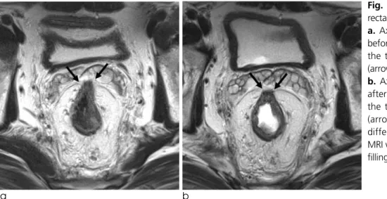
Fig. 5. A 45-year-old man with lower rectal cancer and CRM involvement.
a. Axial T2 weighted image obtained before rectal gel distension shows that the tumor invaded the anterior CRM (arrows).
b. Axial T2 weighted image obtained after rectal gel distension shows that the tumor invaded the anterior CRM (arrows). There were no significantly different assessment scores between MRI with gel filling and MRI without gel filling.
But the curved measurement line and non-distended bowel loop on MRI is more physiologic and it may provide a more accurate tumor distance measurement from the anal verge.
The main limitation of our study was the small number of patients. Data collection period was only 5 months, because twice MR exams in each patient were performed during this period (because of restraint of scan time and patient’s inconvenience). Now all MR exams for rectal cancer evaluation are performed with only gel filling. In addition, we limited the enrollment of patients to only rectal cancer patients with cancer in the lower rectum. In particular, the number of patients for the prediction of T and N staging and prediction of sphincter involvement was small.
Patients who did not receive preoperative CCRT demonstrated lower tumor staging compared with that of patients who did undergo preoperative CCRT. This selection bias may affect the results of accuracy of T&N staging in each MRI set. Though this study was small number patients, our study had a unique merit, in which twice MR exams was performed from the same patient. Secondary, the use of different MRI machines may have also influenced our results but we didn’t differ 1.5 T and 3 T MRI during imaging analysis, because we obtained T2 weighed images by turbo spin-echo sequence on 1.5 T and 3T MR machines. Because the clinical information obtained from 3 T and 1.5 T MRI with turbo spin-echo was not much different. Turbo spin-echo sequence of 3T provided a much higher matrix size than that of 1.5T and it provided a much greater resolution of certain anatomic structures but it produced noisier images by peristaltic artifacts (27). Thirdly, during analysis of MR exam sets, tumor location - such as anterior wall, posterior wall, lateral spread, etc - was not considered.
Tumor location possibly affected the analysis of CRM involvement and sphincter involvement. In addition, the pathological type was not considered in this study.
Also radiologists, who analyzed images, had known information of rectal cancer and this information had affected this study by bias.
In conclusion, MR with gel filling improved tumor depiction. And also MR with gel filling revealed same
References
1. Leo E, Belli F, Andreola S, et al. Sphincter-saving surgery for low rectal cancer. The experience of the National Cancer Institute, Milano. Surg Oncol 2004;13:103-109
2. Holzer B, Urban M, Ho¨lbling N, et al. Magnetic resonance imaging predicts sphincter invasion of low rectal cancer and influences selection of operation. Surgery 2003;133:656-661 3. Iafrate F, Laghi A, Paolantonio P, et al. Preoperative staging of
rectal cancer with MR imaging: correlation with surgical and histopathologic findings. Radiographics 2006;26:701-714 4. Goh JS, Goh JP, Wansaicheong GK. Methylcellulose as a rectal
contrast agent for MR imaging of rectal carcinoma. AJR Am J Roentgenol 2002;178:1145-1146
5. Kim MJ, Lim JS, Oh YT, et al. Preoperative MRI of rectal cancer with and without rectal water filling: an intraindividual compari- son. AJR Am J Roentgenol 2004;182:1469-1476
6. Wallengren NO, Holtas S, Andren-Sandberg A, Jonsson E, Kristoffersson DT, McGill S. Rectal carcinoma: double-contrast MR imaging for preoperative staging. Radiology 2000;215:108- 114
7. Panaccione JL, Ros PR, Torres GM, Burton SS. Rectal barium in pelvic MR imaging: initial results. J Magn Reson Imaging 1991;1:605-607
8. Urban M, Rosen HR, Ho¨lbling N, et al. MR imaging for the preoperative planning of sphincter-saving surgery for tumors of the lower third of the rectum: use of intravenous and endorectal contrast materials. Radiology 2000;214:503-508
9. Chassang M, Novellas S, Bloch-Marcotte C, et al. Utility of vaginal and rectal contrast medium in MRI for the detection of deep pelvic endometriosis. Eur Radiol 2010;20:1003-1010 10. Kikuchi I, Takeuchi H, Kuwatsuru R, et al. Diagnosis of
complete cul-de-sac obliteration (CCDSO) by the MRI jelly method. J Magn Reson Imaging 2009;29:365-370
11. Kim SH, Lee JM, Lee MW, Kim GH, Han JK, Choi BI.
Sonography transmission gel as endorectal contrast agent for tumor visualization in rectal cancer. AJR Am J Roentgenol 2008;191:186-189
12. Arnoletti JP, Bland KI. Neoadjuvant and adjuvant therapy for rectal cancer. Surg Oncol Clin N Am 2006;15:147-157
13. Peschaud F, Cuenod CA, Benoist S, et al. Accuracy of magnetic resonance imaging in rectal cancer depends on location of the tumor. Dis Colon Rectum 2005;48:1603-1609
14. Slater A, Halligan S, Taylor SA, Marshall M. Distance between the rectal wall and mesorectal fascia measured by MRI: Effect of rectal distension and implications for preoperative prediction of a tumour-free circumferential resection margin. Clin Radiol 2006;61:65-70
15. Kuo LJ, Chern MC, Tsou MH, et al. Interpretation of magnetic resonance imaging for locally advanced rectal carcinoma after preoperative chemoradiation therapy. Dis Colon Rectum 2005;48:23-28
16. Chen CC, Lee RC, Lin JK, Wang LW, Yang SH. How accurate is magnetic resonance imaging in restaging rectal cancer in patients receiving preoperative combined chemoradiotherapy?
cancer correlates with a favourable prognosis. Eur J Cancer 2003;39:192-195
18. Vliegen RF, Beets GL, Lammering G, et al. Mesorectal fascia invasion after neoadjuvant chemotherapy and radiation therapy for locally advanced rectal cancer: accuracy of MR imaging for prediction. Radiology 2008;246:454-462
19. Beets-Tan RG, Beets GL. Rectal cancer: review with emphasis on MR imaging. Radiology 2004;232:335-346
20. Brown G, Richards CJ, Newcombe RG, et al. Rectal carcinoma:
thin-section MR imaging for staging in 28 patients. Radiology 1999;211:215-222
21. Gagliardi G, Bayar S, Smith R, Salem RR. Preoperative staging of rectal cancer using magnetic resonance imaging with external phase-arrayed coils. Arch Surg 2002;137:447-451
22. Blomqvist L, Machado M, Rubio C, et al. Rectal tumor staging:
MR imaging using pelvic phasedarray and endorectal coils vs endoscopic ultrasonography. Eur Radiol 2000;10:653-660
23. Kim JH, Beets GL, Kim MJ, Kessels AG, Beets-Tan RG. High resolution MR imaging for nodal staging in rectal cancer: are there any criteria in addition to the size? Eur J Radiol 2004;52:78-83
24. Brown G, Richards CJ, Bourne MW, et al. Morphologic predic- tors of lymph node status in rectal cancer with use of high- spatial-resolution MR imaging with histopathologic comparison.
Radiology 2003;227:371-377
25. Piscatelli N, Hyman N, Osler T. Localizing colorectal cancer by colonoscopy. Arch Surg 2005;140:932-935
26. Baatrup G, Bolstad M, Mortensen JH. Rigid sigmoidoscopy and MRI are not interchangeable in determining the position of rectal cancers. Eur J Surg Oncol 2009;35:1169-1173
27. Choi JY, Kim MJ, Chung YE, et al. Abdominal applications of 3.0-T MR imaging: comparative review versus a 1.5-T system.
Radiographics 2008;28:e30
통신저자 : 김주희, (135-720) 서울시 강남구 도곡동 언주로 211, 연세대학교 강남세브란스병원 영상의학과 Tel. (02) 2019-3510 Fax. (02) 3462-5472 E-mail: [email protected]
하부직장암 환자에서 초음파겔 삽입 전후의 골반 자기공명영상의 비교
1연세대학교 강남세브란스병원 영상의학과
2차의과대학 분당차병원 영상의학과
3연세대학교 세브란스병원 영상의학과
김대중1, 2∙김주희1∙임준석3∙정재준1∙유정식1∙김명진3∙김기황3
목적: 하부직장암 환자에서 수술 전 평가를 위한 골반 자기공명영상에서 직장 내 초음파 겔 삽입 효과를 알아보고자 하였다.
대상과 방법: 2008년 9월부터 2009년 2월까지 하부직장암 환자 25명을 대상으로 하였으며 이들은 모두 수술 전 골반 자기공명영상을 시행하였으며 직장 내 초음파 겔 삽입 전후로 하여 두 차례 시행하였다. 두 명의 영상의학과 의 사가 독립적으로 그리고 후향적으로 각 환자의 두 번의 골반 자기공명영상에서 항문 조임근 침범, 절제 범위 침범, 그 리고 종양 묘사에 대하여 분석하여 각 항목에 대하여 5단계로 점수를 기록하였다. 그리고 위 두 명의 영상의학과 의사 가 합동으로 각 환자의 두 번의 골반 자기공명영상을 바탕으로 항문 피부선에서 종양까지의 거리와 종양의 T항목과 N 항목의 병기를 후향적으로 분석하였으며 내시경상 길이와 조직학적 병기를 기준으로 하였다.
결과: 초음파 겔을 삽입한 골반 자기공명영상이 종양 묘사 점수가 통계학적으로 유의하게 높았다 (p < 0.001). 항문 조임근 침범과 절제범위 침범의 점수는 두 검사간 통계학적으로 유의하게 차이가 있지 않았다 (p > 0.05). 항문 피부 선에서 종양까지의 거리는 초음파 겔을 삽입한 골반 자기공명영상이 내시경과 통계학적으로 유의하게 차이가 있었다 (6.8±1.6 cm vs. 5.8±1.6 cm, p=0.001). 병리적 병기를 기준으로 두 검사간의 영상의학적 병기는 통계학적으 로 유의하게 차이가 있지 않았다.
결론: 초음파 겔을 이용한 골반 자기공명영상은 젤을 이용하지 않는 골반 자기공명영상에 비하여 종양 묘사를 향상시 키며 또한 항문 조임근 침범과 절제범위 침범 판단에 같은 능력을 보여 주었다.
대한자기공명의과학회지 18:323-331(2014)