Vo1. 15, No. 1, May, 2006
※통신저자: 정 윤 규
강원도 원주시 일산동 162
연세대학교 원주의과대학 성형외과학교실
Tel: 82-33-741-0611, Fax: 82-33-732-4022, E-mail: [email protected]
다양한 형태의 생 비골 이식술을 이용한 경골의 재건
연세대학교 원주의과대학 성형외과학교실, 한림대학교 의과대학 성형외과학교실*
남상현∙김범진∙정윤규∙고성훈*
─
─ Abstract ──
Reconstruction of Tibial Defects in Lower Extremity With Various Versions of Vascularized Fibula Transfer
Sang Hyun Nam, M.D., Bom Jin Kim, M.D., Yoon Kyu Chung, M.D., Sung Hoon Koh, M.D.*
Department of Plastic & Reconstructive Surgery Yonsei University Wonju College of Medicine, Wonju, Korea, Department of Plastic & Reconstructive Surgery Hallym University College of Medicine, Seoul, Korea*
Twelve cases in eleven patients with segmental bone defects were treated with contralateral fibula free flap and ipsilateral island fibula flap in an antegrade, retrograde or bidirectional flow fashion. Five cases were managed with free flaps and seven were with ipsilateral fibula island transfer. Among seven cases, antegrade fashion was three, retrograde was three, and bidirectional was one. All patients were related with open tibial fractures and its sequelae except one who had open foot bone fracture. According to Gustilo's classification, ten patients were type IIIb and one was type IIIc. Basically, antegrade-flow flaps based on the peroneal vessels as in the conventional free flap were used for the proximal or middle one-third tibial defects. On the contrary, retrograde- flow flaps based on the communicating branch between the peroneal and posterior tibial vessels were used for the middle or distal one-third of the tibia. Bidirection-flow flap based on intact per- oneal vessels were used for the middle portion of the tibia. The patients who have undergone ipsi- lateral fibula island flap had one of the following problems: a previously failed free flap, below- knee amputation of the opposite leg because of open tibial fracture, refusal to use the contralateral sound leg, or poor general condition to stand a lengthy operation. Six of the patients who have got ipsilateral fibula island flap also had an associated fibula fracture on the same leg, which was ulti- mately used as one of the osteotomy sites. The follow-up period was from 1 to 10 years. Two cases of free flap were failed: one patient had below-knee amputation and the other patient had
Ⅰ. 서 론
심한 외상이나 만성 골수염 등에 의한 광범위한 경골 결손에 대한 치료에는 대측 비골(contralater- al fibula)을 이용한 유리피판술이 가장 좋은 치료 법으로 알려져 있다. 이 방법의 주된 장점은 전이된 비골이 신선골절(fresh fracture)처럼 치유되어 재 형성(remodeling)과 골유합이 신속하게 이루어지는 것이다. 그러나, 유리피판술이 모든 환자에게서 가 능한 것은 아니며, 비록 높은 성공률이 보고되고 있
으나 여전히 혈전증에 대한 두려움은 존재하고 있 다. 또한 하지는 신체의 다른 부위에 비하여 해부학 적으로나 임상적으로 유리피판술의 실패율이 높다.
더욱이, 어떤 경우에는 대측의 비골을 유리피판의 공여부로 사용할 수 없거나, 환자의 전신 상태가 장 시간의 수술에 적합하지 않을 수 있다. 이러한 경우 에 대안으로서 동측(환측)의 비골을 이용할 수 있 다. 일부에서는 경골의 재건에 동측 비골을 사용해 서는 안된다고 주장하고 있으나,4,5비록 단경(single pedicle)에 기초한 진정한 도서형 피판(island ipsilateral fibula transfer. Other cases were successful and excellent hypertophy of the transferred fibula was achieved. Time to bone union ranged from 4 to 11 months. Time to full weight bearing was from 5 to 13 months after surgery. All of the transferred fibulas showed hypertrophy after weight bearing. In one case, stress fracture was developed during ambulation, which was healed conservatively. Nonunion occurred in two cases, which were treated with a long leg cast and can- cellous bone graft, respectively.
Length discrepancy of the legs was noted. The limb was shorter by an average 0.5 cm in three cases, longer by 1.1 cm in one case. In the case of island fibula transfer, limited arc of rotation was not a problem. Other disabling complications were not seen.
We believe that these diverse modalities using a vascularized fibula will make us more comfort- able to handle major bone defects.
Key Words: Tibia defect, Antegrade-flow, Retrograde-flow, Bidirectional-flow, Vascularized fibula flap, Ipsilateral fibula island flap, Lower extremity reconstruction
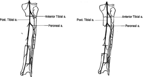
Fig. 1. Antegrade-flow pedicle based on the peroneal vessels for proximal defect.
Fig. 2. Advancement flap based on the peroneal vessels.
flap)을 사용한 예는 적지만,6,8 동측 비골을 사용하 여 경골을 재건한 증례는 많이 보고되었다.1-3 본 저 자들은 광범위한 경골 결손이 있는 11명의 환자에서 결손부의 상태와 위치에 따라 대측의 비골을 이용한 유리이식술 또는 동측의 비골을 이용한 다양한 방법 의 도서형 피판으로 경골을 재건하였으며 각각의 방 법들의 적응증과 장∙단점을 경험하였다.
Ⅱ. 재료 및 방법
11명의 환자에서 12례의 생 비골을 이용한 경골 재건을 시행하였다. 5례에서 대측의 비골을 공여부 로 하여 유리피판술을 시행하였고(Fig. 4), 7례는 동측의 비골을 이용한 도서형 피판술로 치료하였다.
환자들의 나이는 16세에서 52세(평균 나이 36세)였 고, 1명을 제외하고 모두 남자였다. 수상 기전은 교 통 사고, 추락 또는 무거운 물체에 의한 압궤상으로 Gustilo씨 IIIb형 또는 IIIc형에 해당되었다(Table 1). 11명 중 9명은 경골 골절, 1명은 경골 골절 후 불유합, 나머지 1명은 족부의 쐐기뼈(cuneiform bone)와 중족골(metatarsal bone)의 결손을 가지 고 있었다. 수술은 수상 후 37일부터 415일로 평균 105일 째 시행하였다.
도서형 피판은 다음 세 가지 유형 중 하나를 선택 하여 시행하였다. 첫째, 순행성(antegrade-flow) 피판은 보통의 유리피판술처럼 혈관경을 근위부 비 골동맥에 기초를 두었고(Fig. 1), 둘째, 역행성 (vetrograde-flow) 피판은 비골동맥과 후경골동맥 을 연결하는 소통분지(communicating branch)를 혈관경으로 하였으며(Fig. 2), 셋째, 전진형 피판 (advancement)은 비골동맥의 근위부 및 원위부를 절단하지 않고 단지 절골술과 비골 혈관의 골격화 (skeletonization)를 통해 이동시킨 피판이다(Fig.
3). 어떤 피판을 선택하느냐는 원칙적으로 결손부의 위치와 동반된 비골 골절의 유무에 따라 결정하였다.
순행성 피판은 근위부 1/2의 결손에 사용하였고
Table 1. Summary of the patients
Case Age/Sex Cause Type of Fx (Gustilo's) Defect Mode of flap
01 52/M Cultivator overturn Tibiofibular (IIIb) Proximal half Free
02 26/M Fall -down Tibia (IIIb) Proximal half Free+Antegrade*
03 42/M Pedestrian injury Infected Non-union (IIIb) Distal half Retrograde�
04 49/M Tram overturn Tibiofibular (IIIc) Proximal half Free
05 51/M Tram overturn Tibiofibular (IIIb) Proximal half Antegrade*
06 50/M Motorcycle collision Tibiofibular (IIIb) Proximal half Retrograde�
07 29/M Hit by heavy object Tibia (IIIb) Distal half Free
08 24/M Car collision Tibiofibular (IIIb) Distal half Retrograde�
09 26/M Car collision Tibia (IIIb) Middle Advancement
10 36/M Cultivator overturn Tibiofibular (IIIb) Proximal half Antegrade*
11 16/M Motorcycle collision Cuneiform (IIIb) Foot Free
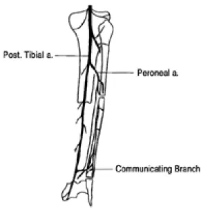
* : Antegrade fibula island flap, �: Retrograde fibula island flap Fig. 3. Retrograde-flow pedicle based on the
Communicating branches between the peronial and posterior tibial vessels
(Fig. 5), 역행성 피판은 원위부 1/2 결손에 사용하 였다. 중간부 결손은 동반된 비골의 골절의 위치에 따라 피판의 유형을 결정하였다. 결손부위가 근위부 또는 원위부로 심하게 편위되어 있지 않고 전반적으 로 중간에 위치해 있을 경우에는 먼저 비골의 양 끝 을 절골하고 비골 혈관을 골격화한 후 피판의 이동성 을 확보하면 전진형 혈관경 피판으로 사용할 수 있었 다(Fig. 6). 그러나 한 증례에서는 결손위치가 근위 부에 있었지만 역행성 피판을 사용하였는데, 이 때에 는 거상한 피판의 위,아래를 바꾸어 수혜부에 삽입하 였다.
근위부에 혈관경을 가진 순행성 도서형 비골 피판 은 유리피판술에서와 마찬가지로 동일한 방법으로
수술하였다. 비골동맥과 동반정맥은 피판의 이동성 을 극대화하기 위하여 경골비골체간 (tibioperoneal trunk)의 분기점까지 박리하였다.
원위부에 혈관경을 가진 역행성 도서형 비골피판 의 거상도 기본적으로 순행성 피판과 동일하나, 혈 관경을 자르기 전 일시적으로 비골동맥을 결찰하여 피판의 혈류 상태를 점검한 후 생존력(viability)이 확인되면 근위부 혈관을 결찰하고 필요한 만큼의 이 동성을 확보하기 위해 좀 더 골격화 시켰다.
결손부로 이동된 피판을 1 cm 내지 2 cm 중첩 시켜 수혜부 경골의 양쪽 끝의 골수강 내에 삽입했 다. 만약 피판의 양 끝을 삽입하기가 어려우면 경골 의 전면 한쪽에 홈을 파서 끼워준 후 하나 내지 두
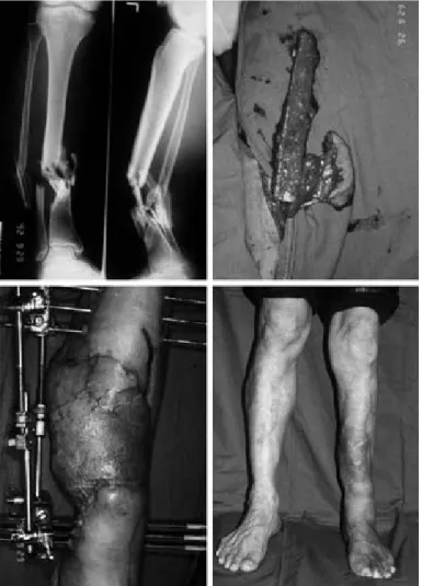
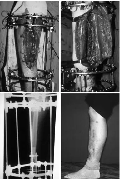
Fig. 4. A 49-year old male patient sustained from open tibiofibular fracture on the left lower leg. Preoperative X-ray finding. (Above left). Soft tissue reconstruction with latissimus dorsi at the first stage. (Above right) Elevation of free vascularized fibular free flap from the contralateral leg. (Below left) Five years after surgery. (Below right)
개의 나사 또는 K 강선으로 고정하였다. 환자의 골 절 치료를 위해 이미 부착되어 있는 외고정기는 추가 적인 안정성을 확보하고 변위를 예방하기 위해 계속 유지시켰다. 비골두(fibula head) 근처에 골절이 있었던 2례를 제외한 모든 환자에서 피판을 거상할 때 총비골신경의 손상과 족관절의 불안정성을 예방 하기 위해 비골의 상단 및 하단 8 cm는 보존하였다.
한편, 어떤 유형의 피판을 선택하던 간에 비골의 근 위 1/3과 중간 1/3의 원위부는 피판의 생존력을 확 보하기 위해 피판에 꼭 포함시켰다. 일단, 창상이 치 유되고 이식된 피판이 안정되면 외고정기를 제거하
고 long leg cast 또는 tendon-bearing cast를 하 여 골유합이 된 후 부분적 체중부하를 시작하였다.
Ⅲ. 결 과
총 12례 중 유리피판술 5례, 순행성 도서형 피판 3례, 역행성 도서형 피판 3례, 나머지 1례는 전진형 피판을 시행하였다(Table 1). 유리피판술 2례를 제 외하고 10례의 피판은 모두 완전 생존하였다. 실패 한 유리피판술 2례는 정맥 혈전증으로 인한 괴사였 다. 이 중 1례는 환자가 더 이상의 치료를 거부하여
Fig. 5. A 36-year old male patient sustained from cultivator overturn. Preoperative phtograph. (Abocve left) Insetting of vascularized fibula with an antegrade fashion. (Above right) Preoperative angiography. (Below left) Nine years after surgery. (Below right)
슬관절 아래에서 절단하였으며, 다른 한 명은 순행성 도서형 피판으로 다시 재건하였다. 골 결손의 길이는 6 cm에서 12 cm로 평균 9.4 cm였다. 이식한 비골 의 길이는 9.5 cm에서 16 cm로 평균 13.3 cm 이 었으며 2례에서는 이중통(double barrel)으로 사용 하였다. 추적 기간은 1년에서 10년으로 평균 3년 7 개월이었다(Table 2). 골유합은 절단한 1례를 제외 한 모든 환자에서 이루어졌다. 골유합에 걸린 기간은 4개월에서 11개월로 평균 6개월이었다. 불유합은 2 명에서 있었으며 각각 석고 부목과 해면골 이식으로 치료하였다. 또한 한 예에서 보행 중 피로 골절이 발
생하였으나 부목고정을 통한 보존적 치료로 치유되 었다. 골수염은 없었다. 완전 체중부하는 수술 후 5 개월에서 13개월부터 평균 9개월째 시작하였다. 재 건된 쪽의 다리는 전반적으로 건강한 다리보다 평균 0.5 cm 짧아져 있었으나, 1례에서는 오히려 1.1 cm가 길어져 있었다.
족관절의 운동범위는 모든 환자에서 제약이 있었 으며, 특히 경골의 근위부에 가깝게 골절을 입은 환 자일수록 운동할 때 슬관절의 통증이 많았다. 11명 중 단지 2명만이 수술 후 원래의 직업인 운전수로 복귀하였다.
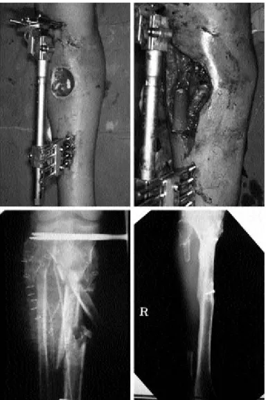
Fig. 6. A 26-year old male patient sustained from traffic accident with type IIIb open tibia fracture. Immediate photo- graph after debridement. (Above left) Elevation of the flap with intact peroneal vessels. (Above right) Postoperative X-ray view. (Below left) Postoperative 3 years. (Below right)
Ⅳ. 고 찰
생 비골 이식술은 결손된 경골을 재건할 때 대측 의 비골을 공여부로 하는 유리피판술의 형태로 많이 이용되고 있다. 그러나, 모든 환자에게 유리피판술을 시행할 수는 없으며 이러한 경우 동측의 비골을 이용 할 수 있다. 경골의 재건에 동측의 비골을 사용하는 방법은 새로운 것이 아니다. 예전에는 우회(bypass) 또는 muscle pedicled graft의 형태로서 이용되었
다.2,3,9-11 동측 비골을 사용할 경우 하지에 대한 보조
적인 지지가 더욱 약화되어 하지를 더욱 불안정하게 만든다는 비판이 있었으나,4 비록 제한적이지만 도서 형 피판술은 미세혈관문합술을 피하고, 수술 시간을 줄일 수 있으며, 다치지 않은 대측 하지를 손상시키 지 않고 유리피판술의 모든 장점을 얻을 수 있다. 피 판술이 실패하였을 경우라도 유리피판술 또는 Ilizarov 골연장술 등으로 해결할 수 있다.3,6,7 저자 들은 동측의 도서형 피판을 첫째, 이전에 대측의 비 골을 공여부로 사용하였거나 양측성 경골 및 비골 골 절로 인하여 대측의 비골을 사용할 수 없는 경우, 둘 째, 환자가 대측의 비골을 공여부로 사용하기를 거절
하는 경우, 셋째, 환자의 전신상태가 좋지 않은 경우 에 사용하였다. 그러나 골절된 다리에 심각한 근육의 손상이 동반되었을 경우에는 사용해서는 안 되는데, 그 이유는 피판을 거상할 때 근육에 추가적인 손상을 주게 되어 수술 후 다리나 발에 심각한 기능적 장애 를 일으키기 때문이다. 또, 후경골동맥이 절단된 경 우 혈관조영술을 시행하여 비골동맥이 발의 주요 공 급 동맥으로 확인되면 절대 사용해서는 안된다. 또 다른 도서형 피판술의 주요 단점은 회전 반경이 제한 되어 있는 것이다.5,6 순행성 도서형 피판의 회전축은 비골동맥이 후경골동맥으로부터 분리되는 지점이다.
회전 반경은 비골동맥의 영양분지가 비골로 들어가 기 전까지의 길이에 따라 달라진다. 비골동맥은 대부 분 평균적으로 비골두 아래 7.5 cm내지 7.9 cm에 서 후경골동맥으로부터 갈라지며, 이 지점부터 영양 동맥의 길이는 평균 6.9 cm가 된다. 그러나, 비골 동맥이 분지되는 위치가 다양하므로 수술 전 혈관조 영술이 도움이 될 수 있다.
원위부에 혈관경을 가진 역행성 도서형 피판 (distally based retrograde island flap)은 후경 골동맥으로 가는 비골동맥의 소통분지(communi- cating branch)를 혈관경으로 한다. 소통분지는
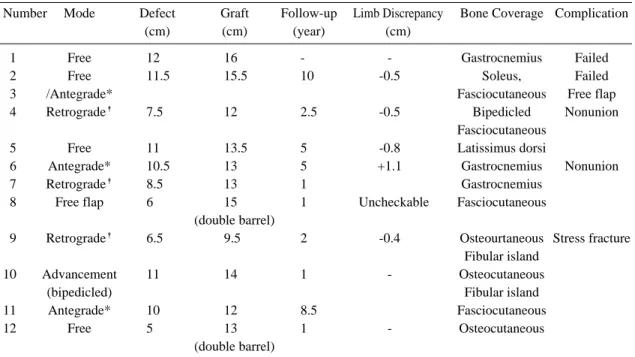
Table 2. Results
Number Mode Defect Graft Follow-up Limb Discrepancy Bone Coverage Complication
(cm) (cm) (year) (cm)
01 Free 12 16.0 - - Gastrocnemius Failed
02 Free 11.5 15.5 1000. -0.5 Soleus, Failed
03 /Antegrade* Fasciocutaneous Free flap
04 Retrograde� 7.5 12.0 2.5 -0.5 Bipedicled Nonunion
Fasciocutaneous
05 Free 11 13.5 500. -0.8 Latissimus dorsi
06 Antegrade* 10.5 13.0 500. +1.1 Gastrocnemius Nonunion
07 Retrograde� 8.5 13.0 100. Gastrocnemius
08 Free flap 6 15.0 100. Uncheckable Fasciocutaneous
(double barrel) 0
09 Retrograde� 6.5 9.5 200. -0.4 Osteourtaneous Stress fracture
Fibular island
10 Advancement 11 14.0 100. - Osteocutaneous
(bipedicled) Fibular island
11 Antegrade* 10 12.0 8.5 Fasciocutaneous
12 Free 5 13.0 100. - Osteocutaneous
0 (double barrel)
* : Antegrade fibula island flap, �: Retrograde fibula island flap
외측복사골(lateral malleolus) 상방 6 cm 또는 13 cm에 위치한다3,14. 회전 반경을 증가시키기 위하 여 소통분지까지 박리해 들어갈 수 있으나, 저자들 의 증례에서는 필요하지 않았다. 회전 반경은 어떠 한 형태의 혈관경을 선택하느냐에 따라 달라질 수 있으며, 또한 피판의 상∙하를 바꿔(upside down) 더욱 증가시킬 수 있다.
혈관경의 형태는 결손부의 위치와 동반된 비골 골 절에따라 결정된다. 경골의 근위부 결손에는 순행성 도서형 피판을 원위부 결손에는 역행성 도서형 피판 을 사용한다. 그러나, 중간부 결손에서는 동반된 비 골 골절의 위치와 영양 동맥의 위치를 고려하여 선 택한다. 또한 결손 부위가 경골의 중간 1/3에 위치 해 있는 경우에는 비골동맥을 절단하지 않고 혈관을 골격화(skeletonization)하면 골피판을 결손부에 충분히 위치시킬 수 있다.
피판을 거상 할 때에는 영양동맥에 손상을 주지 않기 위하여 비골 중간부 1/3의 원위부와 중앙부를 반드시 피판에 포함시켰다. 그러나, 비골의 중간부 에 골절이 있을 경우에는 피판의 거상이 어려워 질 수 있다.
동측의 도서형 피판은 이미 손상 받은 구역에서 비골을 거상해야 하고, 비골 골절의 동반된 경우 염 증∙반흔 및 골막 열상으로 인해 피판의 거상은 더 욱 어렵지만, 저자들의 증례에서 두 유형의 혈관경 은 모두 강력하게 박동하였으며 절골된 골단에서 신 선한 출혈을 항상 관찰할 수 있었다.
골피판을 올바르게 삽입하는 것은 매우 중요하다.
거상한 비골은 최대 압박선 (line of maximal compression)에 놓이게 하여야 한다.8 즉, 골수강 내에 위치시키는 것이 가장 좋다. 저자가 경험한 증 례에서 기술적인 어려움 때문에 비골피판을 경골의 가장자리에 삽입시킨경우가 있었는데, 비록 골은 비 대해졌지만 만족스러운 수준까지는 도달하지 못하였 다.
하지의 단축(limb shortening)이 대부분의 증례 에서 관찰되었다 그러나 심각한 기능적 문제를 보이 지는 않았다. 하지 단축을 극복하기 위하여 삽입시 킨 비골피판의 양 끝과 경골의 접촉부위에 해면골 이식을 해주거나, 비골을 이중통(double-barrel) 으로 이식하였다. 그러나, 이중통으로 이식한 증례 에서 대측 하지가 절단되어 양측하지의 길이를 비교
할 수 없었다.1례에서는 대측에 비해 1.1 cm가 길 어져 있었는데, 이것은 외고정기를 잘못 부착시켜 이로 인한 경골의 결손 길이를 오산했기 때문으로 판단된다.
저자들은 생 비골 유리피판술(vascularized fibula free flap)을 환자의 여건이 허락하는 한 광 범위한 경골 결손의 재건에 가장 우선적으로 고려하 고 있다. 그러나, 기술한 바와 같이 유리피판술을 시행하는데 어려움이 있는 환자에게는 동측의 도서 형 비골피판이 가장 유용한 술식으로 생각된다.
Ⅴ. 결 론
광범위한 경골 결손을 재건하는데 있어서 생 비골 이식술은 가장 좋은 방법이다.
생 비골은 유리피판술의 형태로 주로 사용되고 있 으나 유리피판술이 가능하지 않은 경우에는 동측의 비골을 세 가지 형태의 도서형 피판으로 전이할 수 있다. 이러한 방법을 환자의 여건에 따라 적절히 사 용하면 경골의 결손을 재건하는데 있어 많은 유연성 을 가질 수 있을 것이다.
REFERENCES
01) Chacha PB, Ahmed M, Daruwalla JS: Vascular pedicle graft of the ipsilateral fibula for non-union of the tibia with a large defect. J Bone Joint Surg 63B: 244, 1981.
02) Weiland AJ, Daniel RK: Congenital pseudoarthro- sis of the tibia: Treatment with vascularized auto- genous fibular grafts. A preliminary report. Johns Hopkins Med J 147: 89, 1980.
03) Uchida Y, Kojima T, Sugioka Y: Vascularized fibular graft using reverse of the tibia: Long-term results. J Bone Joint Surg 73B: 846, 1991.
04) Swartz WM, Mears DC: Management of difficult lower extremity fractures and nonunions. Clin Plast Surg 13: 633, 1986.
05) May JW, Jr, Jupiter JB, Weiland AT, Byrd HS:
Clinical classification of posttraumatic tibial osteomyelitis. J Bone Joint Surg 71A: 1422,1989.
06) Minami A, Itoga H, Suzuki K: Reverse -flow vascu- larized fibular graft. A new method. Microsurgery 11: 278,1990.
07) YK Chung, JP Hong, SY Kang, SW Kim: A vascu-
larized osteocutaneous fibular free flap for recon- struction of a complex injury of the foot. Annals of plastic surgery 45(5),541, 2000.
08) Townsend PLG: Vascularized fibular graft using reverse peroneal flow in the treatment of congenital pseudoarthrosis of the tibia. Br J Plast Surg 43:261,1990.
09) Manoylovic R, Cheng JC, Levinsohn DG, Gordon L: Free vascularized fibula transfer in the manage- ment of congenital pseudoarthrosis of the tibia.
Microsurgery 12:170,1991.
10) Bos KE, Besselaar PP, van ver Eyken JW, Taminiau AH, Verbout AJ: Reconstruction of con- genital pseudoarthrosis of the tibia by microsugical
fibula transplants. Microsurgery 14: 558, 1993.
11) Zumiotti A, Ferreira MC: Treatment of congenital pseudoarthrosis fo the tibia by microsurgical fibula transfer. Microsurgery 15: 37, 1994.
12) Beppu MB, Hanel DP, Johnston GH, Carmo JM, Tsai TM: The osteocutaneous fibula flap. An anatomic study J Reconstr Microsurg 8: 215, 1992 13) McKee NH, Haw P, Vettese T: Anatomic study of
the nutrient foramen in the shaft of the fibula. Clin orthop 184: 141, 1984.
14) Yosimura M, Shimada T, Hosokawa M, The vascu- lature of the peroneal tissue transfer. Plast Reconstr Surg 85: 917, 1990.