∙Address for correspondence Keun-Bae Lee, M.D., Ph.D.
Department of Orthopedic Surgery Chonnam National University Hospital
8 Hak-dong, Dong-gu, Gwangju, 501-757, Korea Tel: +82-62-227-1640 Fax: +82-62-225-7794 E-mail: [email protected]
009
거골 경부 골절 및 탈구의 수술적 치료 후 발생한 무혈성 괴사
전남대학교 의과대학 정형외과학교실 공일규⋅서형연⋅이근배
Avascular Necrosis after Operative Treatment for Fracture and Dislocations of the Talar Neck
Il-Kyu Kong, M.D., Hyoung-Yeon Seo, M.D., Ph.D., Keun-Bae Lee, M.D., Ph.D.
Department of Orthopaedic Surgery, Chonnam National University Medical School and Hospital, Gwangju, Korea
=Abstract=
Purpose: To evaluate the incidence of avascular necrosis (AVN), prognostic reliability of the Hawkins sign, and clinical outcomes after operative treatment of fracture and dislocations of the talar neck.
Materials and Methods: We analysed 16 patients with fracture and dislocations of the talar neck which were treated by open reduction and internal fixation and followed up for more than 2 years. The postoperative radiographs were examined for Hawkins sign and avascular necrosis was confirmed by bone scan. The assessment of clinical results was based on the Hawkins scoring system.
Results: AVN was occurred in 2 of 16 cases (12.5%) only in type III. Hawkins sign was found 11 of 16 cases (68.8%), which included 8 cases in type II, 2 cases in type III and 1 case in type IV. The Hawkins sign was not observed in two cases with AVN. In contrast, only 2 of the 5 cases with a negative Hawkins sign developed AVN. According to Hawkins scoring system, 4 patients (25.0%) was in excellent, 7 patients (43.8%) in good, 4 patients (25.0%) in fair and 1 patient (6.3%) in poor.
Conclusion: Incidence of AVN after operative treatment of fracture and dislocations of the talar neck was lower than that of previous reports. Hawkins sign had a high prognostic reliability, but absence of Hawkins’ sign should not be considered a totally reliable indicator of development of avascular necrosis.
Key Words: Talar neck, Fracture and dislocations, Hawkins sign, Avascular necrosis
서 론
거골 경부 골절 및 탈구는 전체 골절의 0.14%에서 0.32%
의 빈도를 갖는 매우 드문 골절이나12), 거골의 해부학적 특 성상 60% 이상이 관절면으로 이루어져 있고 혈액 공급이
취약하며1,7,13), 대부분 교통사고나 추락 등의 고에너지에 의한
관절 내 골절로 무혈성 괴사, 불유합, 외상성 관절염 등의 합병증 및 기능장애가 자주 발생하는 것으로 알려져 있다
16,20,21). 특히 무혈성 괴사는 가장 심각한 합병증으로서, 치
료 후에도 약 15%에서 53%까지 발생하는 것으로 보고되고
있다2,3,9,11,14,19,24,27). 하지만 무혈성 괴사의 발생을 조기에
예측하는 것은 쉽지 않고, 현재까지는 수상 후 약 6주에서
Table 1. Correlation between Hawkins Sign and Avascular Necrosis following Operative Treatment for Fracture and Dis- locations of the Talar Neck
Hawkins
type Cases Hawkins sign Avascular necrosis Type II 9 (57.3%) 8 (88.9%) 0 Type III 6 (36.5%) 2 (33.3%) 2
Type IV 1 (6.3%) 1 (100%) 0
Total 16 11 (66.7%) 2 (12.5%)
Table 2. Clinical Results evaluated by Hawkins Criteria Hawkins type
Total
II III IV
Excellent 3 1 4 (25.0%)
Good 4 2 1 7 (43.8%)
Fair 2 2 4 (25.6%)
Poor 1 1 (6.7%)
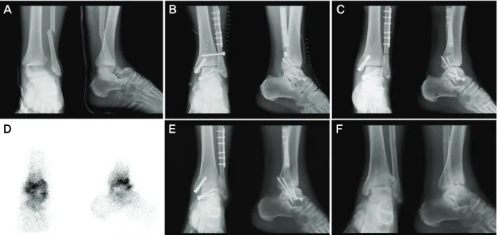
A B C
D E
Figure 1. (A) Initial and (B) postoperative radiograph of a 49-year-old man who had Hawkins type IV talar neck fracture. (C) Radiograph at postoperative 3 months shows Hawkins sign and (D) there is no photon defect of talar body on bone scan. (E) Last follow-up radiograph (postoperative 6 years 5 months) reveals no avascular necrosis and no post-traumatic osteoarthritis.
8주경 단순 방사선 사진 상에 거골 체부의 연골하 음영감소 소견으로 나타나는 Hawkins 징후를 이용하여 향후 무혈성 괴사의 발생 여부를 판단하고 있다9,10).
이에 저자들은 거골 경부 골절 및 탈구의 수술적 치료 후 발생한 거골 체부의 무혈성 괴사의 발생 빈도 및 Hawkins 징후와 무혈성 괴사와의 연관성 그리고 수술적 치료 후의 임상 결과에 대하여 알아보고자 하였다.
대상 및 방법
2000년 2월부터 2005년 8월까지 거골 경부 골절 및 탈구 로 Hawkins II, III, IV형 골절에 대해 수술적 치료를 받은 환자 중 최소 2년 이상 추시가 가능하였던 16명 16예를 대 상으로 하였다. 발생 원인은 교통사고가 10예, 추락사고가 6예였으며, Canale과 Kelly의 변형된 Hawkins의 분류상 제 2형은 9예(56.3%), 제 3형이 6예(37.5%), 그리고 제 4형 이 1예(6.3%)였다. 수술 당시 평균 연령은 32.2세(범위, 19~52세)로 10대 1명, 20대 7명, 30대 4명, 40대 3명, 50대 1명의 분포를 보였다. 평균 추시기간은 41.7개월(범위, 29~89개월)이었다. 거골 단독 골절만 발생한 경우는 5예 였으며 동반 손상으로는 족관절 내과 골절이 가장 흔하였다.
3예는 개방성 골절이었다.
수술 시기는 가능한 조기에 관혈적 정복 및 내고정을 시행하는 것을 원칙으로 하였으며, 평균 수상 후 2일(범위, 0~12일)에 시행하였다. 16예 중 12예는 수상 당일 또는 다음날 수술적 치료를 시행하였으며 나머지 4명의 환자는 타과적인 동반 손상으로 수술이 5일에서 12일까지 지연되 었다. 관혈적 정복 시에는 환자의 골절 상태에 따라 전내측 도달법과 전외측 도달법을 단독 또는 함께 사용하여 해부학 적 정복 및 내고정을 시행하였다. 술 후 족관절 중립 위치에 서 단하지 석고 부목 고정을 1주간 시행하였으며, 그 후 능 동적인 관절 운동을 시행하였다. 술 후 약 10주경부터 부분 체중부하 보행을 시행하였으며, 방사선 소견상 골유합이 관찰된 후에 완전 체중부하를 시행하였다.
A B C
D E F
Figure 2. (A) Initial and (B) postoperative radiograph of a 53-year-old man who had Hawkins type III talar neck fracture. (C) Radiograph at postoperative 3 months shows no Hawkins sign and (D) photon defect of talus body is revealed on bone scan. (E) Radiograph shows joint space narrowing and sclerotic change at postoperative 12 months. (F) Last follow-up radiograph (postoperative 24 months) shows collapse of talus body due to avascular necrosis.
Figure 3. The Hawkins sign was not observed in any cases without avascular necrosis. But only 2 of the 5 cases with a negative Hawkins sign developed avascular necrosis.
방사선학적인 평가는 수술 후와 추시 중 단순 방사선 사진상 Hawkins 징후와 무혈성 괴사 여부를 평가하였으며 술 후 3개월, 6개월, 1년 시점에 시행한 골 주사 검사를 이용하여 무혈성 괴사 여부를 확인하였다. 임상적 평가는 통증, 절뚝거림, 거골하 관절 및 족관절의 운동범위에 따라 판정하는 Hawkins의 방법9)으로 평가하였다.
통계적 분석은 SPSS 11.0 통계 프로그램을 사용하였으며 paired T-test를 이용하여 수술시기, 동반 손상에 따른 무 혈성 괴사 발생 유무 및 임상적 결과의 차이를 통계학적으로 비교하였으며 신뢰도는 95%로 하였다.
결 과
무혈성 괴사는 16예 중 2예(12.5%)에서 발생하였으며, 모두 제 3형이었다.
Hawkins 징후는 11예(68.8%)에서 관찰되었으며, 제 2형 에서 9예 중 8예(88.9%), 제 3형에서 6예 중 2예(33%) 그 리고 제 4형의 1예(100%)에서 나타났다(Table 1). 또한, Hawkins 징후가 관찰된 11예에서는 모두 무혈성 괴사가 발생하지 않았으며, Hawkins 징후가 나타나지 않은 5예 중 2예에서만 무혈성 괴사가 발생하였다(Fig. 1, 2, 3). 거골 무혈성 괴사와 Hawkins 징후와의 연관성을 비교하였을 때 민감도는 78.6%, 특이도는 100%, 양성예측도는 100% 그
리고 음성예측도는 40%를 나타내었다.
Hawkins 평가 방법에 따른 임상 결과는 우수 4예 (25.0%), 양호 7예 (43.8%), 보통 4예(25.0%), 그리고 불량 1예(6.3%) 를 보였으며, 무혈성 괴사를 보인 2예는 각각 보통과 불량의 결과를 보였다(Table 2).
수술 시기 또는 동반 손상에 따른 무혈성 괴사의 발생 빈 도 및 임상 결과에 미치는 영향은 통계학적으로 의미 없게 나타났다(p>0.05). 합병증으로는 무혈성 괴사 2예, 족관절 관절염 2예 및 표재성 감염이 1예에서 발생하였다.
Table 3. Literature Reviews of the Incidence of Avascular Necrosis after Treatment of Fracture and Dislocations of the Talar Neck Percentage of avascular necrosis (Number of cases)
Author Type I Type II Type III Type IV Total
Hawkins9) 0 (6) 42 (24) 91 (27) 6 (32) 53% (57)
Canale and Kelly2) 13 (15) 50 (30) 84 (19) 50 (2) 52% (66)
Penny and Davis19) 0 (5) 20 (11) 100 (11) - 48% (27)
Inokuchi et al.12) 17 (23) 40 (5) 55 (20) 100 (3) 39% (51)
Sohn et al.24) 0 (4) 18 (9) 100 (1) - 33% (14)
Lee et al.15) 0 (4) 17 (6) 83 (6) - 38% (16)
Choi et al.3) 0 (2) 19 (16) 12 (8) 100 (2) 21% (28)
Tezval et al.27) 17 (6) 0 (17) 43 (7) 33 (3) 15% (33)
Present study - 0 (9) 33 (6) 0 (1) 13% (16)
고 찰
거골의 혈액 공급은 매우 특이하고 취약하며, 이에 대한 정확한 근원과 분포에 대해서 많은 연구가 이루어져 왔다
8,14,18). 그 결과 거골 체부의 주된 혈관은 족배 동맥의 분지,
거골관 동맥 및 이와 문합하는 거골동 동맥 그리고 거골관 동맥의 삼각분지라고 알려졌다. 특히 McKeever 등17)은 골외 혈액 공급은 전후 경골 동맥과 천 비골 동맥이 망상 조직을 이루고 거골동 동맥 및 거골관 동맥이 문합을 이루어 혈액을 공급하고, 골내 혈액 공급으로는 두부는 전경골 동맥 및 족근 동맥에 의해서, 체부는 주로 거골관 내의 문합 동맥으로부 터 공급받는다고 보고하였다. 이러한 해부학적 특성 때문에 전위 혹은 탈구가 동반된 골절에서는 무혈성 괴사가 쉽게 발생할 수 있으며, 특히 손상의 정도가 심할수록 높은 빈도 를 보인다고 하였다. 따라서 가능한 조기에 전위된 골절의 해부학적 정복을 이루면 보다 좋은 결과를 얻을 수 있다고 보고되고 있다1-3,5,6,11,13-15,22,24). 그러나 실제로 거골 골절 및 탈구는 고에너지 손상에 의해 발생되는 경우가 대부분이 어서 신체의 타 부위 및 동측 하지의 동반 손상이 많으며, 이로 인하여 거골의 수술적 치료가 지연되는 경우가 많다.
본 연구에서도 16예 중 4예(25.0%)에서 타과적 문제로 인 하여 수술적 치료가 지연되었으나, 수술 시기에 따른 무혈 성 괴사의 발생 및 임상 결과는 통계학적으로 연관성이 없 는 것으로 나타났다. 이에 대한 더 많은 증례를 통한 연구가 필요하리라 생각된다.
거골 골절 및 탈구의 치료 후 발생한 무혈성 괴사의 발생 률에 대해 1970년 Hawkins9)는 약 53%, 1978년 Canale와 Kelly2)는 약 52%를 보고하였다. 하지만 그 이후 발표된 국 내외 논문들에서는 평균 약 30%(범위, 15~48%) 정도의 무 혈성 괴사의 발생률을 보고하고 있으며, 점차 감소하는 양 상을 보이고 있다(Table 3). 본 연구에서도 약 13%의 낮은 발생률을 보였으며, 이러한 감소는 조기에 관혈적 정복 및
내고정술을 시행하였을 뿐 아니라, 골내 재혈관화를 촉진시 키는 지연 나사를 이용한 압박 고정 등의 수술 기법의 발전에 의한 것으로 사료된다2,5,6,15,23,25).
또한 본 연구에서 거골 무혈성 괴사의 발생에 대하여 Hawkins 징후는 100%의 양성 예측도를 보였는데 이는 이전에 보고된 Ingelfinger 등22)의 80%, Tezval 등27)의 100%와 비슷한 결과를 보였다. 따라서 Hawkins 징후 양성 은 거골 무혈성 괴사의 발생을 배제할 수 있는 유용한 예 후 지표라 하겠다9,10). 하지만 Hawkins 징후를 보이지 않았 던 5예 중에서 단 2예만이 무혈성 괴사를 보이고, 나머지 3예에서는 무혈성 괴사가 발생하지 않았으므로 Hawkins 징후 음성이라고 해서 모두 거골 무혈성 괴사가 발생한다 고 판단하는 것은 무리가 있을 것으로 보인다2,19,27). 그러 므로 Hawkins 징후가 음성일지라도 임상적으로 또는 방 사선학적으로 무혈성 괴사가 의심되는 경우엔 자기 공명 영상 검사나 골주사 검사를 조기에 시행하는 것이 필요하 다고 생각한다25,26).
본 연구는 이전에 보고된 연구 결과들과 비슷하게 수술 시기 또는 동반 손상에 따른 임상 결과의 차이는 통계학적 으로 의의를 보이지 않았으며4,15,27), 무혈성 괴사와 임상결 과의 연관성도 보이지 않았다. 하지만 무혈성 괴사가 단 2예에서만 발생하여 그 통계학적 의의를 찾기는 어려우며, 추후 더 많은 증례와 장기적인 추시를 통한 임상 결과의 평가가 필요하리라 생각한다.
결 론
거골 경부 골절 및 탈구 환자에서 술 후 무혈성 괴사의 발생 빈도는 약 13%로 지금까지 보고된 결과에 비하여 낮았 다. 그리고 Hawkins 징후는 무혈성 괴사를 조기에 예측할 수 있는 유용한 방사선학적 소견이라 할 수 있으나, 음성이 라 하여 모두 무혈성 괴사가 발생하는 것은 아니며, 무혈성
괴사의 임상 결과는 더 많은 증례와 장기 추시가 필요할 것 으로 생각한다.
REFERENCES
1. Adelaar RS: Fractures of the talus. Instr Course Lect, 39: 147-156, 1990.
2. Canale ST and Kelly FB: Fractures of the neck of the talus. J Bone Joint Surg, 60-A: 143-156, 1978.
3. Choi HR, Lee JG, Choi HH and Choi SW: Surgical treatment for fractures of the talus. J Korean Fracture Soc, 16: 67-73, 2003.
4. Comfort TH, Behrens F, Gaither DW, Denis F and Sigmond M: Long-term results of displaced talar neck fractures. Clin Orthop, 199: 81-87, 1985.
5. Daniels TR and Smith JW: Talar neck fractures. Foot Ankle, 14: 225-234, 1993.
6. Dennis MD and Tullos HS: Blair tibiotalar arthrodesis for injuries to the talus. J Bone Joint Surg, 62-A: 103-107, 1980.
7. Goldie I: Talar and peritalar injuries. In: Helal B and Wilson D ed. The Foot. 1st ed. London, Churchill Living- stone: 916-931, 1988.
8. Haliburton RA, Sullivan CR, Kelly PJ and Peterson LF:
The extra-osseous and intra-osseous blood supply of the talus. J Bone Joint Surg, 40-A: 1115-1120, 1958.
9. Hawkins LG: Fractures of the neck of the talus. J Bone Joint Surg, 52-A: 991-1002, 1970.
10. Hendrich V: Fractures and dislocations of the talus.
Unfallchirurg, 92: 110-116, 1989.
11. Inokuchi S, Ogawa K, Usami N and Hashimoto T: Long- term follow up of talus fractures. Orthopedics, 19: 477- 481, 1996.
12. Kenwright J and Taylor RG: Major injuries of the talus.
J Bone Joint Surg, 52-B: 36-48, 1970.
13. King RE and Powell DF: Injury to the talus. In: Jahss MH ed. Disorders of the Foot and Ankle. 2nd ed. Philadelphia, WE Saunders Co: 2293-2325, 1991.
14. Kleiger B: Fractures of the talus. J Bone Joint Surg, 30-A:
735-744, 1948.
15. Lee BH, Ha SH and Choi MH: Treatment of fracture- dislocation of talar neck or body. J Korean Fracture Soc, 12: 307-313, 1999.
16. Lorentzen JE, Christensen SB, Krogsoe O and Sneppen O:
Fractures of the neck of the talus. Acta Orthop Scand, 48:
115-120, 1977.
17. McKeever FM: Treatment of complications of fractures and dislocations of the talus. Clin Orthop, 30: 45-52, 1963.
18. Mulfinger GL and Trueta J: The blood supply of the talus.
J Bone Joint Surg, 52-B: 160-167, 1970.
19. Pearce DH, Mongiardi CN, Fornasier VL and Daniels TR:
Avascular necrosis of the talus: a pictorial essay. Radio- graphics, 25: 399-410, 2005.
20. Penny JN and Davis LA: Fractures and fracture-dis locations of the neck of the talus. J Trauma, 10: 1029-
1037, 1980.
21. Peterson L, Goldie IF and Irstam L: Fracture of the neck of the talus. Acta Orthop Scand, 48: 696-706, 1977.
22. Schulze W, Richter J, Russe O, Ingelfinger P and Muhr G:
Prognostic factors for avascular necrosis following talar fracture. Z Orthop Ihre Grenzgeb, 140: 428-434, 2002.
23. Sneppen O, Christensen SB, Krogsoe O and Lorentzen J:
Fracture of the body of the talus. Acta Orthop Scand, 48:
317-324, 1977.
24. Sohn SK, Kim BH and Park SJ: Treatment for fracture of neck of the talus. J Korean Foot Surg Soc, 3: 40-48, 1999.
25. Szyszkowitz R, Reschauer R and Seggl W: Eighty-five talus fractures treated by ORIF with five to eight years of follow-up study of 69 patients. Clin Orthop, 199: 97-107, 1985.
26. Thordarson DB, Triffon MJ and Terk MR: Magnetic resonance imaging to detect avascular necrosis after open reduction and internal fixation of talar neck fractures.
Foot Ankle Int, 17: 742-747, 1996.
27. Trezval M, Dumont C and Stumer KM: Prognostic re- liability of the Hawkins sign in fractures of the talus.
J Orthop Trauma, 21: 538-543, 2007.