Journal of Bacteriology and Virology 2009. Vol. 39, No. 1 p.11 – 19
Isolation and Identification of Lactic Acid Bacteria Inhibiting the Proliferation of Propionibacterium acnes and
Staphylococcus epidermidis
Mi-Sun Kang, Hyun-Ju Oh, Hyun-Chul Lee and Jong-Suk Oh
*Department of Microbiology, School of Medicine, Chonnam National University, Gwangju, Korea
Propionibacterium acnes is the most common causative agent of acne. Staphylococcus epidermidis is another major bacterial strain to be found in acne lesions. Two strains of lactic acid bacteria (LAB) were isolated from normal inhabitants of humans, which inhibited the proliferation of P. acnes and S. epidermidis. The growth of P. acnes and S.
epidermidis was decreased by 4-log scales after incubation for 24 h with LAB isolates, whereas the growth rate of selected LAB isolates were not affected by these pathogenic bacteria. This antibacterial activity of LAB isolates was related to lactic acids, hydrogen peroxide and bacteriocin-like compound production. Two LAB isolates efficiently adhered to human keratinocytes HaCaT and were identified by API 50 CHL medium kit and 16S rDNA partial sequencing analysis. The similarity of 16S rDNA sequences between one isolate and Lactobacillus salivarius subsp. salicinius was 100%, which suggests that they were L. salivarius subsp. salicinius. On the other hand, 16S rDNA sequence similarity between the other isolate and Lactobacillus fermentum was 99.04%, which indicates that it was L. fermentum. In conclusion, these results demonstrate that the two LAB strains isolated from human body were identified as L. salivarius subsp. salicinius and L. fermentum, which inhibit the proliferation of P. acnes and S. epidermidis.
Key Words: Isolation, Identification, Lactobacillus, Propionibacterium acnes, Staphylococcus epidermidis
서 론
여드름은 모낭-피지선에서 발생하는 피부질환이다.
여드름은 피지선에서 피지 분비가 증가하거나 피지선의 모공이 좁아지든지 막혀서 피지가 배출되지 못함에 따 라 세균이 증식하여 염증이 생기는 것이다. 여드름은 주 로 사춘기 나이의 사람에게서 많이 발생하는데 이는 사 춘기 나이에 분비되기 시작하는 안드로젠이라는 남성호 르몬 때문이다. 안드로젠은 피지 분비를 촉진시키고, 표 피의 과각화를 일으킨다. 피지의 분비 증가와 표피 과 각화로 모낭-피지선에서 피지가 정체되어 모낭이 막힘 에 따라 모낭내부가 Propionibacterium acnes를 비롯한 혐
기성 세균이 잘 자랄 수 있는 환경이 된다 (1, 2). 동시에 Staphylococcus epidermidis와 같은 다른 세균들이 모낭주 위에서 여드름과 여드름 합병증을 일으키는데 역할을 한 다 (3). 여드름 발생의 병리 조직학적인 기전은 P. acnes 의 효소, 사이토카인 및 보체와 중성구, 손상된 각질형성 세포에서 분비된 cytokine 등이 염증을 일으키는데 관여 하는 것으로 알려져 있으나 (4), 아직 정확한 기전에 대 해서는 알려진 바 없다.
P. acnes와 S. epidermidis 등의 균들이 염증 반응을 유발 하는데 주된 역할을 하게 되므로 염증성 여드름의 치료 에 항생제가 사용되고 있다 (5). Triclosan, benzoyl peroxide, azelaic acid (6), retinoid, tetracycline, erythromycin, macrolide, clindamycin 등의 항생제가 사용되고 있으나, 부작용이 알 려져 있다. Benzoyl peroxide와 retinoid는 피부건조증이나 과민증을 유발하고 (7), tetracycline, erythromycin, macrolide, clindamycin은 항생제에 대한 내성 발생으로 인하여 지속 적인 사용이 어렵고 간독성이 심하며, 칸디다증과 같은 기회감염증이 나타날 수 있다 (8, 9). 또한, triclosan의 경
11 Received: February 13, 2009/ Revised: March 6, 2009
Accepted: March 11, 2009
*Corresponding author: Jong-Suk Oh, M.D., Ph.D. Department of Microbiology, School of Medicine, Chonnam National University, 5 Hak-Dong, Dong-Gu, Gwangju 501-746, Republic of Korea.
Phone: +82-62-220-4134, Fax: +82-62-228-7294, e-mail: [email protected]
우 빛에 노출되었을 때 환경호르몬으로 바뀌어 심각한 환경오염을 일으킬 수 있다. 따라서, 많은 연구자들이 항 균효과가 있으면서 부작용이 없는 여드름 치료제를 개 발하려고 노력 중이다 (10, 11).
유산균은 탄수화물을 발효하여 최종 대사산물로 유산 을 생산하는 세균을 말한다. 유산균은 인간과 동물의 장 및 질에 존재하며 (12, 13), 통상적으로 김치 또는 요구르 트 등 발효식품의 제조과정에 활용되고 있다. 식품에 활 용되는 유산균으로는 Carnobacterium, Enterococcus, Lacto- bacillus, Lactococcus, Leuconostoc, Oenococcus, Pediococcus, Streptococcus, Tetragenococcus, Vagococcus, Weissella 등이 있다 (14, 15). 이와 같이 이로운 방향으로 작용하는 정상 세균 중 유산막대균인 Lactobacillus는 대표적인 유산균으 로 유해균 발육을 억제하는 효과가 강하여 발효식품과 유산균 제재, 의약품 등으로 사용되고 있으며, 병원성 세 균을 억제하는 Lactobacillus의 분리 및 동정이 연구되고 있다 (16~19).
본 연구의 목적은 세균을 이용한 여드름 치료제를 연 구하기 위한 첫 과정으로, P. acnes와 S. epidermidis의 증 식을 억제하며 각질세포에 대한 부착능이 있는 유산균을 선발하여 생화학적 성질 및 16S rDNA partial sequencing analysis에 의한 동정을 실시하고자 하였다.
재료 및 방법
공시세균 및 배양
공시세균으로는 P. acnes ATCC11828와 S. epidermidis ATCC 12228 (Rockville, MD, USA)를 이용하였다. P. acnes 는 Actinomyces broth (BBL, Sparks, MD, USA)에 접종하여 37℃ 혐기조건 (85% N2, 10% H2, 5% CO2)에서 1일간 배 양하였으며, S. epidermidis는 Brain Heart Infusion broth (BHI, Difco, Detroit, MI, USA)에 접종하여 37℃ 호기조건에서 16시간 배양하였다. 각각의 균은 실험에 이용하기 전에 본 배지에서 2회 계대배양한 후 실험에 이용하였다.
시료채취 및 유산균의 분리
인체 구강, 장, 질로부터 시료를 채취하여 0.9% NaCl로 계단 희석하여 유산막대균 분리용 배지인 Rogosa 우무배 지 (Difco)에 접종하고 37℃에서 48시간 배양한 후 집락 을 취하였다. 이것을 다시 De Man, Rogosa, Sharpe (MRS) 우무배지 (Difco)에 접종, 배양하여 약 3000주의 유산균
을 분리하였다. Actinomyces 우무배지와 MRS 우무배지 가 동량 섞어진 배지 표면 전체에 P. acnes를 도말 접 종한 다음, 분리균주를 점점이 떨어뜨려 37℃ 배양기에 서 24시간 배양하여 투명환 (clear zone)을 나타내는 균주 를 1차 선발하였다. 선발된 균주들은 다시 P. acnes와 S.
epidermidis와의 상호작용을 통하여 최종적으로 유산막대 균 2주를 선발하여 lactic acid bacteria (LAB) 1, LAB 2로 명명하였다. 분리균주 2주는 MRS 액체배지에 배양한 후 글리세롤의 최종농도가 20% (w/v) 되도록 첨가하여 -80℃
에 냉동 보관하면서 필요에 따라 접종, 배양하여 실험에 사용하였다.
분리 유산균의 과산화수소 생성능 측정
분리균주의 과산화수소의 생성능력을 보기 위하여 0.25 mg/ml TMB (3,3',5,5'-tetramethyl benzidine, Sigma, St. Louis, MO, USA) 0.01 mg/ml peroxidase (Sigma)가 첨가된 MRS 우무배지에 각 분리균주를 3 μl씩 접종하여 37℃에서 혐 기배양한 후 꺼내어 호기상태에서 집락의 색깔을 관찰 하여 집락의 색깔이 청색을 띠면 과산화수소 생성 양성 (positive)으로 판단하였고 색깔에 따라 감청색은 strongly positive, 청색은 positive, 약청색은 weakly positive, 색깔의 변화가 없으면 음성 (negative)으로 정하였다 (20).
분리 유산균의 P. acnes와 S. epidermdis에 대한 항균 작용
분리 유산균의 P. acnes에 대한 항균력을 알아보기 위 하여 MRS broth와 Actinomyces broth가 동량 섞어진 배지 에 접종량이 각각 1.0 × 10⁶ CFU가 되도록 P. acnes와 분 리 유산균을 단독 또는 병합으로 접종하였다. 37℃에서 8시간 및 24시간 혐기배양 후 배양액을 희석하여 MRS 우무배지와 Actinomyces 우무배지상에 접종하여 48시간 배양한 다음, 분리 유산균과 P. acnes의 생균수를 산정하 였다. 또한, S. epidermidiis에 대한 항균력을 알아보기 위 하여 MRS broth와 BHI broth가 동량 섞어진 배지에 접종 량이 각각 1.0 × 10⁶ CFU가 되도록 S. epidermidis와 분리 유산균을 단독 또는 혼합으로 접종하였다. 37℃에서 8시 간 및 24시간 호기배양 후 배양액을 희석하여 MRS 우 무배지와 BHI 우무배지상에 접종하여 48시간 배양한 다 음, 분리 유산균과 S. epidermidis의 생균수를 산정하였다.
분리 유산균 배양 상청액의 P. acnes와 S. epidermdis 에 대한 항균작용
분리 유산균 배양 상청액의 분비물질 중에서 어떤 성 분에 의한 항균효과인지를 알아보기 위하여 분리 유산균 을 MRS broth에서 24시간 배양한 다음 원심분리 (4,000 rpm, 20 min, 4℃)한 것을 여과하여 균을 완전히 제거하였 다. 유산에 의한 항균력인지 알아보기 위하여 상청액에 proteinase K (0.1 mg/ml)와 catalase (0.5 mg/ml)를 처리하였 으며, 과산화수소에 의한 항균력인지 알아보기 위하여 상 청액을 pH 6.3으로 중화시키고 proteinase K를 처리하였 다. 또한, 상청액을 pH 6.3으로 중화시키고 catalase를 처 리하여 박테리오신 유사물질 (bacteriocin-like compound) 에 의한 항균력인지 알아보고자 하였다. P. acnes와 S.
epidermidis 배양액을 각각 흡광도 600 nm에서 0.05가 되 도록 희석하여 96 well plate에 100 μl 접종하고 배양 상청 액을 100 μl 첨가하였으며, 대조군으로는 MRS broth를 100 μl 첨가하였다. 37℃에서 24시간 혐기 및 호기배양 후 microplate reader를 이용하여 600 nm에서 흡광도를 측 정하였다.
분리 유산균의 human keratinocytes HaCaT에 대한 부 착능
분리 유산균의 human keratinocytes에 대한 부착능을 알 아보기 위해 실험에 이용한 세포주는 각질형성세포주인 HaCaT (epithelial cell line from adult human skin)으로서, 10%
fetal bovine serum (FBS)과 penicillin (10 U/ml)/streptomycin (10 μg/ml)을 첨가한 Dulbecco's modified Eagle's medium (DMEM, GibcoBRL, Braunschweig, Germany) 배지를 사용 하여 37℃, 5% CO2 배양기에서 배양하였다. 분리 유산균 의 부착실험은 Scaletsky 등 (21)의 방법에 따라 수행하 였다. 4-well Lab-Tek II chamber slide system (Nalge Nunc International, Naperville, IL, USA)에 well당 105 세포를 18시 간 배양한 후, PBS로 세포를 2회 세척하고 multiplicity of infection (MOI)이 250이 되도록 분리 유산균 (109 bacteria/
ml)을 25 μl 가하여 37℃, 5% CO2 배양기에서 30분간 배 양하였다. PBS로 세포를 3회 세척한 후 methanol로 고정 하고 30분간 Giemsa 염색한 후, PBS로 다시 세척하여 공기 중에 건조시켜서 각각의 세포에 대한 부착력을 광 학현미경 (BX51, Olympus, Tokyo, Japan)을 이용하여 관찰 하였으며, 100개의 세포에 부착한 세균의 평균 개수를
측정하였다.
분리 유산균의 동정
분리 유산균의 1차 동정법으로는 API 50 CHL kit (BioMérieux, Marcy, l'Etoile, France)를 사용하여 동정 프 로그램인 API LAB plus로 검사 결과를 분석하여 생리적 특성을 검토하였으며, 2차 동정법으로는 16S rDNA 염기 서열을 분석하여 결정하였다. 16S rDNA 염기서열 분석을 하기 위하여 Rochelle 등 (22)의 방법으로 단일 콜로니에 서 DNA를 분리하였으며, 27F (5'-AGAGTTTGATCMTG- GCTCAG-3')와 1522R (5'-AAGGAGGTGWTCCARCC-3') primer를 사용하여 16S rDNA를 PCR 증폭하였다 (23).
PCR 산물은 Wizard PCR Preps DNA purification system (Promega, Madison, WI, USA)을 이용하여 정제하였으며, 염기서열 분석은 BigDyeTM Terminator Cycle Sequencing Ready Reaction Kit (Applied Biosystems, Foster City, CA, USA)와 ABI PRISMTM 310 Genetic Analyzer (Applied Bio- systems)을 이용하여 염기서열을 결정하였다. 얻어진 염 기서열 결과는 NCBI의 GenBank 프로그램을 사용하여 상동성을 조사하였다.
통계처리
통계적 처리는 SPSS 통계분석 프로그램 (SPSS version 12.0)을 사용하였으며, Mann-Whitney test로 분석하였다.
결 과
분리 유산균의 과산화수소 생성능력
혐기성 세균은 과산화수소에 예민하므로 분리 유산균 의 과산화수소 생성능력을 측정한 결과, LAB 1과 LAB 2 는 과산화수소를 생성하여 감청색의 집락을 형성하였다 (Table 1).
P. acnes에 대한 분리 유산균의 항균력
P. acnes에 대한 분리 유산균의 항균력을 보기 위하여 Table 1. Hydrogen peroxide-producing LAB isolates from normal
inhabitants of humans
Isolates Source H2O2-productivity
LAB 1 Saliva Positive
LAB 2 Saliva Strongly positive
P. acnes와 분리 유산균의 혼합배양 후 생균수 검사 결과, 24시간 배양 후 두 분리 유산균 모두 P. acnes 생육을 유 의하게 감소시켰다 (p < 0.05). 8시간 혼합배양 후 P. acnes 에 대한 분리 유산균의 항균력은 거의 없었으나, 24시간 배양 후 단독배양한 P. acnes 생균수는 3.6 × 10⁹ ± 8.0 × 10⁸ CFU/ml이었으며, LAB 1과 병합으로 배양시에는 P.
acnes 생균수는 4.0 × 10⁶ ± 7.0 × 10⁴ CFU/ml으로 감소하 였다. 또한, LAB 2와 병합시 2.0 × 10⁴ ± 3.0 × 10³ CFU/ml 으로 크게 감소하였다. 따라서, 두 분리 유산균 중 P.
acnes에 대한 항균력은 LAB 2가 더 좋았다. 반면, 두 분
리 유산균을 혼합배양하였을 때 생균수에 대한 변화가 거의 없었다 (Fig. 1).
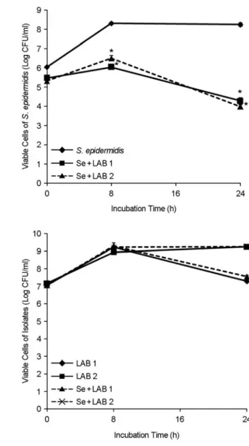
S. epidermidis에 대한 분리 유산균의 항균력
S. epidermidis와 분리 유산균의 혼합배양 후 생균수 검 사 결과, 두 분리 유산균 모두 8시간부터 S. pidermidis 생육을 유의하게 감소시키기 시작하였으며, 24시간 후에 는 S. pidermidis 생육을 104 CFU/ml까지 감소시켰다 (p
< 0.05). 24시간 배양 후 S. epidermidis 생균수는 1.7 × 10⁸
Figure 1. Viable cells of P. acnes and the LAB isolates in the mixed cultures over time. Pa, P. acnes. *p < 0.05 for coculture versus monoculture. Values are means ± standard deviations of three independent experiments.
Figure 2. Viable cells of S. epidermidis and the LAB isolates in the mixed cultures over time. Se, S. epidermidis. *p < 0.05 for coculture versus monoculture. Values are means ± standard devia- tions of three independent experiments.
± 3.0 × 10⁷ CFU/ml이었으며, LAB 1과 병합으로 배양시 에는 S. epidermidis 생균수는 2.0 × 10⁴ ± 7.0 × 10³ CFU/
ml으로 감소하였고, LAB 2와의 병합시 1.0 × 10⁴ ± 2.0 × 10³ CFU/ml으로 감소되었다. 따라서, 두 분리 유산균 중 S. epidermidis에 대한 항균력도 LAB 2가 더 좋았다. 반면, 두 분리 유산균 모두 혼합배양하였을 때 생균수에 대한 변화가 거의 없었다 (Fig. 2).
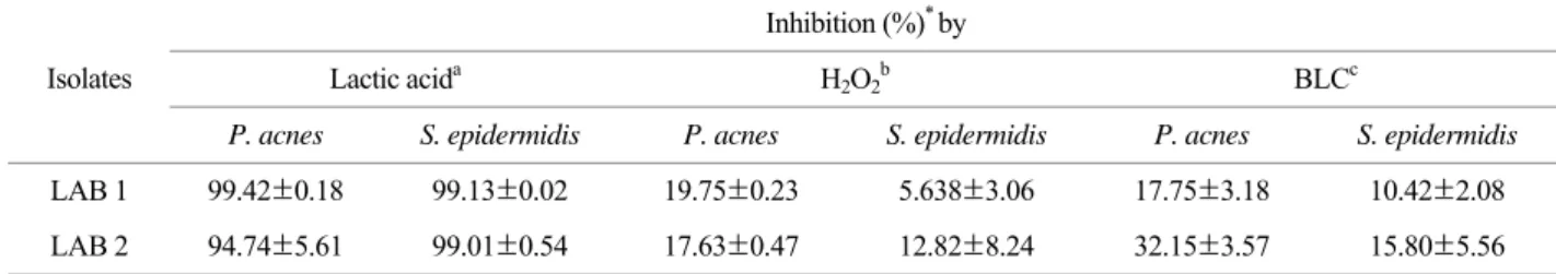
P. acnes 및 S. epidermidis에 대한 분리 유산균 배양 상청액의 항균력
P. acnes 및 S. epidermidis에 대한 분리 유산균 배양 상 청액의 항균력을 조사한 결과, 분리 유산균 2주 모두 유 산에 의하여 약 95% 이상 항균효과를 나타내었으며, 과 산화수소 및 박테리오신 유사물질에 의해서도 P. acnes의 생육을 각각 18~19%와 18~32% 억제하였다 (Table 2).
Human keratinocytes HaCaT에 대한 유산균의 부착력
각질형성세포에 대한 분리 유산균의 부착능을 알아 본 결과, 분리 유산균 모두 각질형성세포주인 HaCaT 세 포에 부착이 잘 되었으며, 1개의 HaCaT 세포당 부착된 LAB 1과 LAB 2 평균 개수는 각각 91.0 ± 9.6.5, 40.0 ± 2.0 개로서, LAB 1이 부착력이 더 우수하였다 (Fig. 3).
유산균의 동정
탄소원의 이용성 차이로 동정하는 생화학적 방법인 API 50 CHL kit로 1차 동정을 한 결과, LAB 1은 L.
salivarius (가능성 99.9%), LAB 2는 Lactobacillus cellobiosus (가능성 92.9%), Lactobacillus delbrueckii subsp. delbrueckii (가능성 3.1%)로 동정되었다.
한편 LAB 1는 16S rDNA 991 bp를 직접 염기서열 분 석하여 (Fig. 4), NCBI의 GenBank 프로그램을 사용하여 상동성을 조사하여 본 결과, 비교한 991개의 염기 중 L. salivarius subsp. salicinius JCM 1230 (accession no.
AB289295)과 100% 상동성을 보여 L. salivarius subsp.
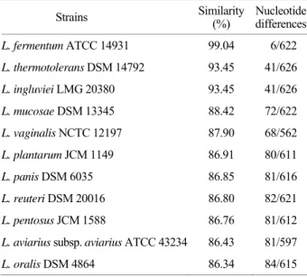
salicinius로 동정되었다 (Table 3). LAB 2는 Lactobacillus fermentum ATCC 14931 (accession no. AB017345)과 유사 치가 99.04%로 가장 높아 L. fermentum으로 동정되었다 (Table 4).
Table 2. Antibacterial activity of lactic acid, hydrogen peroxide (H2O2) and bacteriocin-like compound (BLC) in cultured supernatants of two LAB isolates against P. acnes and S. epidermidis
Inhibition (%)* by
Lactic acida H2O2b BLCc
Isolates
P. acnes S. epidermidis P. acnes S. epidermidis P. acnes S. epidermidis
LAB 1 99.42±0.18 99.13±0.02 19.75±0.23 5.638±3.06 17.75±3.18 10.42±2.08
LAB 2 94.74±5.61 99.01±0.54 17.63±0.47 12.82±8.24 32.15±3.57 15.80±5.56
* Values are means ± standard deviations of three independent experiments.
a Inhibition of growth by lactic acid was measured following treatment of the sterilized supernatants with proteinase K (0.1 mg/ml) and catalase (0.5 mg/ml).
b H2O2-dependent activity was evaluated using the neutralized and proteinase K-treated supernatants.
c Inhibition of growth by BLC-dependent activity was evaluated using catalase-treated and neutralized supernatants
Figure 3. Microscopic observations and numbers of adhered of LAB isolates to HaCaT cells. A, LAB 1 vs. HaCaT cells; B, LAB 2 vs. HaCaT cells. Magnification, × 1000. Values are means ± standard deviations of three independent experiments.
A B
고 찰
여드름은 가장 흔한 피부질환 중 하나로, 모낭-피지선 단위의 자기 국한성 만성 염증성 질환으로 면포, 홍반성 구진, 농포 등을 형성하는 것을 특징으로 하며, 드물게 결절 혹은 가성낭종이 발생하고 활동성 병변의 후유증으 로 소와성 혹은 비후성 반흔을 남기기도 한다. 여드름의 임상 양상은 비염증성인 면포와 염증성인 표재성 병변과 심재성 병변으로 나타나는데, 심재성 병변인 결절은 주 로 남자에서 나타나며 결절사이에 진피를 통한 루 (sinus)
가 형성되어 농양 및 낭종으로 커지게 되고 치유된 후에 는 흔히 영구적으로 위축성 혹은 켈로이드성 반흔을 남 긴다. 여드름의 원인 세균 중에서 가장 대표적이라 할 수 있는 P. acnes는 통성 혐기성 그람 양성 간균으로 호지성 인 특성이 있어서 피지 분비가 많은 부위인 얼굴, 목, 등, 가슴에 호발한다. P. acnes는 여드름 환자의 혈액 단핵구 로부터 IL-8의 분비를 유도하거나 균으로부터 lipase가 Table 3. 16S rDNA similarity of LAB 1 to other bacteria
Strains Similarity
(%) Nucleotide differences L. salivarius subsp. Salicinius
JCM 1230 100.00 0/991
L. salivarius T 99.90 1/985
L. salivarius subsp. Salivarius
ATCC 11741 99.80 2/990
L. aviarius T 94.95 49/970
L. agilis T 92.27 75/970
L. acidipiscis FS60-1 91.68 82/985
L. animalis T 91.58 81/962
Table 4. 16S rDNA similarity of LAB 2 to other bacteria
Strains Similarity
(%) Nucleotide differences L. fermentum ATCC 14931 99.04 6/622 L. thermotolerans DSM 14792 93.45 41/626
L. ingluviei LMG 20380 93.45 41/626
L. mucosae DSM 13345 88.42 72/622
L. vaginalis NCTC 12197 87.90 68/562
L. plantarum JCM 1149 86.91 80/611
L. panis DSM 6035 86.85 81/616
L. reuteri DSM 20016 86.80 82/621
L. pentosus JCM 1588 86.76 81/612
L. aviarius subsp. aviarius ATCC 43234 86.43 81/597
L. oralis DSM 4864 86.34 84/615
Figure 4. Nucleotide sequence of the partially amplified 16S rDNA gene from LAB 1 by PCR.
분비되어 호중구가 피부 병변에 유입된다 (24, 25). 그리 고 P. acnes와 분비되는 coproporphyrin III에 의해 각질형 성세포가 자극되어 IL-1, TNF-α와 GM-CSF와 같은 cyto- kine이 분비되어 여드름의 염증에 관여하는 것으로 알려 져 있다 (26, 27).
여드름 치료에서 항생제는 효과적이지만, 항생제의 장 기간 사용으로 여드름 발생에 관여하는 균주나 피부 상 재균의 항생제 내성이 보고되고 있다 (8). 이런 항생제 내성 문제를 극복하기 위하여 대체 치료법으로 약용 식 물들이 광범위하게 연구되어 왔다. 최근에는 전통 약초 들을 이용한 스킨케어 화장품을 만들어 여드름 치료를 위한 새로운 기능 성분을 찾기 위한 노력이 계속되고 있 다 (10, 28). 또한, 목련 줄기 껍질에서 분리된 마그놀롤 (magnolol)과 호노키올 (honokiol)은 피부질환 치료제로서 이용되어 오고 있는데. 항염증 및 항균효과가 있다고 보 고되었다 (29, 30).
병원성 세균을 억제하는 유산균에 대한 연구는 많이 되어 왔으나, P. acnes에 대한 항균효과를 가진 유산균에 대한 연구는 미비한 편이다 (31). Smith 등 (32)은 거의 모든 유산막대균이 박테리오신을 생성하고, 여성의 질 내 에 상재하는 유산막대균은 과산화수소를 분비하여 항균 력을 가지고 있으며, 병원체와 결합력이 높은 것으로 보 고하였다 (33). 혐기성 세균은 과산화수소에 매우 예민하 므로 과산화수소에 의하여 억제된다 (34, 35). 이러한 항 균물질을 생성하는 세균의 작용은 항생제의 부작용과는 달리 피부 환경의 균형을 파괴시키지 않으면서 효과를 지속적으로 발휘할 수 있어 피부건강의 유지에 중요한 역할을 할 것으로 사료된다. 본 연구에서 분리한 유산막 대균 중에서 L. salivarius subsp. salicinius로 동정된 LAB 1 과 L. fermentum으로 동정된 LAB 2가 과산화수소를 생성 하는 능력이 있었으며, LAB 2가 과산화수소를 더 많이 생성하였다. P. acnes를 단독으로 24시간 배양했을 때에 비해 분리 유산균과 병합시 생균수가 103~105 CFU/ml의 감소를 보였으며, LAB 2가 항균력이 더 우수하였다. 또한, S. epidermidis에 대해서도 분리 유산균과 병합시 약 104 CFU/ml의 감소를 보였으며, P. acnes에 대해서와 마찬가 지로 LAB 2가 항균력이 더 좋았다. 그리고, 분리 유산균 의 배양 상청액 또한 P. acnes와 S. epidermidis의 생육을 거의 억제하였으며, 대부분이 유산에 의해 항균작용을 보였으며, 과산화수소 및 박테리오신 유사물질에 의해서 도 어느 정도 항균력을 나타내었다.
각질형성세포에 부착이 잘 일어나는 세균은 피부의 여 드름 병변에 접종하였을 때, 피부에서 군락화가 되기 쉬 워 세포에 부착이 잘 안되는 세균에 비하여 치료효과가 더 좋을 것으로 사료된다. 따라서, 본 연구에서 각질형성 세포주인 HaCaT 세포에 대한 분리 유산균의 부착 정도 를 본 결과, 분리 유산균 모두 부착이 잘 일어났다.
종래 미생물을 동정하기 위하여 이용하던 배양이나 혈 청학적 방법에 비교하여 미생물에 존재하는 고유한 유전 자를 이용하여 동정하는 방법은 민감도와 정확성에 있어 서 월등하기 때문에 최근에 세균으로부터 추출한 DNA 에 rRNA 유전자 probe를 hybridization시킨 후, 제한효소 로 처리한 결과로 세균을 분류하기 시작하였다 (36). 분 리균주의 16S rDNA 염기서열을 비교하면 세균이 진화과 정상 가까울수록 염기서열은 비슷하고 진화과정상 멀수 록 염기서열의 유사성은 감소하게 된다. 본 연구에서도 분리 유산균 2주를 탄수화물 발효로 검사하는 API 50 CHL medium kit로 동정시험한 결과와 16S rDNA 유전자 와 비교한 결과가 약간의 차이가 있었는데, 분리 유산균 1주는 L. salivarius subsp. salicinius의 유전자와 유사치가 100%를 보여 L. salivarius subsp. salicinius로 동정되었으나, 다른 한 주는 L. fermentum의 유전자와 유사치가 99.04%
로서, API 50 CHL medium kit를 이용한 동정 결과인 L.
cellobiosus (92.9%)와는 상당한 차이가 있음을 알 수 있었 다. 따라서 최종적인 세균의 동정은 유전자 분석에 의해 이루어져야 될 것으로 사료된다.
본 연구에서는 L. salivarius subsp. salicinius와 L.
fermentum으로 동정된 분리 유산균들이 P. acnes와 S.
epidermidis를 억제하는 효과가 있었고, 각질형성세포인 HaCaT 세포에 대한 부착력이 좋았으며, 과산화수소 생성 능력이 우수한 L. fermentum이 P. acnes와 S. epidermdis에 대해서도 가장 억제력이 우수하였다. 향후 이 연구의 결 과가 사람의 여드름 병변에서도 임상적으로 검증된다면 분리 유산균의 여드름 예방과 치료를 위한 프로바이오틱 으로 개발하여 건강의 증진에 기여할 수 있을 것으로 생 각된다. 또한, P. acnes와 S. epidermidis에 대한 분리 유산 균의 억제기전에 대한 추가연구가 필요할 것으로 사료 된다.
참 고 문 헌
1) Harper JC. An update on the pathogenesis and management of
acne vulgaris. J Am Acad Dermatol 2004;51:S36-8.
2) Thiboutot DM. Acne. An overview of clinical research findings.
Dermatol Clin 1997;15:97-109.
3)Nishijima S, Kurokawa I, Katoh N, Watanabe K. The bacteriology of acne vulgaris and antimicrobial susceptibility of Propionibacterium acnes and Staphylococcus epidermidis isolated from acne lesions. J Dermatol 2000;27:318-23.
4) Jeremy AH, Holland DB, Roberts SG, Thomson KF, Cunliffe WJ. Inflammatory events are involved in acne lesion initiation.
J Invest Dermatol 2003;121:20-7.
5) Hughes BR, Murphy CE, Barnett J, Cunliffe WJ. Strategy of acne therapy with long-term antibiotics. Br J Dermatol 1989;
121:623-8.
6) Breathnach AS, Nazzaro-Porro M, Passi S. Azelaic acid. Br J Dermatol 1984;111:115-20.
7) Akhavan A, Bershad S. Topical acne drugs: review of clinical properties, systemic exposure, and safety. Am J Clin Dermatol 2003;4:473-92.
8) Eady EA, Cove JH, Holland KT, Cunliffe WJ. Erythromycin resistant propionibacteria in antibiotic treated acne patients:
association with therapeutic failure. Br J Dermatol 1989;121:
51-7.
9) Wawruch M, Bozekova L, Krcmery S, Kriska M. Risks of antibiotic treatment. Bratisl Lek Listy 2002;103:270-5.
10) Nam C, Kim S, Sim Y, Chang I. Anti-acne effects of oriental herb extracts: a novel screening method to select anti-acne agents. Skin Pharmacol Appl Skin Physiol 2003;16:84-90.
11) Tan HH. Antibacterial therapy for acne: a guide to selection and use of systemic agents. Am J Clin Dermatol 2003;4:307- 14.
12) Ahrné S, Nobaek S, Jeppsson B, Adlerberth I, Wold AE, Molin G. The normal Lactobacillus flora of healthy human rectal and oral mucosa. J Appl Microbiol 1998;85:88-94.
13)Redondo-López V, Cook RL, Sobel JD. Emerging role of lactobacilli in the control and maintenance of the vaginal bacterial microflora. Rev Infect Dis 1990;12:856-72.
14) Choi HJ, Cheigh CI, Kim SB, Lee JC, Lee DW, Choi SW, Park JM, Pyun YR. Weissella kimchi sp. nov., a novel lactic acid bacterium from kimchi. Int J Syst Evol Microbiol 2002;
52:507-11.
15) Martini MC, Lerebours EC, Lin WJ, Harlander SK, Berrada NM, Antoine JM, Savaiano DA. Strains and species of lactic acid bacteria in fermented milks (yogurts): effect on in vivo lactose digestion. Am J Clin Nutr 1991;54:1041-6.
16) Elmer GW. Probiotics: "living drugs". Am J Health Syst Pharm 2001;58:1101-9.
17) Gorbach SL. Lactic acid bacteria and human health. Ann Med 1990;22:37-41.
18) Kaewsrichan J, Peeyananjarassri K, Kongprasertkit J. Selection and identification of anaerobic lactobacilli producing inhibitory compounds against vaginal pathogens. FEMS Immunol Med Microbiol 2006;48:75-83.
19) Mastromarino P, Brigidi P, Macchia S, Maggi L, Pirovano F, Trinchieri V, Conte U, Matteeuzzi D. Characterization and selection of vaginal Lactobacillus strains for the preparation of vaginal tablets. J Appl Microbiol 2002;93:884-93.
20)Eschenbach DA, Davick PR, Williams BL, Klebanoff SJ, Young-Smith K, Critchlow CM, Holmes KK. Prevalence of hydrogen peroxide-producing Lactobacillus species in normal women and women with bacterial vaginosis. J Clin Microbiol 1989;27:251-6.
21) Scaletsky IC, Silva ML, Trabulsi LR. Distinctive patterns of adherence of enteropathogenic Escherichia coli to HeLa cells.
Infect Immun 1984;45:534-6.
22) Rochelle PA, Fry JC, Parkes RJ, Weightman AJ. DNA extrac- tion for 16S rRNA gene analysis to determine genetic diversity in deep sediment communities. FEMS Microbiol Lett 1992;
79:59-65.
23)Weisburg WG, Barns SM, Pelletier DA, Lane DJ. 16S riboso- mal DNA amplification for phylogenetic study. J Bacteriol 1991;173:697-703.
24) Chen Q, Koga T, Uchi H, Hara H, Terao H, Moroi Y, Urabe K, Furue M. Propionibacterium acnes-induced IL-8 production may be mediated by NF-kappaB activation in human mono- cytes. J Dermatol Sci 2002;29:97-103.
25) Lee WL, Shalita AR, Suntharalingam K, Fikrig SM. Neutrophil chemotaxis by Propionibacterium acnes lipase and its inhibition.
Infect Immun 1982;35:71-8.
26)Graham GM, Farrar MD, Cruse-sawyer JE, Holland KT, Ingham E. Proinflammarory cytokine production by human keratinocytes stimulated with Propionibacterium acnes and P.
acnes GroEL. Br J Dermatol 2004;150:421-8.
27) Schaller M, Loewenstein M, Borelli C, Jacob K, Vogeser M, Burgdorf WH, Plewig G. Induction of a chemoattractive proinflammatory cytokine response after stimulation of kera- tinocytes with Propionibacterium acnes and coproporphyrin III. Br J Dermatol 2005;153:66-71.
28) Kim SS, Kim JY, Lee NH, Hyun CG. Antibacterial and anti-
inflammatory effects of Jeju medicinal plants against acne- inducing bacteria. J Gen Appl Microbiol 2008;54:101-6.
29) Wang JP, Ho TF, Chang LC, Chen CC. Anti-inflammatory effect of magnolol, isolated from Magnolia officinalis, on A23187-induced pleurisy in mice. J Pharm Pharmacol 1995;
47:857-60.
30) Ho KY, Tsai CC, Chen CP, Huang JS, Lin CC. Antimicrobial activity of honokiol and magnolol isolated from Magnolia officinalis. Phytother Res 2001;15:139-41.
31) Arihara K, Ogihara S, Mukai T, Itoh M, Kondo Y. Salivacin 140, a novel bacteriocin from Lactobacillus salivarius subsp.
salicinius T140 active against pathogenic bacteria. Lett Appl Microbiol 1996;22:420-4.
32) Smith SI, Aweh AJ, Coker AO, Savage KO, Abosede DA, Oyedeji KS. Lactobacilli in human dental caries and saliva.
Microbios 2001;105:77-85.
33) Ocaña VS, Pesce de Ruiz Holgado AA, Nader- Macías ME.
Selection of vaginal H2O2-generating Lactobacillus species for probiotic use. Curr Microbiol 1999;38:279-84.
34) Leke N, Grenier D, Goldner M, Mayrand D. Effects of hydro- gen peroxide on growth and selected properties of Porphyro- monas gingivalis. FEMS Microbiol Lett 1999;174:347-53.
35) Ohwada T, Shirakawa Y, Kusumoto M, Masuda H, Sato T.
Susceptibility to hydrogen peroxide and catalase activity of root nodule bacteria. Biosci Biotechnol Biochem 1999;63:
457-62.
36)Grimont F, Grimont PA. Ribosomal ribonucleic acid gene restriction patterns as potential taxonomic tools. Ann Inst Pasteur Microbiol 1986;137B:165-75.