Background and Purpose Freezing of gait (FOG) is a frustrating problem in Parkinson’s disease (PD) for which there is no effective treatment. Our aim was to find brain stimulation areas showing greater responses for reducing FOG.
Methods Twelve PD patients with FOG were selected for inclusion. We explored the thera- peutic effect of repetitive transcranial magnetic stimulation (rTMS) in the supplementary mo- tor area (SMA) and the motor cortex (MC). We measured the number of steps, completion time, and freezing episodes during the stand-walk-sit test before and after rTMS treatment. We also tested freezing episodes in two FOG-provoking tasks.
Results There was a trend for a greater reduction in freezing episodes with SMA stimulation than MC stimulation (p=0.071). FOG was significantly improved after SMA stimulation (p<0.05) but not after MC stimulation.
Conclusions Our study suggests that the SMA is a more-appropriate target for brain stimula- tion when treating PD patients with FOG. This study provides evidence that stimulating the SMA using rTMS is beneficial to FOG, which might be useful for future developments of therapeutic strategies.
Key Words freezing of gait, Parkinson’s disease, repetitive transcranial magnetic stimulation, supplementary motor area, motor cortex.
Stimulation in Supplementary Motor Area Versus
Motor Cortex for Freezing of Gait in Parkinson’s Disease
INTRODUCTION
Freezing of gait (FOG) is a symptom of Parkinson’s disease (PD) described as “brief, epi- sodic absence or marked reduction of forward progression of the feet despite the intention to walk.”1 FOG interferes with mobility and causes frequent falls,1 and appears in both early- and advanced-PD patients.2,3 It was reported that 80% of advanced-PD patients had FOG, with this also being present in 10–30% of patients in the early stages of PD.3-6
Treating FOG is challenging. Levodopa is generally effective, but it does not completely eliminate FOG, with patients still finding that FOG interferes with walking.7,8 Levodopa was found to reduce the severity and frequency of FOG, but not completely eliminate it in 80% of PD patients.7 Deep brain stimulation (DBS) of the subthalamic nucleus reduced the number of FOG episodes in a small off-medication patient cohort with short-term fol- low-up.9,10 However, it has also been reported that DBS can aggravated FOG.10 Further, the effects of rehabilitation have been inconsistent, possibly due to the smallness of included samples and evaluations being performed during the on-medication state.11 More-effective treatments are therefore needed for FOG in PD.
Repetitive transcranial magnetic stimulation (rTMS) is a noninvasive brain stimulation technique used to modulate brain function. The mechanism underlying the efficacy of rTMS Sang Jin Kima,b
Sung Hwa Paengc Suk Yun Kangd
a Departments of Neurology and
c Neurosurgery, Inje University College of Medicine, Busan, Korea
b Dementia and Neurodegenerative Disease Research Center, Inje University, Busan, Korea
d Department of Neurology, Dongtan Sacred Heart Hospital, Hallym University College of Medicine, Hwaseong, Korea
pISSN 1738-6586 / eISSN 2005-5013 / J Clin Neurol 2018;14(3):320-326 / https://doi.org/10.3988/jcn.2018.14.3.320
Received September 6, 2017 Revised February 1, 2018 Accepted February 5, 2018 Correspondence Suk Yun Kang, MD, PhD Department of Neurology, Dongtan Sacred Heart Hospital, Hallym University College of Medicine, 7 Keunjaebong-gil,
Hwaseong 18450, Korea Tel +82-31-8086-2310 Fax +82-31-8086-2317
E-mail [email protected]
cc This is an Open Access article distributed under the terms of the Creative Commons Attribution Non-Com- mercial License (http://creativecommons.org/licenses/by-nc/4.0) which permits unrestricted non-commercial use, distribution, and reproduction in any medium, provided the original work is properly cited.
JCN
Open Access ORIGINAL ARTICLEKim SJ et al.
JCN
is poorly understood. rTMS is thought to induce cortical ex- citability and synaptic plasticity. Its aftereffects are postulated to be associated with calcium dynamics for cellular process- es, gene activation and regulation, protein expression, ho- meostatic plasticity, and changes in the glial network.12,13 Two recent meta-analyses suggested that rTMS can improve mo- tor function in PD, although FOG was not addressed.14,15 The few studies that have investigated the effects of rTMS on FOG have produced inconsistent results, possibly due to the application of diverse stimulation methods and the unpre- dictability and episodicity of FOG.1 The motor cortex (MC) was suggested as a suitable stimulation site for add-on treat- ment in medicated PD with FOG,16 but this effect was not confirmed in another study.17 Stimulation of the dorsolater- al prefrontal and premotor cortices produced negative re- sults.17,18 The supplementary motor area (SMA) might be ef- ficacious since it is a pivotal area in the basal ganglia-cortical motor loop, is impaired in PD,19 and is activated less in PD with FOG than in PD without FOG.20
Our aim was to identify an effective stimulation area based on objective measurements in PD with FOG. We pro- voked FOG by applying specific methods that were also used in a previous study.21
METHODS
Patients
We enrolled 12 PD right-handed patients (age 68.5±7.1 years, mean±SD; 6 women) with FOG who visited our hospitals from September 2014 to April 2016. PD was diagnosed ac- cording to UK Brain Bank criteria.22 FOG was identified in participating patients at an outpatient clinic by a movement disorder specialist (S.J.K.). All participants could walk with- out assistance. Exclusion criteria were neurological disorders other than PD, previous history of seizure, epilepsy, and intra- cranial or cardiac metallic implants.23 All patients provided written informed consent. The study was approved by our In- ternal Review Board (approval number: 2013-011). The clini- cal trial identifier number is NCT01853150.
Study design
This study employed a pseudorandomized, double-blind, par- allel design to compare between SMA and MC stimulation.
Patients were evaluated based on the Hoehn and Yahr stage (HY stage), the Unified Parkinson’s Disease Rating Scale (UPDRS), Korean version of the Mini Mental State Examina- tion, Beck Depression Inventory (BDI), Festination of Gait Questionnaire (FSG-Q), Freezing of Gait Questionnaire (FOG- Q), and antiparkinsonian medication at the first visit.24,25 The levodopa equivalent daily dose (LEDD) was calculated us-
ing the standard formula.26 All experiments were conducted at the same time point in each patient’s daily treatment cy- cle. The evaluations were performed in the on-medication state.27
rTMS interventions
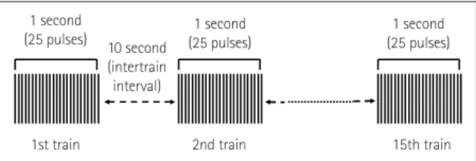
Participants received focal rTMS over two consecutive days, with it being applied via a 70-mm double-air-film coil over the left MC or SMA. The location of rTMS was newly deter- mined at each visit. rTMS was applied with the right first dorsal interosseous muscle at rest. The resting motor thresh- old (RMT) was measured as the lowest stimulus intensity re- quired to produce motor-evoked potentials of at least 50 μV in at least 5 of 10 consecutive trials, and the rTMS stimula- tion intensity was then set at 100% of this RMT. In the MC stimulation group, the MC hand area was stimulated because it is more accessible than the leg area, and several previous studies have shown that several body parts can be simulta- neously affected by stimulation to the distal hand area. Posi- tive effects of rTMS have been reported on gait28-30 and on shoulder bradykinesia with distal hand area stimulation.31 In the SMA stimulation group, the coil was centered on the midline at 4 cm anterior to the vertex (Cz in the Internation- al 10–20 EEG system).32 rTMS was delivered using a Mags- tim Super-Rapid2 stimulator (Magstim, Wales, UK) in a se- ries of four rTMS blocks separated by 10 minutes. Each block consisted of 15 to 25 pulse trains of 1-second duration at 25 Hz, with an intertrain interval of 10 seconds. The inter- train interval refers to the interval between the last pulse of a train to the first pulse of the next train (Fig. 1).31 rTMS was applied in the on-medication state.27
Outcome measurements
Participants were asked to perform the tasks as described be- low before rTMS was applied on the 1st day and immediately after rTMS was applied on the 2nd day. All task performanc- es were recorded on video and were assessed by a blinded rater.
Patients were asked to perform the stand-walk-sit (SWS)
1 second
(25 pulses) 1 second
(25 pulses)
1st train 2nd train
10 second (intertrain interval)
15th train 1 second (25 pulses)
Fig. 1. One block of the protocol for repetitive transcranial magnetic stimulation. The intertrain interval is the interval between the last pulse of a train to the first pulse of the next train. Four blocks were applied with a 10-minute interval during each visit.
Effective Brain Stimulation Area for FOG
JCN
test by standing up, walking 10 m, turning back, and sitting down as quickly as possible. To elicit FOG, patients per- formed 360° turns as rapidly as possible from a standstill within a restricted area with two leftward turns and two right- ward turns in random order. Patients performed another task simultaneously (called the dual task, which involved sub- tracting 7 serially from 100) during the SWS test.21 At the end of the experiment on the 2nd day, the Patient Global Impression and Clinical Global Impression rating scales were applied to assess the effect of rTMS.33 The number of steps and the completion time of the SWS test were measured.
Freezing episodes during the SWS test, rapid-full-turn test, and the dual task during SWS test were analyzed.
Statistical analyses
Demographics, clinical variables, and relative changes in the outcome measurements were compared between the two interventions by Mann-Whitney and Fisher’s exact tests as appropriate. The relative change in each measurement be- tween pre- and postintervention was calculated as [(value at postintervention-value at preintervention)/(value at postint-
ervention +value at preintervention)/2]×100. If both values were 0 at the pre- and postinterventions, 1.0 was added in or- der to prevent the denominator from being 0. The outcome measurements before and after interventions within each group were compared using Wilcoxon signed-rank tests.
Probability values of p<0.05 were considered statistically sig- nificant.
RESULTS
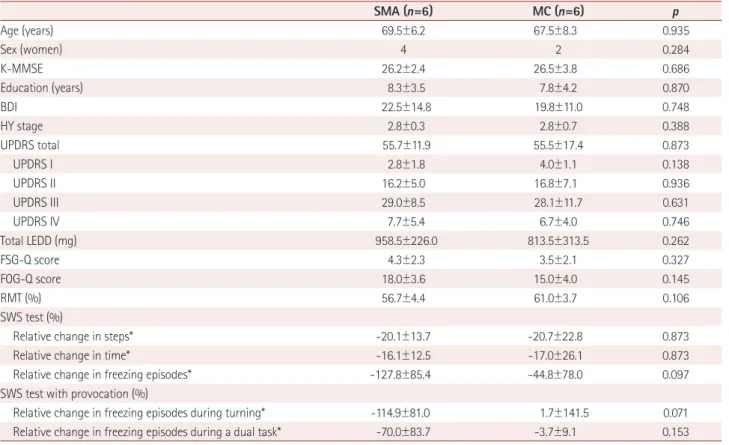
There was no difference in demographics, BDI score, HY stage, UPDRS score, total LEDD, FSG-Q total score, or FOG- Q total score between the two interventions (Table 1). There was also no difference in the relative change in gait or freez- ing variables after rTMS between the SMA and MC groups, but trends for relative changes in freezing episodes during the SWS test (p=0.097) and the rapid-full-turn test (p=0.071) were seen. There were fewer freezing episodes in the SMA group than in the MC group (Table 1). A significant improve- ment in gait and a reduction in the number of freezing epi- sodes from baseline were seen in the SMA group (p<0.05)
Table 1. Clinical features of patients and the relative changes in gait parameters after rTMS
SMA (n=6) MC (n=6) p
Age (years) 69.5±6.2 67.5±8.3 0.935
Sex (women) 4 2 0.284
K-MMSE 26.2±2.4 26.5±3.8 0.686
Education (years) 8.3±3.5 7.8±4.2 0.870
BDI 22.5±14.8 19.8±11.0 0.748
HY stage 2.8±0.3 2.8±0.7 0.388
UPDRS total 55.7±11.9 55.5±17.4 0.873
UPDRS I 2.8±1.8 4.0±1.1 0.138
UPDRS II 16.2±5.0 16.8±7.1 0.936
UPDRS III 29.0±8.5 28.1±11.7 0.631
UPDRS IV 7.7±5.4 6.7±4.0 0.746
Total LEDD (mg) 958.5±226.0 813.5±313.5 0.262
FSG-Q score 4.3±2.3 3.5±2.1 0.327
FOG-Q score 18.0±3.6 15.0±4.0 0.145
RMT (%) 56.7±4.4 61.0±3.7 0.106
SWS test (%)
Relative change in steps* -20.1±13.7 -20.7±22.8 0.873
Relative change in time* -16.1±12.5 -17.0±26.1 0.873
Relative change in freezing episodes* -127.8±85.4 -44.8±78.0 0.097
SWS test with provocation (%)
Relative change in freezing episodes during turning* -114.9±81.0 1.7±141.5 0.071
Relative change in freezing episodes during a dual task* -70.0±83.7 -3.7±9.1 0.153
Data are n or mean±SD values.
*Negative value indicates that the value of the gait parameter decreased after rTMS.
BDI: Beck Depression Inventory, FOG-Q: Freezing of Gait Questionnaire, FSG-Q: Festination of Gait Questionnaire, HY stage: Hoehn and Yahr stage, K- MMSE: Korean version of the Mini Mental State Examination, LEDD: levodopa equivalent daily dose, RMT: resting motor threshold, rTMS: repetitive transcranial magnetic stimulation, SWS test: stand-walk-sit test, UPDRS: Unified Parkinson’s Disease Rating Scale.
Kim SJ et al.
JCN
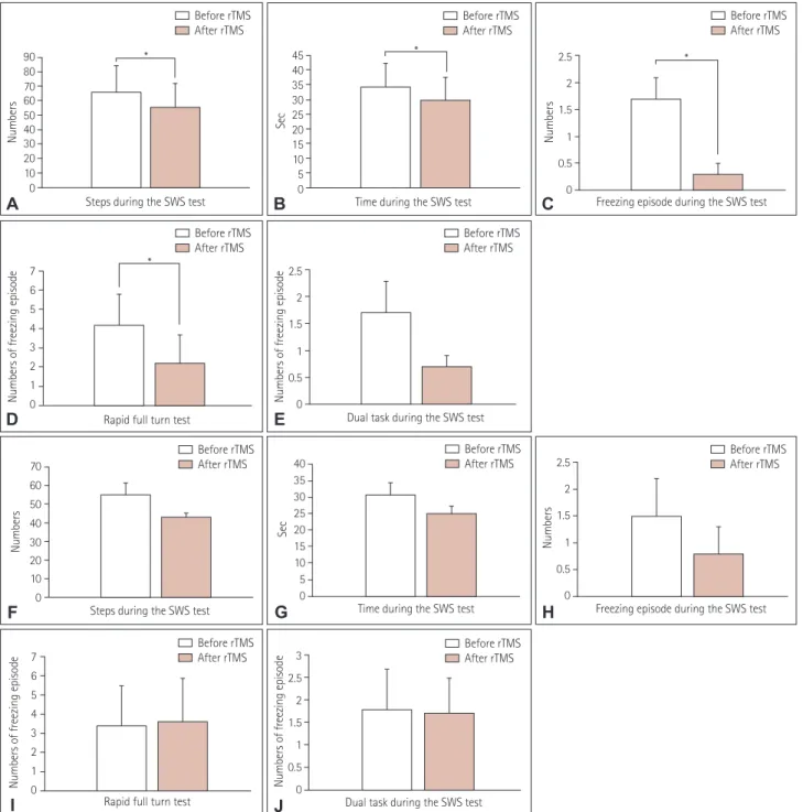
(Fig. 2A-E), whereas there were no significant changes from baseline in the MC group (Fig. 2F-J).
DISCUSSION
Our results suggest that the SMA is a more-appropriate site for rTMS in PD patients with FOG. SMA stimulation im- proved FOG, with there being fewer steps, a shorter walk time,
and fewer freezing events during the SWS test in the SMA group than in the MC group, although the differences were not statistically significant. Compared with prestimulation find- ings, the number of freezing episodes clearly decreased signif- icantly after SMA stimulation but not after MC stimulation.
Previous imaging studies suggest that the SMA is closely associated with FOG. During gait motor imaging, brain ac- tivity was decreased in the SMA and increased in the mesen-
90 80 7060 50 4030 20 10 0
Numbers
Steps during the SWS test
*
Before rTMS After rTMS
A
7 6 5 4 3 2 1 Numbers of freezing episode 0
Rapid full turn test
*
Before rTMS After rTMS
D
40 35 30 25 20 15 10 5 0
Sec
Time during the SWS test Before rTMS After rTMS
G
3 2.5 2 1.5 1 0.5 Numbers of freezing episode 0
Dual task during the SWS test Before rTMS After rTMS
J
45 40 35 30 25 20 15 10 5 0
Sec
Time during the SWS test
*
Before rTMS After rTMS
B
2.5 2 1.5 1 0.5 Numbers of freezing episode 0
Dual task during the SWS test Before rTMS After rTMS
E
2.5 2 1.5 1 0.5 0
Numbers
Freezing episode during the SWS test Before rTMS After rTMS
H
2.5 2 1.5 1 0.5 0
Numbers
Freezing episode during the SWS test
*
Before rTMS After rTMS
C
70 60 50 40 30 20 10 0
Numbers
Steps during the SWS test Before rTMS After rTMS
F
7 6 5 4 3 2 1 Numbers of freezing episode 0
Rapid full turn test
Before rTMS After rTMS
I
Fig. 2. Comparison of gait parameters before and after stimulation of the SMA (A-E) and after MC stimulation (F-J). Gait parameters were im- proved and freezing episodes were reduced from baseline in the SMA group, whereas there were no significant changes in gait and freezing epi- sodes from baseline in the MC group. *p<0.05. MC: motor cortex, rTMS: repetitive transcranial magnetic stimulation, SMA: supplementary motor area, SWS: stand-walk-sit.
Effective Brain Stimulation Area for FOG
JCN
cephalic locomotor region in PD with FOG.20 Those brain areas that were active during cognitive loading, which is as- sociated with FOG, showed less recruitment in PD with FOG (bilateral anterior insula, ventral striatum, and pre-SMA).34
The SMA is located anterior to the MC leg area. Recent anatomical studies suggest that the SMA comprises two dis- tinct parts: the pre-SMA (rostral part) that is linked to the prefrontal cortex, and the SMA proper (caudal part) that is joined directly to the MC, dorsal premotor cortex, and spi- nal cord.35,36 The SMA is important in several types of motor processes and is activated before movement initiation.19,36 It is active during voluntary and externally triggered actions, sequential movements, learning processes, and executive control.19 During movements, the SMA participates in action preparation, the initiation and selection of actions, motor learning, inhibition, conditional action, action control, and monitoring of action outcomes;36 however, its precise roles in these various processes remain uncertain. It was recently sug- gested that the SMA participates in cognitive processes under- lying sensorimotor integration, and its role might be to com- bine individual actions into a sequential process.35
Because the SMA reportedly adjusts anticipatory postures by shifting the body weight during gait, it might coordinate postural adjustment and stepping. The SMA might be unable to regulate submovements in FOG, which would hinder the performing of sequential movements during automatic gait.37
Only one previous study has investigated rTMS thera- peutic effects over three different regions (MC, SMA, and dorsolateral prefrontal cortex) in heterogeneous disease pop- ulations showing parkinsonism, including patients with PD, vascular parkinsonism, multiple-system atrophy, and Lewy- body disease.38 Those authors reported that MC stimulation was effective for FOG and gait, whereas SMA stimulation was not. It is difficult to compare that study with ours due to differences in their designs in terms of stimulation frequen- cies, disease groups, and methods of FOG evaluation. FOG is generally difficult to measure because it occurs episodically in specific situations such as turning or passing through a narrow walkway. This means that the specific task used to provoke FOG is important. A previous study found that 360°
turns were more effective than 180° turns,21 and so the for- mer were used in the present study. We also used another provocation task (the dual task) to substantiate our findings.
rTMS has recently been shown to exert therapeutic effects in several neurological and psychiatric disorders.12 Despite the positive results obtained when applying rTMS, greater standardization may be needed in order to facilitate its im- plementation in clinical practice. Many different types of stim- ulation protocols have been used previously, with some taking a long time (due to the numbers of stimulations or the num-
ber of visits required for treatment) and also the stimulation sites used being inconsistent even in the same disease.14-18 Because patients with neurological and psychiatric diseases may be fragile both physically and mentally, it is essential to apply rTMS in the most effective way possible and in as a short time as possible. Our study suggests that the SMA is a more-effective therapeutic target for FOG, and hence should be considered the primary target area for FOG treatments.
This study was subject to some limitations. First, the sam- ple was small and we did not include a control group. We had difficulty registering patients for several reasons, includ- ing the poor mobility associated with FOG, the distance from home to our hospitals the requirement for several visits, and the application of rather strict exclusion criteria. Patients with dementia were initially excluded since cognition can af- fect gait, and patients with cognitive impairment might also be less cooperative due to their impaired understanding.
However, this adversely affected the sample size since most PD patients with FOG are in an advanced state, and they of- ten exhibit cognitive impairment. Second, we targeted the MC hand area due to ease of accessibility. The left-hemi- sphere MC was also stimulated, whereas SMA stimulation was presumed to affect both hemispheres simultaneously.39 Bilateral MC stimulation should be addressed in future stud- ies. We do not know whether stimulation of the dominant or bilateral hemisphere is more effective. The same number of rTMS pulses should have been applied to a specific area in the MC and SMA groups, but this is impossible in bilateral MC stimulation if the total number of pulses must be con- stant in each group. Also, we had to conduct our experiment as rapidly as possible, because participants with advanced PD are more likely to tire quickly. Bilateral MC stimulation lengthens the duration of its experiment, which would have resulted in bias. Third, we did not use a computerized navi- gation system to localize the SMA, instead targeting the area based on a previous study.32 The closeness of the SMA to the MC for the leg may make it difficult to discern if the rTMS effects came from the SMA or the leg-area MC stimulation.
This issue could be addressed by performing objective navi- gation to confirm the stimulation site or including a control group that receives leg MC rTMS. Fourth, we did not assess later time points after rTMS (e.g., 24 hours). Because some studies have shown greater improvement of motor function at later times,16,40 including measurements at later time points would have been helpful for drawing definitive conclusions.
All of these limitations mean that further studies are needed to confirm the present findings, including larger sham-con- trolled trials, the use of navigation systems for accurately identifying the location of stimulation targets, and more-ex- tended exploration for determining possible candidates in-
Kim SJ et al.
JCN
cluding the dorsolateral prefrontal cortex, leg area MC, and bilateral MC stimulation.
Our study found that SMA stimulation improved the gen- eral gait pattern and FOG in PD, whereas MC stimulation did not. These results suggest that SMA stimulation is a more-appropriate target in PD patients with FOG. The re- sults of our study provide evidence that stimulating the SMA using rTMS can exert beneficial effects on FOG, which might be useful for future developments of therapeutic strat- egies.
Conflicts of Interest
The authors have no financial conflicts of interest.
Acknowledgements
This work was supported by a grant from University Research Park Proj- ect of Busan National University funded by Busan Institute of S&T Eval- uation and Planning.
REFERENCES
1. Nutt JG, Bloem BR, Giladi N, Hallett M, Horak FB, Nieuwboer A.
Freezing of gait: moving forward on a mysterious clinical phenome- non. Lancet Neurol 2011;10:734-744.
2. Giladi N, McDermott MP, Fahn S, Przedborski S, Jankovic J, Stern M, et al. Freezing of gait in PD: prospective assessment in the DATATOP cohort. Neurology 2001;56:1712-1721.
3. Macht M, Kaussner Y, Möller JC, Stiasny-Kolster K, Eggert KM, Krüger HP, et al. Predictors of freezing in Parkinson's disease: a sur- vey of 6,620 patients. Mov Disord 2007;22:953-956.
4. Contreras A, Grandas F. Risk factors for freezing of gait in Parkinson’s disease. J Neurol Sci 2012;320:66-71.
5. Ou R, Guo X, Song W, Cao B, Yang J, Wei Q, et al. Freezing of gait in Chinese patients with Parkinson disease. J Neurol Sci 2014;345:56-60.
6. Perez-Lloret S, Negre-Pages L, Damier P, Delval A, Derkinderen P, Destée A, et al. Prevalence, determinants, and effect on quality of life of freezing of gait in Parkinson disease. JAMA Neurol 2014;71:884- 7. Fietzek UM, Zwosta J, Schroeteler FE, Ziegler K, Ceballos-Baumann 890.
AO. Levodopa changes the severity of freezing in Parkinson’s disease.
Parkinsonism Relat Disord 2013;19:894-896.
8. Nonnekes J, Snijders AH, Nutt JG, Deuschl G, Giladi N, Bloem BR.
Freezing of gait: a practical approach to management. Lancet Neurol 2015;14:768-778.
9. Schlenstedt C, Shalash A, Muthuraman M, Falk D, Witt K, Deuschl G.
Effect of high-frequency subthalamic neurostimulation on gait and freezing of gait in Parkinson's disease: a systematic review and meta- analysis. Eur J Neurol 2017;24:18-26.
10. Xie T, Kang UJ, Warnke P. Effect of stimulation frequency on imme- diate freezing of gait in newly activated STN DBS in Parkinson’s dis- ease. J Neurol Neurosurg Psychiatry 2012;83:1015-1017.
11. Okuma Y. Practical approach to freezing of gait in Parkinson’s dis- ease. Pract Neurol 2014;14:222-230.
12. Cirillo G, Di Pino G, Capone F, Ranieri F, Florio L, Todisco V, et al.
Neurobiological after-effects of non-invasive brain stimulation. Brain Stimul 2017;10:1-18.
13. Henrich-Noack P, Sergeeva EG, Sabel BA. Non-invasive electrical brain stimulation: from acute to late-stage treatment of central ner- vous system damage. Neural Regen Res 2017;12:1590-1594.
14. Chou YH, Hickey PT, Sundman M, Song AW, Chen NK. Effects of re- petitive transcranial magnetic stimulation on motor symptoms in Par-
kinson disease: a systematic review and meta-analysis. JAMA Neurol 2015;72:432-440.
15. Chung CL, Mak MK. Effect of repetitive transcranial magnetic stimu- lation on physical function and motor signs in Parkinson’s disease: a systematic review and meta-analysis. Brain Stimul 2016;9:475-487.
16. Kim MS, Chang WH, Cho JW, Youn J, Kim YK, Kim SW, et al. Effi- cacy of cumulative high-frequency rTMS on freezing of gait in Par- kinson’s disease. Restor Neurol Neurosci 2015;33:521-530.
17. Rektorova I, Sedlackova S, Telecka S, Hlubocky A, Rektor I. Repeti- tive transcranial stimulation for freezing of gait in Parkinson’s disease.
Mov Disord 2007;22:1518-1519.
18. Tard C, Devanne H, Defebvre L, Delval A. Single session intermittent theta-burst stimulation on the left premotor cortex does not alleviate freezing of gait in Parkinson’s disease. Neurosci Lett 2016;628:1-9.
19. Nachev P, Kennard C, Husain M. Functional role of the supplementa- ry and pre-supplementary motor areas. Nat Rev Neurosci 2008;9:856- 869.
20. Snijders AH, Takakusaki K, Debu B, Lozano AM, Krishna V, Fasano A, et al. Physiology of freezing of gait. Ann Neurol 2016;80:644-659.
21. Snijders AH, Haaxma CA, Hagen YJ, Munneke M, Bloem BR. Freezer or non-freezer: clinical assessment of freezing of gait. Parkinsonism Relat Disord 2012;18:149-154.
22. Hughes AJ, Daniel SE, Kilford L, Lees AJ. Accuracy of clinical diagno- sis of idiopathic Parkinson’s disease: a clinico-pathological study of 100 cases. J Neurol Neurosurg Psychiatry 1992;55:181-184.
23. Rossi S, Hallett M, Rossini PM, Pascual-Leone A; Safety of TMS Con- sensus Group. Safety, ethical considerations, and application guide- lines for the use of transcranial magnetic stimulation in clinical prac- tice and research. Clin Neurophysiol 2009;120:2008-2039.
24. Giladi N, Shabtai H, Rozenberg E, Shabtai E. Gait festination in Par- kinson’s disease. Parkinsonism Relat Disord 2001;7:135-138.
25. Giladi N, Shabtai H, Simon ES, Biran S, Tal J, Korczyn AD. Construc- tion of freezing of gait questionnaire for patients with Parkinsonism.
Parkinsonism Relat Disord 2000;6:165-170.
26. Tomlinson CL, Stowe R, Patel S, Rick C, Gray R, Clarke CE. Systematic review of levodopa dose equivalency reporting in Parkinson’s disease.
Mov Disord 2010;25:2649-2653.
27. Hamada M, Ugawa Y, Tsuji S; Effectiveness of rTMS on Parkinson’s Disease Study Group, Japan. High-frequency rTMS over the supple- mentary motor area for treatment of Parkinson’s disease. Mov Disord 2008;23:1524-1531.
28. Du J, Tian L, Liu W, Hu J, Xu G, Ma M, et al. Effects of repetitive tran- scranial magnetic stimulation on motor recovery and motor cortex excitability in patients with stroke: a randomized controlled trial. Eur J Neurol 2016;23:1666-1672.
29. Lefaucheur JP, Drouot X, Von Raison F, Ménard-Lefaucheur I, Cesaro P, Nguyen JP. Improvement of motor performance and modulation of cortical excitability by repetitive transcranial magnetic stimulation of the motor cortex in Parkinson’s disease. Clin Neurophysiol 2004;115:
2530-2541.
30. von Papen M, Fisse M, Sarfeld AS, Fink GR, Nowak DA. The effects of 1 Hz rTMS preconditioned by tDCS on gait kinematics in Parkin- son’s disease. J Neural Transm 2014;121:743-754.
31. Kang SY, Wasaka T, Shamim EA, Auh S, Ueki Y, Lopez GJ, et al.
Characteristics of the sequence effect in Parkinson’s disease. Mov Dis- ord 2010;25:2148-2155.
32. Arai N, Lu MK, Ugawa Y, Ziemann U. Effective connectivity between human supplementary motor area and primary motor cortex: a paired- coil TMS study. Exp Brain Res 2012;220:79-87.
33. Guy W. ECDEU Assessment Manual for Psychopharmacology. Rock- ville (MD): U.S. Department of Health, Education, and Welfare, Pub- lic Health Service Alcohol, Drug Abuse, and Mental Health Adminis- tration, 1976.
34. Shine JM, Matar E, Ward PB, Bolitho SJ, Pearson M, Naismith SL, et al. Differential neural activation patterns in patients with Parkinson’s
Effective Brain Stimulation Area for FOG
JCN
disease and freezing of gait in response to concurrent cognitive and motor load. PLoS One 2013;8:e52602.
35. Cona G, Semenza C. Supplementary motor area as key structure for do- main-general sequence processing: a unified account. Neurosci Biobehav Rev 2017;72:28-42.
36. Lima CF, Krishnan S, Scott SK. Roles of supplementary motor areas in auditory processing and auditory imagery. Trends Neurosci 2016;
39:527-542.
37. Fling BW, Cohen RG, Mancini M, Carpenter SD, Fair DA, Nutt JG, et al. Functional reorganization of the locomotor network in Parkinson
patients with freezing of gait. PLoS One 2014;9:e100291.
38. Lee SY, Kim MS, Chang WH, Cho JW, Youn JY, Kim YH. Effects of repetitive transcranial magnetic stimulation on freezing of gait in pa- tients with Parkinsonism. Restor Neurol Neurosci 2014;32:743-753.
39. Koch G, Brusa L, Caltagirone C, Peppe A, Oliveri M, Stanzione P, et al. rTMS of supplementary motor area modulates therapy-induced dyskinesias in Parkinson disease. Neurology 2005;65:623-625.
40. Park JE. Repetitive transcranial magnetic stimulation for limb-kinet- ic apraxia in Parkinson’s disease. J Clin Neurol 2018;14:110-111.