Background and PurposezzThe aim of this study was to determine the efficacy and tolera- bility of granulocyte colony-stimulating factor (G-CSF) in subjects with amyotrophic lateral sclerosis (ALS).
MethodszzForty subjects with ALS were randomly assigned to two groups, which received ei- ther subcutaneous G-CSF (5 μg/kg/q12h) or placebo for 5 days. The subjects were then fol- lowed up for 3 months using the ALS Functional Rating Scale-Revised (ALSFRS-R), manual muscle testing, ALS Assessment Questionnaire-40, and nerve conduction studies. CD34+/
CD133+ cell count and monocyte chemoattractant protein-1 (MCP-1) levels were evaluated at baseline.
ResultszzThe rate of disease progression did not differ significantly between the two groups.
The reduction in ALSFRS-R scores was greater in female subjects in the G-CSF group than in their counterparts in the placebo group. There was a trend toward a positive correlation be- tween baseline CSF MCP-1 levels and the change in ALSFRS-R scores in both groups (Spear- man’s ρ=0.370, p=0.070).
ConclusionszzWith the protocol implemented in this study, G-CSF is not a promising option for the treatment of ALS. Furthermore, it may accelerate disease progression in females.
Key Wordszz amyotrophic lateral sclerosis, ALS Functional Rating Scale, granulocyte colony-stimulating factor, CD34+/CD133+ cells,
monocyte chemoattractant protein-1, compound motor action potential.
Granulocyte Colony-Stimulating Factor for Amyotrophic Lateral Sclerosis: A Randomized, Double-Blind,
Placebo-Controlled Study of Iranian Patients
INTRODUCTION
The effect of granulocyte colony-stimulating factor (G-CSF) on the course of neurological diseases has drawn considerable attention over the past decade. G-CSF mobilizes hemato- poietic (CD34+) stem cells and—with repeated administration—recruits immature CD34+/
CD133+ stem cells that are capable of differentiating into neuronal progenitors.1,2 G-CSF receptor expression may play an autocrine protective role in various cells of the nervous system,3 and G-CSF may protect against ischemic neuronal damage by inhibiting apopto- sis and inflammation, mobilizing stem cells, or enhancing neuronal differentiation.3,4
Recent studies have suggested a role for G-CSF in the treatment of amyotrophic lateral sclerosis (ALS).5-9 In a mouse model of ALS, G-CSF significantly improved motor perfor- mance and motoneuron survival, and reduced denervation atrophy.10 A study involving subjects with ALS found significantly reduced G-CSF receptor expression in motoneurons and increased G-CSF expression in reactive astrocytes.8 The authors suggested that re- duced G-CSF receptor expression on motor neurons might account for the pathophysiol- Nasibeh Amirzagara
Shahriar Nafissib Abbas Tafakhoria
Amirhossein Modabberniac Aliakbar Amirzargard Majid Ghaffarpoura Bahaddin Siroosa
Mohammad Hossein Harirchiana
a Iranian Center of Neurological Research, Imam Khomeini Hospital, Tehran University of Medical Sciences, Tehran, Iran
b Neurology Department, Tehran Shariati Hospital, University of Medical Sciences, Tehran, Iran
c Department of Psychiatry,
Icahn School of Medicine at Mount Sinai, New York, NY, USA
d Department of Immunology,
Faculty of Medicine, Tehran University of Medical Sciences, Tehran, Iran
pISSN 1738-6586 / eISSN 2005-5013 / J Clin Neurol 2015;11(2):164-171 / http://dx.doi.org/10.3988/jcn.2015.11.2.164
Received September 15, 2014 Revised November 18, 2014 Accepted November 18, 2014 Correspondence
Mohammad Hossein Harirchian, MD Iranian Center of Neurological Research, Imam Khomeini Hospital, Tehran University of Medical Sciences, Keshavarz Blvd, Tehran 1419635493, IranTel +9821662424
Fax +982166581558
E-mail [email protected]
cc This is an Open Access article distributed under the terms of the Creative Commons Attribution Non-Com- mercial License (http://creativecommons.org/licenses/by-nc/3.0) which permits unrestricted non-commercial use, distribution, and reproduction in any medium, provided the original work is properly cited.
JCN
Open Access ORIGINAL ARTICLEAmirzagar N et al.
JCN
JCN
Open Accessogy of ALS. Simultaneously the levels of monocyte chemoat- tractant protein-1 (MCP-1) were increased, suggesting that reduced–CSF receptors can exacerbate the ALS course.
Most studies of G-CSF efficacy in subjects with ALS have been limited in several ways. A nonrandomized study in- volving 13 subjects with ALS demonstrated a significantly lower monthly reduction in ALS Functional Rating Scale- Revised (ALSFRS-R) scores and compound motor action potential (CMAP) amplitude following 5 days of G-CSF treat- ment.9 Another uncontrolled study involving 24 individuals did not show any significant effect of G-CSF on clinical out- comes.11 In a placebo-controlled study of G-CSF treatment in ten subjects with ALS, G-CSF treatment failed to improve any of the clinical outcomes, although it significantly reduced the fractional anisotropy on diffusion tensor imaging.12 Final- ly, a double-blind study involving 39 subjects found no sta- tistically significant benefit with G-CSF, although there was a trend toward slowing of disease progression following two cycles of G-CSF treatment.13 One problem with that study was that more than 50% of the subjects were lost to 1-year fol- low-up.
The present study was a randomized double-blind, place- bo-controlled trial of G-CSF with assessment of various clinical, functional, electrophysiological, and molecular out- comes, the aim of which was to provide a thorough view of the efficacy of G-CSF and its tolerability in Iranian subjects with ALS, and to address some of the caveats noted in previ- ous studies.
METHODS
Trial design and setting
This was a single-center, 3-month, randomized, double-blind, placebo-controlled, and parallel-group study that was con- ducted in a tertiary referral center affiliated with Tehran Uni- versity of Medical Sciences between November 2012 and No- vember 2013.
Participants
Male and female subjects with a diagnosis of probable or defi- nite ALS based on the revised El Escorial World Federation of Neurology criteria were screened for inclusion in the study.
The following inclusion criteria were applied: aged 18–85 years, symptom history not exceeding 2 years, and ALSFRS- R score ≥20. Although ethnicity was not an inclusion crite- rion, all of the subjects were ethnically Iranian. Riluzole treat- ment was not an exclusion criterion if the patient had been stable on the therapeutic protocol for at least 30 days prior to study entry. The following exclusion criteria were applied:
ALS in first-degree relatives; pregnancy or lactation; history
of neoplasia, myeloproliferative, or any other disorders that could be exacerbated by G-CSF; active immunological dis- ease; spleen diameter ≥180 mm; severe heart, kidney, or liv- er disease, positive HIV status, forced vital capacity ≤50% of that predicted; cognitive disorders that interfere with the study procedure; and history of hypersensitivity reaction to G- CSF or Escherichia-coli-derived proteins.
After receiving a complete explanation of the study pro- cedures, all subjects (or their representatives) provided writ- ten informed consent to participate prior to study entry. The study protocol was approved by the Ethics Committee and the Institutional Review Board of Tehran University of Med- ical Sciences (approval no. 91-01-54-17265). The trial was conducted in accordance with the last revision of the Decla- ration of Helsinki and was registered at ClinicalTrials.gov (registration no. NCT01825551).
Study procedures
The subjects underwent a complete physical and neurologic examination, peripheral blood smear, complete blood count with differentials, liver enzymes, serum lactate dehydroge- nase (LDH), uric acid, CD34+ and CD133+ cell count, serum and cerebrospinal fluid (CSF; in a subset of subjects) MCP- 1 levels, abdominal ultrasound (if the spleen was palpable), and pulmonary function testing (if significant respiratory dysfunction was present). Scores for the ALS Assessment Questionnaire-40 (ALSAQ-40), ALSFRS-R, and manual muscle testing (MMT) were assessed every month. Nerve con- duction velocity (NCV) studies were performed by an expert (A.T.) at baseline and at the study endpoint. The white blood cell (WBC) count was measured every day during the treat- ment phase. WBC differentials, liver enzymes, and CD34+
and CD133+ cell counts were measured on days 4 and 6, and LDH and uric acid levels were assessed on day 4 of the trial.
MCP-1 concentration was only tested at baseline.
Subjects were randomly assigned to receive either subcuta- neous G-CSF (containing PDgrastim, filgrastim, 300-μg re- combinant G-CSF, equal to 30,000,000 IU of filgrastim, man- nitol, and sodium acetate; Pooyesh Darou, Tehran, Iran) 5 μg/kg/q12h or normal saline for five consecutive days. The subjects were hospitalized and closely monitored for any pos- sible serious adverse events during the first few days. Treat- ment was discontinued if the leukocyte count rose to more than 50,000/μL; the remaining doses were administered when levels had returned to lower values (below 15,000/μL). The participants were then followed up in the outpatient clinic.
Assessments
Amyotrophic lateral sclerosis Functional Rating Scale-Re- vised is a 12-item (total score of 0–48) physician-administered
G-CSF for ALS
JCN
measure of ALS severity that assesses function in three major domains: bulbar, motor, and respiratory. Each item is scored on a five-point scale (0–4), with higher scores reflecting better function.14
Amyotrophic lateral sclerosis Assessment Questionnaire-40 is a 40-item subjective measure of health status for subjects with ALS that is categorized into five domains: eating/drink- ing, communication, activities of daily living/independent activities of daily living, physical mobility, and emotional func- tioning. The total ALSAQ-40 score and those of its domains are converted into a 100-point scale, with lower scores reflect- ing a better health status.15,16
Manual muscle testing was based on the examination of 34 muscles, converted to a 10-point scale. The final MMT score is the mean of the scores for all 34 muscles. MMT appears to be the preferred method for measurement of global strength.17
The NCV studies involved a belly-tendon montage of CMAP amplitude recordings from the median (abductor pollicis brevis), ulnar (abductor digiti minimi), tibial (abductor hal- lucis brevis), and common peroneal nerves (extensor digito- rum brevis). CMAPs were recorded in response to supra- maximal stimulation with 0.2-ms duration.
MCP-1, CD34+, and CD34+/CD133+ cells
The method for evaluating CD34+, CD34+/CD133+, and MCP-1 has been described in detail elsewhere.1,18
Outcome measures
The primary outcome measure was the monthly rate of de- cline in ALSFRS-R score. Secondary outcome measures were the changes from baseline in the ALSAQ-40 and MMT scores, and the CMAP amplitude of the nerves. Furthermore, the cor- relations between MCP-1 levels, counts of CD34+, CD133+, and CD34+/133+ cells, and ALSFRS-R scores were calculat- ed. Safety issues were systematically assessed using both a checklist for clinical symptoms and laboratory values.
Randomization, allocation concealment, and blinding
A randomization list was prepared using a computerized random-number generator in a 1:1 ratio and block size of four.
Allocation concealment was achieved using sequentially numbered and opaque envelopes. Treatment allocation, evalu- ation of side effects, and possible changes in treatment pro- tocol, clinical rating, and electrophysiological assessment of the subjects were conducted by separate researchers. The sub- jects, the evaluator, the person responsible for administer- ing the intervention, and the statistician were all blind to the treatment allocation.
Statistical analysis
STATA version 12.0 (StataCorp, College Station, TX, USA) was used for the data analysis. All analyses were carried out on the data from participants with at least one postbaseline measurement. Per-protocol and linear mixed model analy- ses were both used for comparison of outcomes. Repeated- measures linear mixed model analysis was performed using the STATA module for analyzing repeated-measures data (xtmixed), which can account for unbalanced data. Except where stated otherwise, the data are presented as mean±SD values, and the cutoff for statistical significance was set at p<0.05.
The sample size was calculated based on a between-group difference of three (with a standard deviation of three) for the change in ALSFRS-R scores from baseline. This yielded a sample size of 16 in each group, and accounting for a poten- tial 20% loss to follow-up, a sample size of 40 was reached.
RESULTS
Baseline characteristics and laboratory values
Sixty-seven subjects were screened for eligibility criteria and 40 subjects were randomized into either the G-CSF (n=20) or placebo (n=20) group. All subjects had at least one post- baseline measurement, and 35 subjects completed the study (n=18 and 17 for the G-CSF and placebo groups, respective- ly). The baseline characteristics of the subjects are summa- rized in Table 1. One patient experienced transient fever and chills at day 4 of treatment.The WBC count in the G-CSF group rose from 7,892±
2,009/μL at baseline to 49,689±14,613/μL at day 6 (p<0.001, repeated-measures ANOVA), whereas no significant change was observed in the placebo group (7,041±1,613/μL and 6,862±1,312/μL at baseline and day 6, respectively; p=0.985).
The numbers of neutrophils, CD34+, CD133+, and CD34/
133+ cells increased significantly in the G-CSF group (p<0.01 for all), but not the placebo group. Of the laboratory tests, alkaline phosphatase and LDH levels increased significantly in the G-CSF group.
ALSFRS-R scores
Both groups exhibited a progressive decline in the ALSFRS- R score (p<0.0001 for time effect). In the mixed model anal- ysis, no significant time-treatment interaction was observed (Fig. 1, Table 2). The per-protocol analysis (Table 3) revealed that the monthly reduction in ALSFRS-R scores did not differ significantly between the G-CSF group (1.53 points/month) and the placebo group [1.61 points/month; mean difference (95% CI)=0.074 (-0.952 to 1.101), t (34)=0.146, p=0.884].
Amirzagar N et al.
JCN
ALSAQ-40 and MMT
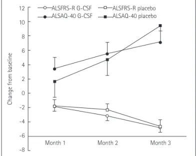
A progressive change in the ALSAQ-40 and MMT scores was observed in both groups, corresponding to a worsened health status (p<0.0001 for time effect). The change in either outcome did not differ significantly between the two groups (Fig. 1, Table 2 and 3).
NCV
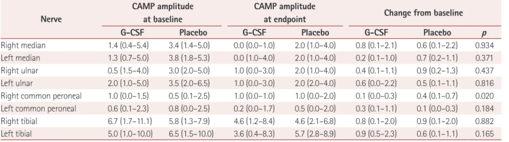
Except for one of the measurements (right common perone- al), changes from baseline did not differ significantly be- tween the two groups (Table 4).Association between CD34+ (CD133+) cell count, MCP-1 level, and ALSFRS-R score
In the G-CSF group, changes in the CD34+ cell count in the first 6 days were negatively correlated with the ALSFRS-R score reduction (r=-0.485, p=0.041). Further analysis revealed that those with large changes in CD34+ (>100/μL) cell count did not differ significantly from the placebo group in terms of reduction in ALSFRS-R scores. The elevation in the CD34+
cell count was lower in the females than the males of the G- CSF group, with the difference tending toward significance (55.9±45.7/μL vs. 106.2±61.8/μL; p=0.062, Mann-Whitney U-test). Further exploratory analysis demonstrated that female Table 1. Baseline characteristics of the patients in the two groups
Variable G-CSF (n=20) Placebo (n=20)
Gender, male (n, %) 13 (65) 12 (60)
Age (years) 51.3 (8.6) 52.5 (11.6)
Duration of disease (months) 17.0 (6.4) 15.7 (6.3)
Time since diagnosis (months) 9.7 (8.2) 9.8 (7.6)
History of riluzole treatment (n, %) 14 (70) 14 (70)
Duration of riluzole treatment (months) 5.5 (6.7) 5.4 (6.1)
History of smoking (n, %) 7 (35) 6 (30)
History of alcohol use (n, %) 3 (15) 0 (0)
BMI (kg/m2) 24.3 (4.7) 25.1 (4.5)
Baseline ALSFRS-R score 33.3 (7.9) 36.6 (4.6)
Bulbar domain 9.6 (2.9) 10.5 (1.9)
Motor domain 11.8 (6.8) 14.4 (4.6)
Respiratory domain 11.9 (0.3) 11.6 (0.7)
Baseline ALSAQ-40 score 63.0 (17.5) 60.3 (12.1)
Physical mobility domain 71.7 (20.2) 72.2 (19.0)
ADL/IADL 69.6 (25.1) 66.0 (22.4)
Eating disorder 44.7 (30.4) 33.7 (19.0)
Communication 52.3 (31.5) 50.1 (29.0)
Emotional functioning 60.9 (26.6) 57.7 (18.6)
Baseline MMT score 6.6 (2.5) 7.9 (1.2)
Baseline WBC count (/μL) 7,892 (2,009) 7,041 (1,613)
Baseline neutrophil count (/μL) 4,605 (1,320) 4,139 (1,129)
Baseline lymphocyte count (/μL) 2,442 (887) 2,193 (793)
Baseline aspartate aminotransferase (U/L) 26.9 (7.3) 23.9 (8.2)
Baseline alanine aminotransferase (U/L) 30.0 (13.0) 28.0 (17.0)
Baseline alkaline phosphatase (U/L) 174.6 (55.3) 160.4 (50.0)
Baseline lactate dehydrogenase (U/L) 336.7 (83.2) 317.8 (69.5)
Baseline CD34+ cell count (/μL) 5.9 (4.7) 4.6 (1.2)
Baseline CD133+ cell count (/μL) 3.2 (1.4) 3.0 (1.5)
Baseline CD34+/133+ cell count (/μL) 0.2 (0.2) 0.4 (0.2)
Baseline serum MCP-1 (pg/mL) 95.0 (32.3) 91.5 (40.2)
Baseline CSF MCP-1 (pg/mL) 162.6 (30.9) 144.1 (42.5)
Except where indicated otherwise, the data are presented as mean (SD) values.
ADL: activities of daily living, ALS: amyotrophic lateral sclerosis, ALSAQ-40: ALS Assessment Questionnaire-40, ALSFRS-R: ALS Functional Rating Scale-Revised, BMI: body mass index, CSF: cerebrospinal fluid, G-CSF: granulocyte colony-stimulating factor, IADL: independent activities of daily liv- ing, MCP-1: monocyte chemoattractant protein-1, MMT: manual muscle testing, WBC: white blood cell.
G-CSF for ALS
JCN
subjects in the G-CSF group tended toward a greater reduc- tion in ALSFRS-R scores than their counterparts in the pla- cebo group (p=0.073). Changes in CD133+ (r=-0.102, p=0.687) or CD133+/CD34+ (r=-0.323, p=0.190) cell counts were not significantly associated with ALSFRS-R reduction.
Baseline CSF MCP-1 levels were positively correlated with the change in ALSFRS-R scores in both groups (Spearman’s ρ=0.370, p=0.070). No significant correlation was observed between the serum levels of MCP-1 and ALSFRS-R scores.
DISCUSSION
In the present study, although G-CSF treatment increased
the WBC (and CD34+/CD133+ cells) count to the expected range, it failed to improve any of the study outcomes in the subjects. Furthermore, an elevation in the CD34+ and CD133+
counts was not correlated with better outcomes. These find- ings, together with most previous studies demonstrating lit- tle clinical advantage with G-CSF use in subjects with ALS, suggest that G-CSF administration with current protocols is unlikely to be of any clinical benefit in subjects with ALS.
An important question is thus raised as to why, despite promising preliminary data, G-CSF failed to improve clinical outcomes in subjects with ALS. G-CSF can pass through the blood-brain barrier (BBB) in rats, but human data are not yet available.13,19 However, the neuroradiological changes ob- served following G-CSF administration in humans could be taken as evidence of the ability of this growth factor to exert its effects beyond the BBB.12 Furthermore, the findings of a separate line of studies suggest that G-CSF has direct neuro- protective properties through immunomodulation or coun- teraction of apoptotic pathways.5,10 Another reason for the lack of a G-CSF effect might be inadequacy of the duration or dose of treatment.12 All clinical studies to date have used one or only a few cycles of G-CSF with up to 1 year of follow- up, and found only minor differences in clinical outcomes.
Therefore, within the range of currently routine protocols, it is unlikely that extended follow-up periods will yield ad- ditional clinical benefits of G-CSF administration. Con- versely, studies of G-CSF administration in a mouse model of ALS employed much higher doses (up to 100 μg/kg body weight), which may account for the observed difference in the outcomes between human and animal studies.7,10
In the present study, the elevation in CD34+ cell count was positively correlated with a better clinical response. In
12 10 8 6 4 2 0 -2 -4 -6 -8
Change from baseline
ALSFRS-R G-CSF ALSAQ-40 G-CSF
ALSFRS-R placebo ALSAQ-40 placebo
Month 1 Month 2 Month 3
Fig. 1. Changes from baseline (mean±standard error of mean) in ALSFRS-R and ALSAQ-40. ALSAQ-40: ALS Assessment Question- naire-40, ALSFRS-R: ALS Functional Rating Scale-Revised, G-CSF:
granulocyte colony-stimulating factor.
Table 2. Results of mixed-effects analyses
Variable Coefficient Standard error Z p>|z| 95% CI p† joint group×time
ALSFRS-R total 0.964
G-CSF group×month 1* 0.00 1.10 0.00 1.000 -2.15–2.15
G-CSF group×month 2 -0.38 1.12 -0.34 0.734 -2.58–1.81
G-CSF group×month 3 0.22 1.14 0.18 0.856 -2.03–2.44
ALSAQ-40 0.378
G-CSF group×month 1 1.8 2.4 0.74 0.461 -3.0–6.6
G-CSF group×month 2 0.5 2.5 0.19 0.847 -4.4–5.4
G-CSF group×month 3 -2.6 2.5 -1.01 0.311 -7.5–2.4
MMT 0.771
G-CSF group×month 1 0.11 0.16 0.71 0.478 -0.20–0.43
G-CSF group×month 2 0.16 0.16 0.97 0.333 -0.16–0.48
G-CSF group×month 3 0.04 0.17 0.26 0.792 -0.28–0.37
*In the first column “G-CSF group×month 1” means the interaction of being in the G-CSF group versus the placebo group×measurement at month 1 versus at baseline visit, †The p value of the last column combines the p values from all interactions.
ALS: amyotrophic lateral sclerosis, ALSAQ-40: ALS Assessment Questionnaire-40, ALSFRS-R: ALS Functional Rating Scale-Revised, G-CSF: granulocyte colony-stimulating factor, MMT: manual muscle testing.
Amirzagar N et al.
JCN
Table 3. Comparison of outcomes between the two groups
Variable Month 1 Month 2 Month 3 Change from baseline
at endpoint t p
ALSFRS-R -0.147 0.884
G-CSF 31.5 (8.8) 30.8 (8.9) 29.0 (9.6) 4.8 (4.2)
Placebo 34.8 (7.0) 34.6 (4.3) 32.5 (5.5) 4.6 (4.9)
ALSFRS-R bulbar domain 0.000 1.000
G-CSF 9.2 (3.6) 9.2 (3.8) 8.9 (4.1) 0.8 (1.7)
Placebo 10.0 (2.2) 10.0 (2.0) 9.8 (2.6) 0.8 (1.3)
ALSFRS-R motor domain 0.666 0.510
G-CSF 11.0 (6.5) 10.6 (5.7) 9.4 (5.6) 3.5 (3.5)
Placebo 13.6 (4.6) 12.9 (3.9) 11.3 (4.5) 2.8 (2.4)
ALSFRS-R respiratory domain -1.358 0.183
G-CSF 11.3 (1.5) 11.0 (2.0) 10.7 (2.5) 0.3 (1.2)
Placebo 11.3 (2.2) 11.6 (0.7) 11.4 (1.0) 1.2 (2.5)
ALSAQ-40 -0.681 0.501
G-CSF 66.4 (17.9) 67.4 (18.6) 69.0 (19.5) -7.1 (8.7)
Placebo 61.9 (12.4) 64.3 (14.8) 69.4 (17.4) -9.4 (11.7)
ALSAQ-40 physical mobility domain 0.508 0.618
G-CSF 75.5 (18.7) 78.4 (19.2) 79.2 (19.2) -7.3 (11.6)
Placebo 73.9 (18.5) 75.6 (21.1) 79.2 (20.8) -9.3 (12.0)
ALSAQ-40 ADL/IADL domain 0.627 0.535
G-CSF 76.2 (24.7) 78.8 (24.0) 81.9 (22.2) -12.2 (12.3)
Placebo 66.0 (22.4) 71.1 (24.8) 76.4 (24.6) -9.6 (13.2)
ALSAQ-40 eating/drinking domain -0.834 0.410
G-CSF 47.3 (30.0) 50.5 (31.8) 54.8 (34.4) -8.5 (16.5)
Placebo 38.0 (18.2) 44.6 (22.3) 45.2 (27.6) -14.8 (6.5)
ALSAQ-40 communication domain -0.754 0.456
G-CSF 54.0 (32.7) 54.4 (33.8) 56.7 (34.5) -5.1 (9.0)
Placebo 52.8 (27.6) 51.7 (28.1) 56.7 (33.0) -9.7 (24.3)
ALSAQ-40 emotional functioning domain -1.829 0.076
G-CSF 62.1 (24.7) 59.1 (25.4) 59.0 (25.4) -0.8 (13.9)
Placebo 57.6 (18.2) 61.0 (22.8) 68.7 (23.2) -9.7 (15.3)
MMT 0.122 0.903
G-CSF 6.3 (2.6) 6.2 (2.4) 5.6 (2.6) 20.4 (12.1)
Placebo 7.5 (1.3) 7.3 (1.1) 6.9 (1.2) 20.9 (12.4)
Data are presented as mean (SD) values.
ADL: activities of daily living, ALS: amyotrophic lateral sclerosis, ALSAQ-40: ALS Assessment Questionnaire-40, ALSFRS-R: ALS Functional Rating Scale-Revised, G-CSF: granulocyte colony-stimulating factor, IADL: independent activities of daily living, MMT: manual muscle testing.
Table 4. Comparison of CMAP amplitude between the two groups Nerve
CAMP amplitude at baseline
CAMP amplitude
at endpoint Change from baseline
G-CSF Placebo G-CSF Placebo G-CSF Placebo p
Right median 1.4 (0.4–5.4) 3.4 (1.4–5.0) 0.0 (0.0–1.0) 2.0 (1.0–4.0) 0.8 (0.1–2.1) 0.6 (0.1–2.2) 0.934 Left median 1.3 (0.7–5.0) 3.8 (1.8–5.3) 0.0 (1.0–4.0) 2.0 (1.0–4.0) 0.2 (0.1–1.0) 0.7 (0.2–1.1) 0.371 Right ulnar 0.5 (1.5–4.0) 3.0 (2.0–5.0) 1.0 (0.0–3.0) 2.0 (1.0–4.0) 0.4 (0.1–1.1) 0.9 (0.2–1.3) 0.437 Left ulnar 2.0 (1.0–5.0) 3.5 (2.0–6.5) 1.0 (0.0–3.0) 2.0 (2.0–4.0) 0.6 (0.0–2.2) 0.5 (0.1–1.1) 0.816 Right common peroneal 1.0 (0.0–1.5) 0.5 (0.1–2.5) 1.0 (0.0–1.0) 1.0 (0.0–2.0) 0.1 (0.0–0.3) 0.4 (0.1–0.7) 0.020 Left common peroneal 0.6 (0.1–2.3) 0.8 (0.0–2.5) 0.2 (0.0–1.7) 0.5 (0.0–2.0) 0.3 (0.1–1.1) 0.1 (0.0–0.3) 0.184 Right tibial 6.7 (1.7–11.1) 5.8 (1.3–7.9) 4.6 (1.2–8.4) 4.6 (2.1–6.8) 0.8 (0.1–2.0) 0.9 (0.1–2.0) 0.882 Left tibial 5.0 (1.0–10.0) 6.5 (1.5–10.0) 3.6 (0.4–8.3) 5.7 (2.8–8.9) 0.9 (0.5–2.3) 0.6 (0.1–1.1) 0.165 Data are presented as median (interquartile range) values. Mann-Whitney U test.
CMAP: compound motor action potential, G-CSF: granulocyte colony-stimulating factor.
G-CSF for ALS
JCN
further exploratory analysis it was determined that the lower CD34+ count and lower clinical response in the females of the G-CSF group accounted for this finding. G-CSF did not improve the ALSFRS-R score in the females; indeed, it ap- peared to aggravate it compared to placebo. The lower CD34+
cell count observed in the females is probably an innocent- bystander phenomenon, because healthy females generally have a lower CD34+ cell yield following G-CSF treatment.20 Furthermore, the greater symptom progression in the fe- males of the G-CSF group is unlikely to be related to the pre- dictive role of female gender in disease progression, because similar findings were not observed in the females of the placebo group.21 Together these exploratory findings raise the possibility that G-CSF exacerbates the course of ALS in fe- male subjects. However, our study was not designed to ana- lyze outcomes based on gender subgroups, and therefore further investigation is required to address this finding.
The present study demonstrated a trend toward a faster symptom progression in subjects with higher baseline CSF MCP-1 concentrations. As a multifunctional chemokine, MCP- 1 is involved in several inflammatory, allergic, and immuno- deficiency conditions.22 Importantly, MCP-1 can increase BBB permeability to monocytes and dendritic cells, and can thus exacerbate inflammation.23 Accordingly, elevated MCP- 1 serum and CSF concentrations may hasten neuronal death by enhancing inflammation in ALS.8,23
This study was limited by its short duration. However, find- ings of previous studies suggest that a longer duration of fol- low-up is unlikely to confer any important clinical benefit with currently administered doses of G-CSF. We recruited a rela- tively large number of subjects compared to other published trials of G-CSF in subjects with ALS. However, there were still near-significant p values in some of the secondary out- comes, and it is possible that type II errors were present due to the smallness of the sample.
While this study is not entirely novel, it has some distinctive features. Given that ALS exhibits a diverse risk profile across ethnicities,24 genetic background and therefore response to treatment might also differ across populations. Thus, this study can be considered an account of how Iranian ALS sub- jects respond to G-CSF. This might also be the reason for some of the subtle differences between the findings of this study and others. This study had several strengths. The strin- gent eligibility criteria led to the recruitment of a homoge- neous patient population. Data from all subjects were en- tered into intention-to-treat analysis, and the final dropout rate was only 10%. Although the short study duration is prob- ably one reason for the low number of dropouts, at the same time the short-spaced follow-ups provide a clearer view of the disease response in the short term. Moreover, the assess-
ment of several aspects (e.g., clinical and functional) of ALS in our subjects ensures that the lack of G-CSF efficacy is not due to insensitivity of some of the measurement instruments.
In summary, although short-term G-CSF treatment was found to be safe in this study, there was little evidence of its efficacy in subjects with ALS. If G-CSF is to be tested further in studies of subjects with ALS, it probably should be admin- istered with a different dose or delivery protocol (e.g., high dose or intraspinal delivery) and with greater caution in fe- males.
Conflicts of Interest
The authors have no financial conflicts of interest.
Acknowledgements
This research was supported by Tehran University of Medical Sciences (grant no. 91-01-54-17265). The authors gratefully acknowledge Pooyesh Darou Pharmaceutical Company for kindly providing PDgrastim free of cost and with caution in female subjects.
REFERENCES
1. Tarella C, Rutella S, Gualandi F, Melazzini M, Scimè R, Petrini M, et al.
Consistent bone marrow-derived cell mobilization following repeated short courses of granulocyte-colony-stimulating factor in patients with amyotrophic lateral sclerosis: results from a multicenter prospec- tive trial. Cytotherapy 2010;12:50-59.
2. Zangiacomi V, Balon N, Maddens S, Lapierre V, Tiberghien P, Schli- chter R, et al. Cord blood-derived neurons are originated from CD133+/
CD34 stem/progenitor cells in a cell-to-cell contact dependent man- ner. Stem Cells Dev 2008;17:1005-1016.
3. Solaroglu I, Jadhav V, Zhang JH. Neuroprotective effect of granulo- cyte-colony stimulating factor. Front Biosci 2007;12:712-724.
4. Lu CZ, Xiao BG. Neuroprotection of G-CSF in cerebral ischemia.
Front Biosci 2007;12:2869-2875.
5. Pollari E, Savchenko E, Jaronen M, Kanninen K, Malm T, Wojciechowski S, et al. Granulocyte colony stimulating factor attenuates inflammation in a mouse model of amyotrophic lateral sclerosis. J Neuroinflamma- tion 2011;8:74.
6. Henriques A, Pitzer C, Dittgen T, Klugmann M, Dupuis L, Schneider A. CNS-targeted viral delivery of G-CSF in an animal model for ALS:
improved efficacy and preservation of the neuromuscular unit. Mol Ther 2011;19:284-292.
7. Henriques A, Pitzer C, Dupuis L, Schneider A. G-CSF protects moto- neurons against axotomy-induced apoptotic death in neonatal mice.
BMC Neurosci 2010;11:25.
8. Tanaka M, Kikuchi H, Ishizu T, Minohara M, Osoegawa M, Moto- mura K, et al. Intrathecal upregulation of granulocyte colony stimu- lating factor and its neuroprotective actions on motor neurons in amyotrophic lateral sclerosis. J Neuropathol Exp Neurol 2006;65:816- 9. Zhang Y, Wang L, Fu Y, Song H, Zhao H, Deng M, et al. Preliminary 825.
investigation of effect of granulocyte colony stimulating factor on amyotrophic lateral sclerosis. Amyotroph Lateral Scler 2009;10:430- 10. Pitzer C, Krüger C, Plaas C, Kirsch F, Dittgen T, Müller R, et al. Granu-431.
locyte-colony stimulating factor improves outcome in a mouse model of amyotrophic lateral sclerosis. Brain 2008;131(Pt 12):3335-3347.
11. Chiò A, Mora G, La Bella V, Caponnetto C, Mancardi G, Sabatelli M, et al. Repeated courses of granulocyte colony-stimulating factor in amyotrophic lateral sclerosis: clinical and biological results from a
Amirzagar N et al.
JCN
prospective multicenter study. Muscle Nerve 2011;43:189-195.
12. Duning T, Schiffbauer H, Warnecke T, Mohammadi S, Floel A, Kol- patzik K, et al. G-CSF prevents the progression of structural disinte- gration of white matter tracts in amyotrophic lateral sclerosis: a pilot trial. PLoS One 2011;6:e17770.
13. Nefussy B, Artamonov I, Deutsch V, Naparstek E, Nagler A, Drory VE. Recombinant human granulocyte-colony stimulating factor ad- ministration for treating amyotrophic lateral sclerosis: a pilot study.
Amyotroph Lateral Scler 2010;11:187-193.
14. Franchignoni F, Mora G, Giordano A, Volanti P, Chiò A. Evidence of multidimensionality in the ALSFRS-R Scale: a critical appraisal on its measurement properties using Rasch analysis. J Neurol Neurosurg Psychiatry 2013;84:1340-1345.
15. Jenkinson C, Fitzpatrick R, Brennan C, Bromberg M, Swash M. De- velopment and validation of a short measure of health status for in- dividuals with amyotrophic lateral sclerosis/motor neurone disease:
the ALSAQ-40. J Neurol 1999;246 Suppl 3:III16-III21.
16. Shamshiri H, Eshraghian MR, Ameli N, Nafissi S. Validation of the Persian version of the 40-item amyotrophic lateral sclerosis assess- ment questionnaire. Iran J Neurol 2013;12:102-105.
17. Great Lakes ALS Study Group. A comparison of muscle strength testing techniques in amyotrophic lateral sclerosis. Neurology 2003;
61:1503-1507.
18. Wilms H, Sievers J, Dengler R, Bufler J, Deuschl G, Lucius R. Intra- thecal synthesis of monocyte chemoattractant protein-1 (MCP-1) in amyotrophic lateral sclerosis: further evidence for microglial activa- tion in neurodegeneration. J Neuroimmunol 2003;144:139-142.
19. Zhao LR, Navalitloha Y, Singhal S, Mehta J, Piao CS, Guo WP, et al.
Hematopoietic growth factors pass through the blood-brain barrier in intact rats. Exp Neurol 2007;204:569-573.
20. Martino M, Callea I, Condemi A, Dattola A, Irrera G, Marcuccio D, et al. Predictive factors that affect the mobilization of CD34(+) cells in healthy donors treated with recombinant granulocyte colony-stimu- lating factor (G-CSF). J Clin Apher 2006;21:169-175.
21. Chiò A, Logroscino G, Hardiman O, Swingler R, Mitchell D, Beghi E, et al. Prognostic factors in ALS: A critical review. Amyotroph Lateral Scler 2009;10:310-323.
22. Yadav A, Saini V, Arora S. MCP-1: chemoattractant with a role be- yond immunity: a review. Clin Chim Acta 2010;411:1570-1579.
23. Henkel JS, Engelhardt JI, Siklós L, Simpson EP, Kim SH, Pan T, et al.
Presence of dendritic cells, MCP-1, and activated microglia/macro- phages in amyotrophic lateral sclerosis spinal cord tissue. Ann Neurol 2004;55:221-235.
24. Cronin S, Hardiman O, Traynor BJ. Ethnic variation in the incidence of ALS: a systematic review. Neurology 2007;68:1002-1007.