http://dx.doi.org/10.3988/jcn.2012.8.2.104 J Clin Neurol 2012;8:104-108
Coexisting Carotid Atherosclerosis in Patients with Intracranial Small- or Large-Vessel Disease
Ka Won Jung, Young-Min Shon, Dong Won Yang, Beum Saeng Kim, A-Hyun Cho
Department of Neurology, The Catholic University of Korea College of Medicine, Yeouido St. Mary’s Hospital, Seoul, Korea
Received July 28, 2011 Revised September 14, 2011 Accepted September 14, 2011 Correspondence A-Hyun Cho, MD, PhD Department of Neurology, The Catholic University of Korea College of Medicine,
Yeouido St. Mary’s Hospital, 62 Yeouido-dong, Yeongdeungpo-gu, Seoul 150-713, Korea
Tel +82-2-3779-2433 Fax +82-2-782-8654 E-mail [email protected]
Background and PurposezzThe coexistence of carotid atherosclerosis in ischemic stroke pa- tients with small-vessel disease (SVD) or intracranial large-vessel disease (ICLVD) was inves- tigated using carotid duplex ultrasonography, and whether its coexistence affected the clinical prognosis was determined.
MethodszzIschemic stroke patients with SVD or ICLVD were enrolled (n=103). Risk factors, demographic data, and National Institutes of Health Stroke Scale (NIHSS) scores were ob- tained for all of the subjects. Early neurological progression was defined by an increase in NI- HSS score during the first 7 days. Carotid ultrasonography was performed to measure the inti- ma-media thickness (IMT) and carotid plaques.
ResultszzAmong the 103 patients who were retrospectively enrolled in this study (56 with SVD and 47 with ICLVD), 66 (64.1%) had an atherosclerotic plaque and 23 (22.3%) had in- creased IMT. Increased IMT was observed more frequently in ICLVD than in SVD [15/47 (31.9%) vs. 8/56 (14.3%), p=0.032]. An atherosclerotic plaque was observed on subsequent ca- rotid ultrasonographic examination in 28 (50%) of the 56 patients whose computed tomogra- phy angiography scans of the neck vessels were interpreted as normal. There was no associa- tion between presence of atherosclerotic change and early neurologic progression (p=0.94).
ConclusionszzA coexisting atherosclerotic plaque or increased IMT was observed in 71.8%
of patients with SVD or ICLVD. Whether the coexistence of carotid atherosclerotic change with either of these conditions affects the clinical prognosis remains to be elucidated.
J Clin Neurol 2012;8:104-108 Key Wordszz small-vessel disease, intracranial large-vessel disease, atherosclerosis, plaque,
stroke.
Open Access
cc This is an Open Access article distributed under the terms of the Cre- ative Commons Attribution Non-Commercial License (http://creative- commons.org/licenses/by-nc/3.0) which permits unrestricted non-com- mercial use, distribution, and reproduction in any medium, provided the ori- ginal work is properly cited.
Introduction
Ischemic stroke can be subdivided into four subtypes: small- vessel disease (SVD), intracranial large-vessel disease (ICLVD), extracranial large-vessel disease, and cardioembolism.1 How- ever, since these subtypes have several risk factors in common that largely contribute to the development of atherosclerosis, carotid atherosclerosis may often coexist even with SVD and ICLVD, in which significant extracranial carotid atherosclero-
sis is considered to be absent using current diagnostic tools such as magnetic resonance (MR) angiography or computed tomog- raphy (CT) angiography.1 However, noninvasive ultrasono- graphic examinations of the intima-media thickness (IMT) and carotid plaques can provide precise information regarding early or minimal atherosclerotic change in stroke patients be- fore it advances.2 Comprehensive sonographic examinations of the carotid artery are not routinely performed in acute stroke evaluation unless CT or MR angiography reveals a significant stenosis, which has resulted in little being known about how commonly carotid atherosclerotic change coexists with SVD and ICLVD and, when it does, how severe those changes are and the clinical implications of this coexistence.
The aim of this study was therefore to characterize the carot-
id IMT and plaques in patients with SVD and ICLVD using carotid duplex ultrasonography, and to determine whether the coexistence of carotid atherosclerosis with these two condi- tions affects the clinical prognosis.
Methods
Patients
We retrospectively included 103 consecutive acute ischemic stroke patients within 7 days of stroke onset who had been ad- mitted to Yeouido St. Mary’s Hospital and whose final diagno- sis was either SVD or ICLVD according to the Trial of Org 10172 in Acute Stroke Treatment (TOAST) classification.1 Neurologic examinations, electrocardiography, routine blood tests with lipid profile, chest radiography, CT angiography (n=93) or MR angiography (n=10), carotid duplex ultrasonog- raphy, and MR imaging were performed. Transthoracic echo- cardiography and 24-hour electrocardiography monitoring were also applied to patients who were suspected to have car- dioembolic stroke. The demographic data, initial National In- stitutes of Health Stroke Scale (NIHSS) score, stroke subtype according to the TOAST classification, and risk factors (hy- pertension, hyperlipidemia, diabetes, and smoking) were ob- tained for each patient. Hypertension was defined for a systolic blood pressure of ≥140 mm Hg and/or a diastolic blood pres- sure of ≥90 mm Hg, or treatment with antihypertensive medi- cation. Diabetes mellitus was defined as a fasting plasma glu- cose level of ≥126 mg/dL, a random plasma glucose level of
≥200 mg/dL, an HbA1c level of ≥6.5%, or the reported use of a treatment for diabetes mellitus. Early neurological pro- gression was defined by any increase in NIHSS score during the first 7 days of admission.
SVD was defined as the presence of a supratentorial lesion of less than 20 mm or a 15-mm infratentorial infarcted lesion without other probable source such as cardioembolism or sig- nificant proximal large-vessel disease. ICLVD was defined as the presence of an ischemic infarction resulting from occlu- sion of the intracranial arteries without proximal arterial dis- ease or cardioembolism.
This study was approved by the Institutional Review Board of our institution. The patient’s informed consent to participate was not obtained, and was not required because this study was performed as a retrospective review.
CT angiography and MR angiography protocols Helical [three-dimensional (3D)] CT angiography was per- formed with a LightSpeed VCT system (GE Medical Systems, Milwaukee, WI, USA). 3D CT angiography was performed with a computer workstation (Rapidia, Infinitt, Seoul, Ko- rea). The MR angiography (CVi, GE Medical Systems) pa-
rameters included a flip angle of 20°, a 320×224 matrix, and a field-of-view of 200×200. 3D contrast-enhanced MR angi- ography scans were obtained from the aortic arch to the level of the central skull base with an intravenous, 15-mL bolus injection of gadopentetate dimeglumine, a repetition time of 27 ms, and an echo time of 6.9 ms.
Duplex ultrasonography protocol
Duplex ultrasonography was performed using a General Elec- tronics LOGIQ 7.0 system (GE Medical Systems) to measure IMT and carotid plaques.3 IMT was measured at the far wall of each common carotid artery. A region 1 cm proximal to the bulb was first identified. The measurement was made at three continuous sites separated by approximately 1 cm. If IMT was increased at both sides, we chose the thicker measurement.
‘Increased IMT’ was defined when the average IMT was >1 mm. Plaques were defined as focal structures that encroached upon the arterial lumen by at least 0.5 mm or 50% of the sur- rounding IMT. When a plaque was present, IMT was mea- sured at the nearest plaque-free point. All sonograms were evaluated independently by one investigator (H.-Y. Jee). All of the cases were reviewed by one stroke neurologist (A.-H. Cho) immediately after acquisition, who was blinded to the clinical characteristics as listed previously.
Statistical analysis
Statistical analysis was performed using SPSS 18, with the Mann-Whitney test used for comparing continuous variables and the chi-square test or Fisher’s exact test used for categori- cal variables. The level of statistical significance was set at p<0.05.
Results
Between November 2007 and May 2009, the data of 103 pa- tients were obtained for analysis (56 with SVD and 47 with ICLVD). Among them, 66 (64.1%) patients had an atheroscle- rotic plaque and 23 (22.3%) had increased IMT. Any athero- sclerotic change (presence of a plaque or increased IMT) was observed in 74 patients (Fig. 1). A plaque was observed slight- ly more commonly in ICLVD than in SVD [31/47 (66.0%) vs.
35/56 (62.5%)], but the difference was not significant (p=0.71).
The plaque thickness was 2.7±1.1 mm (mean±SD, range 1.5- 8.0 mm). An increased IMT was observed more frequently in ICLVD than in SVD [15/47 (31.9%) vs. 8/56 (14.3%), p=0.032;
case 3, Fig. 1J]. The risk factors did not differ between SVD and ICLVD (Table 1).
Risk factors and initial NIHSS scores did not differ between patients with and without a carotid plaque. However, patients with a carotid plaque were significantly older (67.5±11.4
years vs. 59.6±11.29 years; p=0.002) (Table 2). Plaques were categorized into five types: 1) echogenic (n=25 patients), 2) isoechoic (n=14), 3) echolucent (n=12), 4) mixed echo- genicity (n=11), and 5) unclassified (n=4).
Regarding the correlation between CT angiographic find- ings and duplex findings (cases 1, 2 and 3; Fig. 1B, E and I, respectively), atherosclerotic plaque on duplex sonographic examinations were observed in 28 of 56 patients (50%) who were regarded as normal according to the CT angiography (e.g., case 1, Fig. 1B and C; case 2, Fig. 1E and F; case 3, Fig.
1I and J). Early neurological progression was observed in 16 patients, of whom a plaque was observed in 10. There was no correlation between carotid artery atherosclerotic change and
early neurologic progression in the acute stroke patients [10/16 (62.5%) vs. 45/73 (61.6%), p=0.94).
Discussion
This is the first study to investigate carotid artery status in ischemic stroke patients with duplex ultrasonography, and to compare this with CT or MR angiographic data. A similar pre- vious study found that a higher IMT was more likely to be as- sociated with nonlacunar stroke than with lacunar stroke,4 and the results of another study that evaluated IMT and plaque scores in stroke patients suggested a relationship between ath- erothrombotic infarction and these two parameters.5 However, (ECG)
(CCA)
Fig. 1. Case 1: A 43-year-old male patient presented with lacunar infarction on the right basal ganglia (A). The carotid artery appeared to be patent on neck computed tomography (CT) angiography (B). However, ultrasonography revealed an increased intima-media thickness (IMT) and a focal plaque on the left common carotid artery (C). Case 2: A 54-year-old male patient with a left basal ganglia infarction had a focal atherosclerotic plaque on the left bulb (D and F), which was not detected on CT angiography (E). Case 3: A 58-year-old man with a right middle cerebral artery (MCA) territorial infarction from a right MCA stenosis appeared to have a patent carotid artery (G-I). However, a diffusely increased IMT was observed on ultrasonographic examination (J).
G H I J
A
D
B
E
C
F Case 1
Case 2
Case 3
those studies did not perform CT or MR angiographic exami- nations in all patients. In our study, 71.8% of patients with SVD or ICLVD (according to the TOAST classification) had coex- isting carotid atherosclerotic changes on duplex ultrasonogra- phy. In addition, CT or MR angiographic findings underesti- mated the presence of a carotid plaque in 50% of patients in whom the angiographic results were interpreted as normal pat- ent vessels. This is probably attributable to the methodological characteristics of CT and MR angiography, such as a flow ar- tifacts and visualization of the artery being restricted to its in- traluminal appearance, which can result in significant but lon- gitudinally located plaques with smooth surfaces easily being overlooked. However, such plaques require long-term treat- ment such as statin use, risk factor modification, and serial fol- low-ups.
Stroke subtype classification is performed in order to clear-
ly define and divide the stroke etiology, but the findings of our study suggest that a clear-cut pattern of one stroke subtype is uncommon. Rather, the coexistence of carotid atherosclerosis with SVD or intracranial artery disease may be more prevalent.
Each stroke victim seems to be at a certain point in the clinical spectrum from SVD to intracranial or extracranial large-artery atherosclerosis because all such victims share similar risk fac- tors and prevention strategies. Although the coexistence of an atherosclerotic plaque presents only a small burden, echolu- cent plaques with irregular ulcerative surfaces are a possible source of thromboembolism leading to another stroke that will require active medical management. Therefore, a comprehen- sive assessment of carotid status through noninvasive ultraso- nography would be useful for identifying carotid plaques or IMT, which can be underestimated by CT or MR angiography.
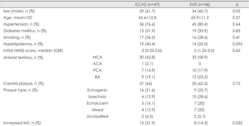
In the present study, increased IMT was more common in Table 1. Clinical characteristics and carotid atherosclerotic status in intracranial large-vessel disease (ICLVD) and small-vessel disease (SVD)
ICLVD (n=47) SVD (n=56) p
Sex (male), n (%) 29 (61.7) 34 (60.7) 0.92
Age, mean±SD 65.6±12.8 63.9±11.2 0.57
Hypertension, n (%) 36 (76.6) 45 (80.4) 0.64
Diabetes mellitus, n (%) 15 (31.9) 19 (33.9) 0.83
Smoking, n (%) 17 (36.2) 16 (28.6) 0.41
Hyperlipidemia, n (%) 19 (40.4) 14 (25.0) 0.095
Initial NIHSS score, median (IQR) 3 (0.25-5.0) 3 (1.25-3.0) 0.63
Arterial territory, n (%) MCA 30 (63.8) 33 (58.9)
ACA 1 (2.1) 0
PCA 7 (14.9) 10 (17.9)
BA 9 (19.1) 13 (23.2)
Carotid plaque, n (%) 31 (66) 35 (62.5) 0.72
Plaque type, n (%) Echogenic 16 (51.6) 9 (25.7)
Isoechoic 4 (12.9) 10 (28.6)
Echolucent 5 (16.1) 7 (20)
Mixed 4 (12.9) 7 (20)
Unclassified 2 (6.5) 2 (5.7)
Increased IMT, n (%) 15 (31.9) 8 (14.3) 0.032
ACA: anterior cerebral artery, BA: basilar artery, IMT: intima-media thickness, IQR: interquartile ratio, MCA: middle cerebral artery, NI- HSS: National Institutes of Health Stroke Scale, PCA: posterior cerebral artery.
Table 2. Comparison of clinical characteristics between patients with and without a carotid plaque
Carotid plaque (-), n=37 Carotid plaque (+), n=66 p
Age (years), mean±SD 59.6±11.3 67.5±11.4 0.002
Sex (male), n (%) 27 (73.0) 36 (54.5) 0.066
Hypertension, n (%) 27 (73.0) 54 (81.8) 0.29
Diabetes mellitus, n (%) 8 (21.6) 26 (39.4) 0.066
Hyperlipidemia, n (%) 10 (27.0) 23 (34.8) 0.41
Smoking, n (%) 15 (40.5) 18 (27.3) 0.17
Initial NIHSS score, median (IQR) 3 (2-5) 2 (1-4) 0.28
Early neurological progression, n (%) 6/34 (17.6) 10/55 (18.2) 0.94 IQR: interquartile ratio, NIHSS: National Institutes of Health Stroke Scale.
patients with ICLVD than in those with SVD. This finding ap- pears to support the atherosclerotic pathogenesis of intracrani- al arterial stenosis. However, there was no difference between ICLVD and SVD regarding the incidence of carotid plaques.
Although IMT and plaques are strongly correlated, they may reflect different biological aspects of atherogenesis.6 A correla- tion between increased IMT and presence of a carotid plaque was not found in a previous related study.7 Moreover, no sig- nificant relationship was found for the contribution of coexist- ing carotid atherosclerosis to early neurological progression in the present study.
There are several limitations to this study. The small sample necessitates prudent interpretation of our data. Furthermore, statin use was not detailed in our results; however, the preva- lence of hyperlipidemia did not differ between SVD and ICLVD. Thus, we do not believe that the results of our descrip- tive study were affected by statin use. Finally, we used the common carotid artery rather than the internal carotid artery for IMT measurement, and the IMT in these two arteries may involve different biological aspects, which may lead to differ- ent results.
Conflicts of Interest
The authors have no financial conflicts of interest.
Acknowledgements
This research was supported by the Basic Science Research Program through the National Research Foundation of Korea (NRF), funded by the Ministry of Education, Science, and Technology (No. 2010-0002537).
REFERENCES
1. Adams HP Jr, Bendixen BH, Kappelle LJ, Biller J, Love BB, Gordon DL, et al. Classification of subtype of acute ischemic stroke. Defini- tions for use in a multicenter clinical trial. TOAST. Trial of Org 10172 in Acute Stroke Treatment. Stroke 1993;24:35-41.
2. Nagai Y, Matsumoto M, Metter EJ. The carotid artery as a noninvasive window for cardiovascular risk in apparently healthy individuals. Ul- trasound Med Biol 2002;28:1231-1238.
3. Touboul PJ, Hennerici MG, Meairs S, Adams H, Amarenco P, Born- stein N, et al. Mannheim carotid intima-media thickness consensus (2004-2006). An update on behalf of the Advisory Board of the 3rd and 4th Watching the Risk Symposium, 13th and 15th European Stroke Conferences, Mannheim, Germany, 2004, and Brussels, Bel- gium, 2006. Cerebrovasc Dis 2007;23:75-80.
4. Cupini LM, Pasqualetti P, Diomedi M, Vernieri F, Silvestrini M, Riz- zato B, et al. Carotid artery intima-media thickness and lacunar versus nonlacunar infarcts. Stroke 2002;33:689-694.
5. Nagai Y, Kitagawa K, Yamagami H, Kondo K, Hougaku H, Hori M, et al. Carotid artery intima-media thickness and plaque score for the risk assessment of stroke subtypes. Ultrasound Med Biol 2002;28:1239- 1243.
6. Johnsen SH, Mathiesen EB. Carotid plaque compared with intima-me- dia thickness as a predictor of coronary and cerebrovascular disease.
Curr Cardiol Rep 2009;11:21-27.
7. Mackinnon AD, Jerrard-Dunne P, Porteous L, Markus HS. Carotid in- tima-media thickness is greater but carotid plaque prevalence is lower in black compared with white subjects. AJNR Am J Neuroradiol 2010;
31:1951-1955.