http://dx.doi.org/10.3988/jcn.2014.10.1.24 J Clin Neurol 2014;10:24-31
Introduction
Centronuclear myopathy (CNM) is a rare congenital disorder of striated muscle. It is characterized pathologically by a high frequency of central nuclei in the muscle fibers.1 Mutations in the gene encoding myotubularinare responsible for the X- linked recessive form, better known as myotubular myopathy (MTM),2 while those of yhe gene encoding dynamin 2 (DNM2) account for 50% of autosomal dominant or sporadic cases of
CNM.3 Some cases with autosomal recessive inheritance are related to mutations in the gene encoding amphiphysin 2 (BIN1).4 Other genetic causes for CNMs with different clini- co-pathological patterns have recently been identified,5 al- though there are still genetically unidentified CNM cases.
X-linked MTM usually presents clinically as severe neona- tal hypotonia and respiratory failure at birth, while CNM of autosomal inheritance is not fatal during the perinatal period and exhibits a predominantly later onset and heterogeneous phenotypes. Typical forms of DNM2-related CNM usually commence in late childhood or early adolescence, and have slowly progressive clinical courses.3 However, severe and neonatal-onset forms with DNM2 mutations and intermediate forms between the two extremes have also been reported,
Clinical and Pathological Features of Korean Patients with DNM2-Related Centronuclear Myopathy
Young-Eun Park,a,b Young-Chul Choi,c Jong-Suk Bae,d Chang-Hoon Lee,b,e Hyang-Suk Kim,f Jin-Hong Shin,a,f Dae-Seong Kima,f
aDepartments of Neurology and ePathology, Pusan National University School of Medicine, Busan, Korea
bMedical Research Institute, Pusan National University Hospital, Busan, Korea
cDepartment of Neurology, Gangnam Severance Hospital, Yonsei University College of Medicine, Seoul, Korea
dDepartment of Neurology, Inje University School of Medicine, Busan, Korea
fResearch Institute for Convergence of Biomedical Research and Technology, Pusan National University Yangsan Hospital, Yangsan, Korea
Received November 14, 2012 Revised July 15, 2013 Accepted July 15, 2013 Correspondence Dae-Seong Kim, MD, PhD Department of Neurology, Pusan National University Yangsan Hospital,
20 Geumo-ro, Mulgeum-eup, Yangsan 626-770, Korea Tel +82-55-360-2450 Fax +82-55-360-2152 E-mail [email protected]
Background and PurposezzCentronuclear myopathy (CNM) is characterized by the presence of central nuclei within a large number of muscle fibers. Mutations of the dynamin 2 gene (DNM2) are common causes of autosomal dominant or sporadic CNM. The aim of this study was to char- acterize the clinical and pathological features of CNM relative to the presence of DNM2 mutations.
MethodszzSix patients with clinical and pathological features of CNM were recruited. Detailed clinical and pathological findings were analyzed according to the presence of DNM2 mutations.
ResultszzWe detected DNM2 mutations in four of the six sporadic CNM patients, and identified the following distinct clinical and pathological features in those patients with DNM2 mutations:
preferential involvement of the distal lower limbs, typical nuclear centralization, and radially dis- tributed sarcoplasmic strands in muscle pathology. In contrast, those without DNM2 mutations exhibited rather diffuse muscular involvement, and nuclear internalization and myofibrillar dis- organization were more pronounced features of their muscle pathology.
ConclusionszzThese findings suggest the presence of specific features in Korean CNM patients.
A detailed clinical and pathological examination of CNM patients would be helpful for molecu- lar genetic analyses of this condition. J Clin Neurol 2014;10:24-31 Key Wordszz centronuclear myopathy, DNM2, muscle involvement, central nuclei,
internal nuclei, sarcoplasmic strands.
Open Access
cc This is an Open Access article distributed under the terms of the Cre- ative Commons Attribution Non-Commercial License (http://creative- commons.org/licenses/by-nc/3.0) which permits unrestricted non-com- mercial use, distribution, and reproduction in any medium, provided the ori- ginal work is properly cited.
broadening the clinical spectrum of CNM.6-8 Recent studies have shown that CNMs associated with mutations of the gene encoding ryanodine receptor 1 or BIN1 exhibit a more severe phenotype than those associated with DNM2 mutations, even to a similar extent to MTM.4,5 They all show common fea- tures of progressive skeletal muscle weakness with varying degrees of facial muscle involvement, ptosis, and external ophthalmoplegia.7 Like other congenital myopathies, the muscle pathology of CNM usually displays type 1 fiber atro- phy/predominance. It may exhibit a radial distribution of sar- coplasmic strands around central nuclei and nonreactive ar- eas for oxidative enzymes.7
In the present study, the DNM2 genotype was sequenced in six patients with sporadic CNM. Mutations were detected in four of these patients. Clinical characteristics that appear to be specific to Korean CNM patients are noted and DNM2-mu- tation-related clinical and pathological features are reported.
The findings of this study will help toward the development of a molecular genetic test for various forms of CNM.
Methods
Patients
Six patients showing clinical features of congenital myopathy with histopathological findings of a high frequency of central nuclei within muscle fibers were recruited for this study. Writ- ten informed consent to participate was obtained from each patient. This study was approved by the Institutional Review Board of Pusan National University Yangsan Hospital.
Mutational analyses of DNM2
Direct sequencing analysis of DNM2 was performed to detect genetic mutations. Genomic DNA was extracted from the pa- tients’ peripheral leukocytes or skeletal muscles. PCR reac- tions were conducted using standard procedures with 20 pairs of primers (available on request) covering 20 exons and the exon-intron boundaries of DNM2. Myotonic discharge on needle EMG was detected in patient 1, leading to Southern blot analysis of the gene encoding dystrophia myotonica pro- tein kinase (DMPK) to exclude the possibility of myotonic dystrophy.
Patient characterization and clinical analysis Clinical information was obtained from the patients regarding the age at CNM onset, affected family members, initial symp- tom, distribution of muscle weakness, current disability, and disease progression. Three of the patients underwent muscle computed tomography (CT), and serum creatine kinase (CK) was measured in all patients.
Muscle pathology
Routine histochemical staining procedures including hema- toxylin and eosin, modified Gomori trichrome, nicotinamide adenine dinucleotide dehydrogenase-tetrazolium reductase (NADH-TR), and adenosine triphosphotase (ATPase) were applied to the biopsied muscle specimens. Muscle specimens were also prepared for electron microscopy by first fixing them with 2% glutaraldehyde in 0.1 M cacodylate buffer. After shaking with a mixture of 4% osmium tetroxide, 1.5% lantha- num nitrate, and 0.2 M s-collidine for 2–3 hours, the samples were embedded in epoxy resin. Semithin (1-µm-thick) sec- tions were cut and then stained with toluidine blue. Ultrathin (50-nm-thick) sections were then cut and stained with uranyl acetate and lead citrate.
Results
Mutational analyses of DNM2
Direct sequencing of DNM2 yielded four kinds of heterozy- gous missense mutations in four of the six patients. The E650K (c.1948G>A) mutation found in patient 1 is located on the GT- Pase effector domain.9 Patient 2 harbored an R522H (c.1565G>
A) mutation in the N-terminal of the Pleckstrin homology (PH) domain.10 Two of the other mutations, R369Q (c.1106G>
A) in patient 3 and E368K (c.1102G>A) in patient 4, are lo- cated in the middle domain. No pathogenic DNM2 mutation was found in patients 5 and 6.
Clinical analyses
The clinical data from six patients are summarized in Table 1.
Three of the patients (patients 1, 3, and 4) experienced their first symptoms during childhood, while the onset in patient 2 was in early adolescence. The other two patients (patients 5 and 6) had motor developmental delay during infancy: patient 5 achieved independent ambulation at the age of 18 months, and patient 6 could stand up around the age of 1 year. None of the patients reported perinatal or antenatal complications.
At the first examination, patients 1, 2, and 4 exhibited distal limb dominant muscle weakness, while patients 5 and 6 ex- hibited proximal dominant limb weakness. In patient 3, mus- cle weakness was evenly distributed between the proximal and distal limbs. Neck flexor weakness was noted in five of the patients. Facial paresis with incomplete ptosis was ob- served in patients 1, 4, and 5, and external ophthalmoplegia was detected in patients 1, 3, 4, and 5. Achilles tendon con- tractures were also common, being present in four of the pa- tients; patient 1 had bilateral Achilles tenorrhaphy at the age of 10 years. None of the patients noted jaw muscle contrac- tures. Patients 3, 4, and 5 exhibited lumbar lordosis, and pa- tient 6 had severe scoliosis requiring a corrective operation.
Respiratory problems were observed in two of the patients.
Patient 5 displayed a mild restrictive pattern on the pulmo- nary function test. Patient 6 began to suffer from dyspnea at the age of 18 years; she was found to have a vital capacity of 32% of normal, and is now dependent on a noninvasive ven- tilator.
Three of the patients underwent muscle CT (patients 1, 4, and 6). In patients 1 and 4, the gastrocnemius and soleus muscles were the most preferentially involved (arrows in Fig.
1B and E), while in patient 1 the biceps femoris and rectus femoris were also involved (arrowheads in Fig. 1A), and in patient 4 the biceps femoris and vasti muscles were also in- volved (arrowheads in Fig. 1D). Patient 6 revealed no prefer- ential involvement of muscles, and exhibited diffuse muscle atrophy in both the anterior and posterior compartments of the lower legs and thigh muscles (Fig. 1G, H, and I). The muscles in the hip girdles were involved in patients 1 and 6, whereas
they were relatively spared in patient 4 (Fig. 1C, F, and I).
Serum CK levels were either normal or mildly elevated (23–454 IU/L) in all patients. Electrophysiological studies revealed normal nerve conduction status and myopathic mo- tor unit potentials in all patients. Myotonic discharge was ob- served in patient 1; genetic testing for DMPK revealed a re- peat sequence of the normal range in this patient.
At a follow-up, patients 4 and 6 are not able to ambulate independently, while the other four patients remain ambulant.
Of these four, patient 6 has been unable to stand up spontane- ously since middle school, and had been using a wheelchair since the age of 20 years.
Muscle pathology
Muscle biopsies from all six patients were reviewed (Figs. 2 and 3). Nuclear misplacement (mostly nuclear centralization) was detected in almost all muscle fibers (99%) from patients Table 1. Clinical data of 4 patients
With DNM2 mutations Without DNM2 mutations
Patient 1 Patient 2 Patient 3 Patient 4 Patient 5 Patient 6
Sex/Age F/35 M/22 F/49 F/34 F/6 F/19
Onset age Childhood Early
adolescence
Childhood Childhood Infant
Developmental delay
Infant Developmental
delay
Family history Not available (-) (-) (-) (-) (-)
Motor power
Neck 4 5 4 3 3 4
Proximal 4+ 5 4 2–3 4 2–3
Distal 4 3–4+ 4 2–4 5 4–5
Facial involvement (+) (-) (-) (+) (+) (-)
Ptosis (+) (-) (-) (+) (+) (-)
Ophthalmoplegia (+) (-) (+) (+) (+) (-)
DTR Absent Absent Preserved Absent Absent Absent
Achilles contracture (+) (+) (-) (-) (+) (+)
Lumbar lordosis (-) (-) (+) (+) (+) Scoliosis
Serum CK (IU/L) 270 454 27 60 23 30
Needle EMG Myopathic MUPs myotonic discharges
Myopathic MUPs
Myopathic MUPs
Myopathic MUPs
Myopathic MUPs
Myopathic MUPs
Muscle CT GC>TA,
post. thigh
ND ND GC>TA,
quadriceps, post. thigh
ND Diffuse atrophy
Current status Ambulant Ambulant Ambulant Wheelchair
bound
Ambulant Respiratory
distress
Wheelchair bound Respiratory
distress DNM2 mutations E650K
(c.1948G>C)
R522H (c.1565G>A)
R369Q (c.1106G>A)
E368K (c.1102G>A)
(-) (-)
CK: creatine kinase, DMN2: dynamin 2 gene, DTR: deep tendon reflex, EMG: electromyography, GC: gastrocnemius, MUP: motor unit potential, NCS: nerve conduction study, ND: not done, TA: tibialis anterior.
1, 2, and 4, and in around 80% of muscle fibers from patient 3 (Fig. 1A and D). Nuclear chains were demonstrated in longi- tudinal sections (Fig. 1B). NADH-TR staining revealed a ra-
dial arrangement of sarcoplasmic strands in patients 1, 3, and 4, and central areas containing nuclei were not reactive for the enzyme (Fig. 2C and E). Some fibers exhibited central ac-
A
D
G
B
E
H
C
F
I
Fig. 1. Muscle CT scans of patients 1, 4, and 6. In patient 1, the rectus femoris and biceps femoris were mildly atro- phied (arrowheads, A), while the poste- rior compartment of the lower legs was the most preferentially involved (arrows, B). The hip girdle muscles in patient 1 were somehow affected (C). In patient 4, muscles in the posterior lower legs were more prominently atrophied (ar- rows, E) and the biceps femoris and vasti muscles were also affected (ar- rowheads, D); however, the hip girdle muscles were relatively spared (F). In patient 6, the muscles in the thighs (G), lower legs (H), and hip girdle (I) were evenly atrophied.
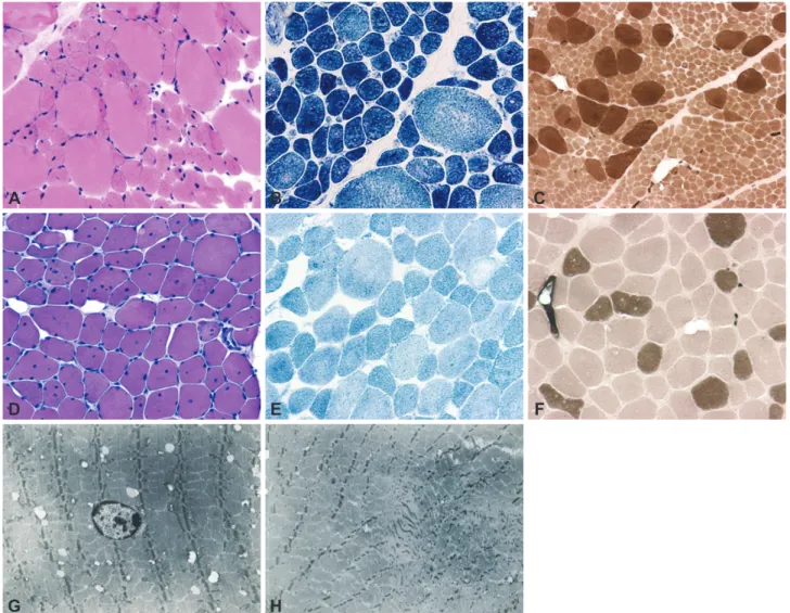
Fig. 2. Muscle pathology in patients 1 and 4. In patient 1, most of the nuclei were placed in the center of the muscle fibers in hematoxylin and eosin (H&E) staining (A), a nuclear chain was highlighted in a longitudinal H&E-stained section (B), and sarcoplasmic strands were ra- dially arranged in nicotinamide adenine dinucleotide dehydrogenase-tetrazolium reductase (NADH-TR) staining (C). In patient 4, most of the nuclei were centrally placed in H&E staining (D), and radial arrangement of the sarcoplasmic strands and nonreactive regions for the enzyme were demonstrated in NADH-TR staining (E). Accumulation of stain and peripheral halos were observed in some fibers (arrows in E). Electron microscopic observation revealed a central nucleus and radial arrangement of strands in patient 4 (F).
A
D
B
E
C
F
cumulation of stains and peripheral halos (arrows in Fig. 2E).
Myofibrils were absent around the nucleus, and radially ar- ranged sarcoplasmic strands around the central nucleus were also found in electron microscopy (Fig. 2F).
Patients 5 and 6 had nuclear misplacement, both central- ization and internalization, in 31.6% and 40.6% of muscle fi- bers, respectively. In patient 5, nuclear centralization and in- ternalization were found in 18.7% (range, 15.7–22.1%) and 25.8% (range, 19.6–32.5%) of muscle fibers, respectively. In patient 6, nuclear centralization and internalization were ob- served in 13.1% (range, 11.1–17.1%) and 17.6% (range, 12.9–
20.5%) of muscle fibers, respectively. Misplaced nuclei were contained mostly in small type 1 fibers in patient 5 (Fig. 3A), and frequently appeared multiply within a single muscle fiber
in patients 5 and 6 (Fig. 3A and D). The areas containing cen- tral nuclei were nonreactive for NADH-TR in some fibers from patient 5 (Fig. 3B), but most of the fibers in patient 6 had no abnormal NADH-TR staining patterns (Fig. 3E). Myofi- brillar ATPase staining revealed that type 1 fibers were pre- dominant and atrophied (Fig. 3C and F), even mimicking the pattern of congenital fiber type disproportion in patient 5 (Fig. 3C). The presence of a central nucleus was not associat- ed with radial arrangement of sarcoplasmic strands (Fig. 3G), but small areas of myofibrillar disorganization were found (Fig. 3H) in patient 6 in electron microscopy.
All of the muscle pathologies were free of dystrophic chang- es. However, the amount of interstitial connective tissue was significantly increased in all patients.
Fig. 3. Muscle pathology in patients 5 and 6. In patient 5, nuclear centralization and internalization were mostly found in the small fibers in H&E staining (A), and nonreactive regions for NADH-TR were observed in some fibers (B). With myofibrillar ATPase stain (pH 10.7), type I fiber atrophy and predominance was noted in patient 5, mimicking the pattern of congenital fiber type disproportion (C). In patient 6, nuclear centralization and internalization were noted, and multiple internal nuclei appeared frequently in H&E staining (D), an abnormal pattern of staining was absent in NADH-TR staining (E), and type 1 fiber predominance was noted in ATPase staining (F; pH 10.7). Radial arrange- ment of sarcoplasmic strands was not observed around a central nucleus (G), but small areas of myofibrillar disorganization were detected (H) in muscle biopsy tissue from patient 6 in electron microscopy. NADH-TR: nicotinamide adenine dinucleotide dehydrogenase- tetrazoli- um reductase, ATPase: adenosine triphosphotase.
A
D
G
B
E
H
C
F
Discussion
This study recruited six patients with clinical features of con- genital myopathy and high frequency of central nuclei within muscle fibers as a main pathological feature. Gene sequenc- ing revealed that four of these patients harbored four differ- ent types of DNM2 mutations. This is consistent with previ- ous results showing that DNM2 is responsible for more than 50% of autosomal dominant or sporadic cases of CNM.11 The mutations of this study were scattered over three different do- mains, as described previously for CNM cases of mild phe- notypes, although the E368K mutation has occasionally been reported in association with infantile or neonatal onset. Two of the mutations in the middle domain, E368K and R369Q, are among the most common DNM2 mutations associated with CNM.11 An initial study about DNM2 mutations found that a mild CNM phenotype was associated with mutations in the middle domain.3 However, many mutations in the other domains have also been described in patients with mild CNM, indicating that there is no correlation between phenotypes and specific domains.10 DNM2 is also responsible for autosomal dominant Charcot-Marie-Tooth disease (CMT), and several mutations in the PH domain have been described in that dis- ease. Some DNM2-related CNM cases exhibit slowed motor conduction velocities on nerve conduction studies, suggesting an overlap between these two diseases. However, none of the patients in the present study with DNM2 mutations had marked nerve conduction abnormalities, which suggests that periph- eral nerve involvement is not consistently present in DNM2- related CNM patients, even when mutations are present in the PH domain. Supporting this notion, one recent study showed that different functional defects caused by each of the CNM- and CMT-related DNM2 mutations are mediated in the de- velopment of the diseases.12
The clinical presentation differed between patients with and without DNM2 mutations in this study. Those with DNM2 mutations suffered from weakness in the lower legs, whereas those without mutations complained of proximal lower limb weakness at the first examination. This pattern was clearly re- produced on muscle CT scans. In patients 1 and 4, the poste- rior lower leg muscles were the most prominently atrophied.
However, no preferential muscular involvement was observed in patient 6. This finding is compatible with a previous report of the posterior compartment of the distal lower legs being the most prominent muscular involvement in DNM2-related CNM.13 However, the thigh and pelvic muscle involvements in DNM2-related CNM appear to vary between studies. The present study demonstrated additional involvement of the bi- ceps femoris and rectus femoris muscles in patient 1 and the biceps femoris and vasti muscles in patient 4; in contrast, these
muscles were comparatively preserved in a previous study.14 Recognizing these patterns of muscle involvement will help to differentiate between congenital myopathies of different genetic causes, with increasing use of muscle imaging as an initial evaluation tool.
Respiration difficulty occurred only in the patients without DNM2 mutations, who already presented with respiratory re- striction at the first examination, and ultimately required re- spiratory rehabilitation or ventilator support during follow-up.
At the last follow-up, none of the patients with DNM2 muta- tions complained of respiratory problems. In fact, many of the published reports on CNM have focused on respiratory distress in DNM2-related CNM patients. Recent results of an Italian and German CNM cohort show that all patients with DNM2 mutations had a restrictive ventilatory defect with varying degrees of severity.15,16 However, in the present study, respiratory restriction was observed exclusively in patients without DNM2 mutations; this may therefore be a Korean- specific CNM characteristic.
Patients with DNM2 mutations experienced onset on child- hood or early adolescence, while in those without DNM2 mu- tations the disease began during infancy with motor develop- mental delay. However, the present results cannot show categorically that the clinical courses of CNM are determined by the presence of DNM2 mutations, since clinical courses vary even among patients with DNM2-related CNM. Neonatal onset and severe courses of CNM were reported with DNM2 mutations in the C-terminal of the PH domain.14
Regardless of DNM2 mutations, all six CNM patients pre- sented slowly progressive limb weakness with or without fa- cial involvement and ophthalmoplegia. Facial paresis and ptosis (3/6, 50%), external ophthalmoplegia (4/6, 66.7%), and neck flexor weakness (5/6, 83.3%) were common, and thus might be dominant features in CNM. It is especially notable that patient 2 harbored the R522H mutation and did not pres- ent with external ophthalmoplegia, in agreement with several other reports of CNM patients with R522H not displaying ophthalmoplegia.10 Joint contractures and scoliosis/lumbar lordosis were also common from earlier stages of the disease.
Accordingly, appropriate physical or surgical corrections can improve motor status and help prevent further complications in CNM patients. However, jaw muscle contractures were to- tally absent in the present series, although according to other reports it appears to be both common and pronounced, in particular with the E368K mutation.15
It is notable that the presence of DNM2 mutations was as- sociated with distinct pathological findings. Frequent central nuclei and type 1 fiber atrophy/predominance were common in all patients. However, in DNM2-related cases, 1) nuclear centralization was the most frequent characteristic (80–99%)
and 2) sarcoplasmic strands were radially distributed around the central nuclei. By contrast, in patients without DNM2 mu- tations, 1) both nuclear internalization and nuclear centraliza- tion were marked, 2) misplaced nuclei were less frequently observed (31.6–40%), and 3) sarcoplasmic strands were not radially arranged. Electron microscopic observation revealed small areas of myofibrillar disorganization as an additional finding in the absence of DNM2 mutations. However, reac- tivity for oxidative enzymes in the center of the muscle fibers was variable, regardless of the presence of DNM2 mutations.
These findings are consistent with previous reports on the pathological features of CNM. A recent study of CNM found that its pathological manifestation vary with the genetic causes.17 Variable pathological features, such as nuclear inter- nalization, necklace fibers,18 and myofibrillar disorganiza- tion,5 suggest other genetic causes of CNM, while radial sar- coplasmic strands are always typical of DNM2-related cases.
Another Japanese report also underlined homogeneous path- ological features in DNM2-related CNM.19 Considering our results and those of previous studies, exclusive centralization of the nuclei and radial arrangement of sarcoplasmic strands may be consistent pathologic markers of DNM2 mutations in CNM patients.
In conclusion, to our knowledge this is the first reported study of Korean CNM patients, and it has shown that DNM2 mutations may be one of the more frequent genetic causes of sporadic CNM in Korea. Clinical presentations revealed the presence of several characteristics in Korean CNM patients, and demonstrated some differences between patients with and without DNM2 mutations. Muscle imaging findings sup- port the notion that specific muscular involvement of distal lower limbs may help distinguish DNM2-related CNM from other genetic causes. It is notable that respiratory restriction was not observed in the present cohort of DNM2-related CNM patients, and that jaw contractures appear to be uncommon in Korean CNM patients regardless of the presence of DNM2 mutations. The pathological features in patients with DNM2 mutations described herein appear to be consistent with the findings of previous studies: nuclear centralization is more marked than in those without DNM2 mutations, and the sar- coplasmic strands are radially arranged. Furthermore, it was found that other pathological features, such as myofibrillar disorganization, can be present in the absence of DNM2 mu- tations. Although genetic causes can be evaluated in patients without DNM2 mutations, the findings of this study imply that careful examination for clinical and pathological findings can guide molecular genetics analyses of CNM.
Conflicts of Interest
The authors have no financial conflicts of interest.
Acknowledgements
This work was supported by the Bio-Scientific Research Grant funded by the Pusan National University (PNU, Bio-Scientific Research Grant) (PNU-2010-101-255).
REFERENCES
1. Spiro AJ, Shy GM, Gonatas NK. Myotubular myopathy. Persistence of fetal muscle in an adolescent boy. Arch Neurol 1966;14:1-14.
2. Laporte J, Hu LJ, Kretz C, Mandel JL, Kioschis P, Coy JF, et al. A gene mutated in X-linked myotubular myopathy defines a new puta- tive tyrosine phosphatase family conserved in yeast. Nat Genet 1996;
13:175-182.
3. Bitoun M, Maugenre S, Jeannet PY, Lacène E, Ferrer X, Laforêt P, et al. Mutations in dynamin 2 cause dominant centronuclear myopathy.
Nat Genet 2005;37:1207-1209.
4. Nicot AS, Toussaint A, Tosch V, Kretz C, Wallgren-Pettersson C, Iwarsson E, et al. Mutations in amphiphysin 2 (BIN1) disrupt interac- tion with dynamin 2 and cause autosomal recessive centronuclear my- opathy. Nat Genet 2007;39:1134-1139.
5. Wilmshurst JM, Lillis S, Zhou H, Pillay K, Henderson H, Kress W, et al. RYR1 mutations are a common cause of congenital myopathies with central nuclei. Ann Neurol 2010;68:717-726.
6. Züchner S, Noureddine M, Kennerson M, Verhoeven K, Claeys K, De Jonghe P, et al. Mutations in the pleckstrin homology domain of dynamin 2 cause dominant intermediate Charcot-Marie-Tooth dis- ease. Nat Genet 2005;37:289-294.
7. Jeannet PY, Bassez G, Eymard B, Laforêt P, Urtizberea JA, Rouche A, et al. Clinical and histologic findings in autosomal centronuclear my- opathy. Neurology 2004;62:1484-1490.
8. Melberg A, Kretz C, Kalimo H, Wallgren-Pettersson C, Toussaint A, Böhm J, et al. Adult course in dynamin 2 dominant centronuclear myopathy with neonatal onset. Neuromuscul Disord 2010;20:53-56.
9. Bitoun M, Durieux AC, Prudhon B, Bevilacqua JA, Herledan A, Sakanyan V, et al. Dynamin 2 mutations associated with human dis- eases impair clathrin-mediated receptor endocytosis. Hum Mutat 2009;
30:1419-1427.
10. Susman RD, Quijano-Roy S, Yang N, Webster R, Clarke NF, Dowl- ing J, et al. Expanding the clinical, pathological and MRI phenotype of DNM2-related centronuclear myopathy. Neuromuscul Disord 2010;
20:229-237.
11. Böhm J, Biancalana V, Dechene ET, Bitoun M, Pierson CR, Schaefer E, et al. Mutation spectrum in the large GTPase dynamin 2, and geno- type-phenotype correlation in autosomal dominant centronuclear my- opathy. Hum Mutat 2012;33:949-959.
12. Koutsopoulos OS, Koch C, Tosch V, Böhm J, North KN, Laporte J.
Mild functional differences of dynamin 2 mutations associated to cen- tronuclear myopathy and Charcot-Marie Tooth peripheral neuropathy.
PLoS One 2011;6:e27498.
13. Fischer D, Herasse M, Bitoun M, Barragán-Campos HM, Chiras J, Laforêt P, et al. Characterization of the muscle involvement in dyna- min 2-related centronuclear myopathy. Brain 2006;129:1463-1469.
14. Catteruccia M, Fattori F, Codemo V, Ruggiero L, Maggi L, Tasca G, et al. Centronuclear myopathy related to dynamin 2 mutations: clini- cal, morphological, muscle imaging and genetic features of an Italian cohort. Neuromuscul Disord 2013;23:229-238.
15. Bitoun M, Bevilacqua JA, Prudhon B, Maugenre S, Taratuto AL, Monges S, et al. Dynamin 2 mutations cause sporadic centronuclear myopathy with neonatal onset. Ann Neurol 2007;62:666-670.
16. Hanisch F, Müller T, Dietz A, Bitoun M, Kress W, Weis J, et al. Phe- notype variability and histopathological findings in centronuclear myopathy due to DNM2 mutations. J Neurol 2011;258:1085-1090.
17. Romero NB. Centronuclear myopathies: a widening concept. Neuro- muscul Disord 2010;20:223-228.
18. Bevilacqua JA, Bitoun M, Biancalana V, Oldfors A, Stoltenburg G, Claeys KG, et al. “Necklace” fibers, a new histological marker of late- onset MTM1-related centronuclear myopathy. Acta Neuropathol 2009;
117:283-291.
19. Mori-Yoshimura M, Okuma A, Oya Y, Fujimura-Kiyono C, Nakajima H, Matsuura K, et al. Clinicopathological features of centronuclear myopathy in Japanese populations harboring mutations in dynamin 2.
Clin Neurol Neurosurg 2012;114:678-683.