ISSN 2288-8101(Print) ISSN 2288-8586(Online)
Case Report
RECEIVED September 13, 2013, REVISED February 17, 2014, ACCEPTED March 14, 2014 Correspondence to Sang-Hwa Lee
Department of Oral and Maxillofacial Surgery, The Catholic University of Korea, Yeouido St. Mary’s Hospital 10 63-ro, Yeongdeungpo-gu, Seoul 150-713, Korea
Tel: 82-2-3779-2148, Fax: 82-2-769-1689, E-mail: [email protected]
Copyright © 2014 by The Korean Association of Maxillofacial Plastic and Reconstructive Surgeons. All rights reserved.
CC
This is an open access article distributed under the terms of the Creative Commons Attribution Non-Commercial License (http://creativecommons.org/licenses/
by-nc/3.0) which permits unrestricted non-commercial use, distribution, and reproduction in any medium, provided the original work is properly cited.
Cervical Necrotizing Fasciitis Caused by Dental Infection
Chi-Woong Song, Hyun-Joong Yoon, Da-Woon Jung, Sang-Hwa Lee
Department of Oral and Maxillofacial Surgery, The Catholic University of Korea, Yeouido St. Mary’s Hospital
Abstract
Necrotizing fasciitis (NF) is defined as rapidly progressive necrosis of subcutaneous fat and fascia. Although NF of the face is rare, its mortality rate is nearly 30%. It usually originates from dental infection and can lead to involvement of the neck, mediastinum, and chest wall. Complications resulting from pre-existing systemic diseases can increase the mortality rate. Known complication factors for NF include diabetes, malnutrition, advanced age, peripheral vascular disease, renal failure, and obesity.
Here, we report a case of NF originating from dental infection in an 88-year-old woman already diagnosed with hypertension, thoracic aortic aneurysm, and renal diseases. Such conditions limited adequate surgical and antibiotic treatment. However, interdisciplinary treatment involving multiple departments was implemented with good results.
Key words: Necrotizing fasciitis, Thoracic aortic aneurysm
Introduction
Necrotizing fasciitis (NF) is defined as rapidly pro- gressive necrosis of subcutaneous fat and fascia. It is a life-threatening infection characterized by progressive, rap- id necrosis of subcutaneous tissues and fascial planes. NF of the face is rare and usually originates from dental in- fection, and the mortality rate is nearly 30%[1]. The proba- bility of death among patients with NF due to infectious causes is 14%, and common causes of death are usually complications related to underlying systemic diseases. Such complications arise due to cardiopulmonary diseases, dia- betes mellitus (DM), cancer, hepatic failure, renal disease, hypertension, Methicillin-Resistant Staphylococcus aureus
(MRSA), and age[2,3]. It is very important to check patients’
medical history when treating oral and maxillofacial infections. Elderly patients are more at risk in terms of underlying systemic diseases and are more likely to devel- op complications. The major complication factor when treating infections is DM. In the case of DM, patients’ meta- bolic regulation can be altered by surgery, and delayed wound healing can increase the risk of infection. Further, the immune systems of older DM patients usually become weak with advanced age. A weak immune system can increase the risk of spreading infections, thus leading to complications[2,3]. Another complication factor is renal failure, as antibiotics are excreted by the kidneys.
Nephrotoxins are present in aminoglycosides and anti-
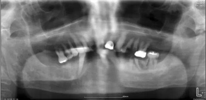
Fig. 1. Dental panoramic view.
Table 1. Results of laboratory investigation
Investigation Result Normal range
Hemoglobin (g/dL) White cell count (×10
9/L) Seg-neutrophils (%) Lymphocytes (%) Platelet count (×10
9/L) Blood urea nitrogen (mg/dL) Creatinine (mg/dL)
C-reactive protein (mg/L)
10.00 19.91 93.90 3.70 143.00 44.80 2.07 360.36
12.0∼16.0 5.0∼10.0 55.0∼75.0 20.0∼44.0 150.0∼450.0 8.0∼23.0 0.5∼1.2 0.1∼5.0
resulting in patient death[5]. Aneurysm of the thoracic aorta causes local enlargement of the thoracic aorta, resulting in ruptures as the disease progresses. Enlargement of the aorta has been shown to be related to higher risk of rup- ture[6]. It is also known that intramural hematomas are caused by ruptures in the Vasa vasorum. These occur natu- rally in elderly patients or in patients with hypertension for the most part[7]. We successfully treated an 88-year-old female patient presenting with several underlying diseases, including aortic aneurysms, vascular wall hemorrhages, and hypertension, who had developed NF as well as my- onecrosis on the left side of the face due to a dental infection. We successfully administered multidisciplinary treatments combining the departments of thoracic surgery, infection, and nephrology. We report this case along with a review of the literature.
Case Report
An 88-year-old female patient presenting with swelling on the left side of the face for several months was referred to the Department of Oral and Maxillofacial Surgery, Yeouido St. Mary’s Hospital. This patient had visited a local dental office but was not able to receive treatment due to the occurrence of systemic diseases. Swelling on the left side of the face had been present since March 30th, 2012. The patient had applied honey to the affected
The patient presented with edema and aches on the left side of the face and cervical region, along with com- plaints of dyspnea, dysphagia, and trismus. Oral examina- tion showed multiple caries, periodontal disease, and gin- gival edema of the left mandible (Fig. 1). Complete blood cell counts, vital check-up, and computed tomography (CT) were carried out. Vital check showed a blood pressure (BP) of 108/54 mmHg, which was controlled since the patient had been taking antihypertension medication. Pulse was 78 times/min. Laboratory investigation revealed in- creased white blood cell counts, C-reactive protein, seg-neutrophils, urea nitrogen, and creatinine levels as well as decreased hemoglobin and lymphocyte levels (Table 1).
Neck CT (enhance) showed abscesses in the left buccal space along with air bubbles in left buccal region’s fascial plane. Masseter and pterygoid muscles in the adjacent left masticator space also showed swelling and necrotizing myositis. Left buccal and masticator spaces contained dif- fuse fat infiltration and cellulitis, whereas the left sub- mandibular gland showed swelling due to increased inflammation. Posterior neck area showed cellulitis, but the airway seemed clean (Fig. 2). The patient was diag- nosed with abscesses, NF, and myositis in the left buccal and pterygomandibular spaces based on physical examina- tion, laboratory findings, and radiological imaging.
2. Treatment
Triple-antibiotic regimens are usually administered for
Table 2. Bacterial culture and identification (pus)
Staphylococcus aureus Identification
Ciprofloxacin Clindamycin Erythromycin Gentamycin Penicillin Vancomycin
Sensitive Resistance Resistance Sensitive Resistance Sensitive
Fig. 2. Necrotizing fasciitis on left