Vol.23 No.1 p90-95, June 2006
INTRODUCTION
Desmoplastic small round cell tumor (DSRCT) is composed of small round tumor cells of uncertain histogenesis, associated with prominent stromal desmoplasia and polyphenotypic differentiation.
1)It was first described by Geral and Rosai
2)in 1989, and has most commonly been reported in children and young adults, with a male-to-female ratio of 4:1. The location for this tumor has primarily
been in the abdominal cavity. It mostly occurs in the abdominal cavity. Other primary sites have been rarely reported, have included the paratesticular region,
3)the pleural serosa,
4)the posterior cranial fossa,
5)soft tissue and bone,
6)ovary,
7)and kidney.
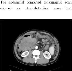
8)We report a case of desmoplastic small round cell tumor that occurred in the abdominal cavity of a 50-year-old man, and review the medical literature.
책임저자:최준혁, 대구광역시 남구 대명동 317-1, 영남대학교 의과대학 병리학교실 Tel: (053) 620-3335, FAX: (053) 656-1429, E-mail: [email protected]