184
ing of bronchovascular bundles, interlobular septa, round- shaped ground-glass opacity (GGO), honeycombing, and bronchiectasis in chest computed tomography (CT), and it is difficult to discern that it comes from other lung diseases
4.
In this paper, we report a case that a patient who came to hospital due to hemoptysis and had lung mass in chest CT was diagnosed IgG4-related disease by biopsy.
Case Report
A 63-year-old man visited the pulmonary clinic complain- ing hemoptysis which has lasted for 5 days. His past medical history were pulmonary tuberculosis (completely healed 20 years ago), diabetes mellitus, hypertension, and chronic kidney disease. He did not smoke and drink. There was no specific family history. Lung sound was clear and heart sound was normal without murmur. There was no palpable mass or lymph node in the neck. Blood pressure was 130/70 mm Hg, body temperature was 36.8
oC, pulse rate was 92 beats per minute, and respiratory rate was 20 breaths per minute. Labo- ratory findings revealed hemoglobin 8.4 g/dL, white blood cell 4,530/mm
3(neutrophils, 64.3%; lymphocytes, 21.7%;
eosinophils, 4.5%), platelets 234,000/mm
3, C-reactive protein 1.10 mg/dL, aspartate aminotransferase 16 IU/L, alanine ami- notransferase 9 IU/L, total bilirubin 0.26 IU/L, creatinine 2.72 mg/dL, blood urea nitrogen 33 mg/dL, and hemoglobin A1c 10.4%. In chest X-ray a simple mass was noted at left upper
Introduction
IgG4-related disease is newly recognized fibroinflamma- tory condition characterized by edema of invaded organ, a dense lymphoplasmacytic infiltration rich in IgG4-positive plasma cells, storiform pattern of fibrosis, and, often but not always, elevated serum IgG4 concentration level
1,2. The most commonly involved organs are pancreas and salivary gland. It may also intrude thyroid, retroperitoneum, kidney, lung, and lymph node, but the invasion of lung is rare
1-3. IgG4-related lung disease may be asymptomatic or has symptoms such as cough, hemoptysis, dyspnea, and chest pain
1-3. IgG4-related lung disease could appear as solitary nodular lesion, thicken-
IgG4-Related Lung Disease without Elevation of Serum IgG4 Level: A Case Report
Min Kyu Kang, M.D., Yongseon Cho, M.D., Minsoo Han, M.D., Sun Young Jung, M.D., Kyoung Min Moon, M.D., Jinyoung Kim, M.D., Ju Ri Kim, M.D., Dong-kyu Lee, M.D., Jun Hyung Park, M.D. and So Hee Chung, M.D.
Division of Pulmonary Medicine, Department of Internal Medicine, Eulji University Hospital, Daejeon, Korea
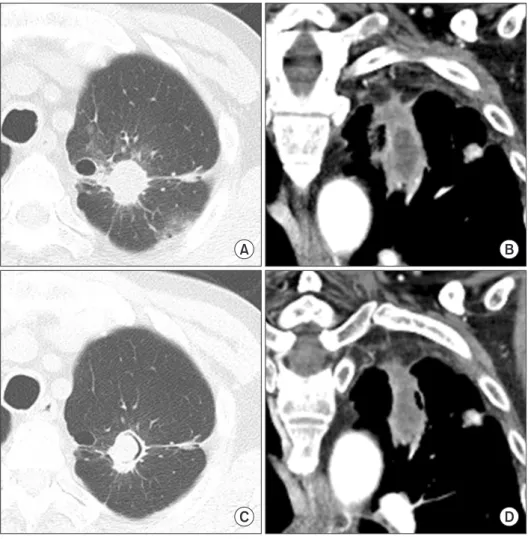
Since IgG4-related pancreatitis was first reported in 2001, IgG4-related disease has been identified in other organs such as salivary gland, gallbladder, thyroid, retroperitoneum and kidney; but lung invasion is rare. A 63-year-old man presented with hemoptysis at the pulmonary clinic and chest computed tomography revealed about 4.1 cm irregular shaped mass with spiculated margin at the left upper lobe. Despite no elevation of serum IgG4 level, he was finally diagnosed as IgG4- related lung disease by transthoracic needle biopsy. After treatment with oral glucocorticoids, hemoptysis disappeared and the size of lung mass was decreased.
Keywords: Immunoglobulins; Lung Diseases; Hemoptysis
Copyright © 2016
The Korean Academy of Tuberculosis and Respiratory Diseases.
All rights reserved.
Address for correspondence: Yongseon Cho, M.D.
Division of Pulmonary Medicine, Department of Internal Medicine, Eulji University Hospital, 95 Dunsanseo-ro, Seo-gu, Daejeon 35233, Korea Phone: 82-42-611-3154, Fax: 82-42-259-1162
E-mail: [email protected] Received: Aug. 10, 2015 Revised: Sep. 15, 2015 Accepted: Sep. 24, 2015
cc
It is identical to the Creative Commons Attribution Non-Commercial License (http://creativecommons.org/licenses/by-nc/4.0/).
CASE REPORT
http://dx.doi.org/10.4046/trd.2016.79.3.184ISSN: 1738-3536(Print)/2005-6184(Online) • Tuberc Respir Dis 2016;79:184-187