Comparative Gene-Expression Analysis of Periodontal Ligament and Dental Pulp in the Human Permanent Teeth
Suk Woo Lee1, Mijeong Jeon1, Hyo-Seol Lee2, Je Seon Song1, Heung-Kyu Son1, Hyung-Jun Choi1, Han-Sung Jung3, Seok-Jun Moon3, Wonse Park4, Seong-Oh Kim1
1
Department of Pediatric Dentistry, College of Dentistry, Yonsei University
2
Department of Pediatric Dentistry, College of Dentistry, Kyunghee University
3
Department of Oral Biology, College of Dentistry, Yonsei University
4
Department of General Dentistry, College of Dentistry, Yonsei University
There is no genetic activity information with the functions of dental pulp and periodontal ligament in human.
The purpose of this study was to identify the gene-expression profiles of, and the molecular biological differences between periodontal ligament and dental pulp obtained from human permanent teeth. cDNA microarray analysis identified 347 genes with a fourfold or greater difference in expression level between the two tissue types 83 and 264, of which were more plentiful in periodontal ligament and dental pulp, respectively. Periodontal ligament exhibited strong expression of genes related to collagen synthesis (FAP), collagen degradation (MMP3, MMP9, and MMP13), and bone development and remodeling (SSP1, BMP3, ACP5, CTSK, and PTHLH). Pulp exhibited strong expression of genes associated with calcium ions (CALB1, SCIN, and CDH12) and the mineralization and formation of enamel and dentin (SPARC/SPOCK3, PHEX, AMBN, and DSPP). Among these genes, SPP1, SPARC/SPOCK3, AMBN, and DSPP were well known in dental research. However, the other genes are the newly found and it may help to find a good source of regenerative therapy if further study is performed.
Key words :
Gene-Expression, Periodontal Ligament, Dental Pulp, Human Permanent Teeth, Korean Abstract
Ⅰ. Introduction
Because of their significance in dental health, dental pulp and PDL have received considerable attention in the field of dental bioengineering
1-3). One of the actively used methods of analysis in this regard is cDNA mi- croarray, since it enables analysis of the expression of thousands of genes and proteins simultaneously and the comparison of the gene-expression profiles of two differ-
ent samples. In case of PDL, Song et al.
4)performed a comparative gene-expression analysis between the PDL of permanent and deciduous teeth. Xie et al.
5)and Lee et al.
6)compared the microRNA profiles of human periodon- tal diseased and healthy gingival tissues. Han and Amar
7)examined the fibroblasts of PDL and gingiva, and de Araujo et al.
8)used cDNA microarray to study PDL cells under mechanical stress. In addition, Paakkonen et al.
9)and McLachlan et al.
10)analyzed the gene-expres-
Corresponding author : Seong-Oh Kim
Department of Pediatric Dentistry, College of Dentistry, Yonsei University, 50-1, Yonsei-ro, Seodaemun-gu, Seoul, 03722, Korea Tel: +82-2-2228-3171 / Fax: +82-2-392-7420 / E-mail: [email protected]
Received May 21, 2015 / Revised November 9, 2015 / Accepted October 27, 2015
※This research was supported by the Basic Science Research Program of the National Research Foundation of Korea (NRF), funded by the Ministry of Education, Science, and Technology (grant nos. 2011-0022160 and 2012R1A1A2041910).
sion patterns of healthy and carious pulp, and Paakkonen et al.
11)further investigated the differences between odontoblasts and dental pulp. Other studies have compared pairs of related dental tissues, such as the ameloblastoma and tooth germ
12), and the PDL and tooth follicle
13). According to these researches, the candi- date genes in specific function or pathologic change have been revealed.
The main functions of the PDL are tooth support, re- generation of periodontal tissues, maintenance of home- ostasis, and provision of the healing process required fol- lowing periodontal disease or mechanical trauma
14). In terms of mechanical trauma, ankylosed teeth cannot be moved by orthodontic force if the PDL is absent
15). The mechanical stress loaded onto a tooth is transferred to the PDL, the cells of which respond to that mechanical stress, regulating the absorption and formation of the bone matrix by signaling the surrounding cells
16). The main functions of dental pulp are the formation of ter- tiary dentin, nerve innervation, and nutrition supply
17). Dental pulp cells also have the ability to differentiate in- to odontoblasts in order to repair dental damage caused by trauma, caries, or dental erosion
18).
However, it is unknown about that the differences are made by which gene expression in spite of the functional differences of PDL and pulp. Each tissue has the specific gene expression pattern, so analysis of gene expression pattern of tissue enables to understanding the function of the tissue clearly. But no direct comparison of gene- expression profiles has yet been made between the healthy normal PDL and dental pulp tissues of human permanent teeth. And, most studies were conducted by gene expression analysis using cultured cells of PDL and dental pulp. Therefore, the purpose of this study is to identify and compare the gene-expression profiles and molecular biological differences between PDL and dental pulp tissues from human normally functioning teeth.
Ⅱ. Materials and Methods 1. Sample collection
The experimental protocol was approved by the Institutional Review Board of the Yonsei University Dental Hospital (approval #2-2011-0050). Written in- formed consent to participate was obtained from all par- ticipants and from their next of kin, caregivers, or guardians on behalf of minors/children. The PDL sam-
ples (n=9; aged 11-19 years) and the pulp samples (n=9; aged 11-19 years) examined in this study were obtained from healthy, mature premolars extracted for orthodontic reasons from healthy persons. The extracted teeth were immediately frozen and stored in liquid nitro- gen. Tissues were obtained carefully from the middle third of the PDL using sterile curettes. The teeth were subsequently crushed with a bolt cutter and the pulp was carefully obtained using sterile tweezers. The PDL and pulp were then immediately submerged in RNA-sta- bilizing reagent (RNAlater, Qiagen, CA, USA).
2. RNA isolation
Total RNA was extracted from the PDL and pulp sam- ples using the RNeasy Fibrous Minikit (Qiagen) accord- ing to the manufacturer’ s instructions. Prior to RNA ex- traction, the tissues were homogenized using a Bullet Blender Bead (Next Advanced, NY, USA). The extract- ed RNA was eluted in 25 μl of sterile water. RNA con- centrations were determined from absorbance values ob- tained at a wavelength of 260 nm using a spectropho- tometer (NanoDrop ND-1000, Thermo Scientific, IL, USA). The RNA samples analyzed in this study had 260/280 nm ratios of ≥1.8.
3. cDNA microarray analysis
The microarray procedure was conducted in triplicate
for each sample, and using three samples from each
group. Global gene expression analyses were conducted
using Affymetrix GeneChip
�Human Gene 1.0 ST
oligonucleotide arrays (Affymetrix Inc., CA, USA). As
recommended by the munafacturer`s protocol, 300 ng
were used. Briefly, 300 ng of total RNA from each sam-
ple was converted to double-strand cDNA, as described
previously
4). Fragmented end-labeled cDNA was hy-
bridized to the GeneChip Human Gene 1.0 ST arrays for
16 hours at 45℃ and 60 rpm, as described in the
GeneChip Whole Transcript Sense Target Labeling
Assay Manual (Affymetrix). After hybridization, the
chips were stained and washed in a GeneChip Fluidics
Station 450 (Affymetrix), and then scanned using
Affymetrix Command Console software (version 1.1,
Affymetrix). The raw file generated through this proce-
dure provides expression intensity data, which were
used for the next step.
4. Gene ontology analysis
Expression data were generated by Affymetrix Expression Console software version 1.1 (Affymetrix).
The robust multichip average algorithm implemented in Affymetrix Expression Console software was used to nor- malize the data. One-way ANOVA was applied to the RMA expression values to determine whether genes were differentially expressed between the three groups.
A multiple testing correction was applied to the p values of the F statistics to adjust for the false discovery rate.
Genes with adjusted F-statistic p values of < 0.05 were extracted. Strongly expressed genes in PDL or pulp that exhibited differences of over 6- or 11-fold relative to the signal value of the control and each test group were se- lected for the further study. In order to classify the coex- pression gene group, for which the expression pattern was similar, hierarchical clustering and k-means cluster- ing were performed using MultiExperiment Viewer soft- ware, version 4.4 (www.tm4.org; Dana-Farber Cancer Institute, MA, USA). The web-based tool Database for Annotation, Visualization, and Integrated Discovery (DAVID) was used to biologically interpret the differen- tially expressed genes (http://david.abcc.ncifcrf.gov/
home.jsp). These genes were then classified based on in- formation on gene function from the gene ontology, Kyoto Encyclopedia of Genes and Genomes (KEGG) Pathway database (heep://david.abcc.ncifcrf.gov/home.
jsp).
This microarray data set was approved by the Gene Expression Omnibus (http://www.ncbi.nlm.gov/geo/);
its GEO accession number is GSE50639.
5. Quantitative polymerase chain reaction
The single-stranded cDNA required for the polymerase chain reaction (PCR) analysis was synthesized using 500 ng of extracted total RNA as a template for reverse transcription (Superscript III Reverse Transcriptase and random primer, Invitrogen, UK), as described previously
4).
The following TaqMan gene-expression assay primers were used (Applied Biosystems, CA, USA): ameloblastin (AMBN), calbindin 1 (CALB1), collagen, type XII, alpha (COL12A), dentin sialophosphoprotein (DSPP), matrix metallopeptidase (MMP)9, MMP20, secreted protein acidic and rich in cysteine (SPARC)/osteonectin 3 (SPOCK3), secreted phosphoprotein 1 (SPP1), and 18S rRNA. The results are plotted versus time, which was
quantified as the cycle number. A precise quantification of the initial target was obtained by examining the am- plification plots during the early log phase of product ac- cumulation above background [the threshold cycle (Ct) number]. Ct values were subsequently used to deter- mine ΔCt values (ΔCt=Ct of the gene minus Ct of the 18S rRNA control), and differences in Ct values were used to quantify the relative amount of PCR product, expressed as the relative change by applying the equa- tion 2
-ΔCt.
6. Immunohistochemical staining
In preparation for immunohistochemical (IHC) stain- ing, permanent teeth were fixed in 10% buffered forma- lin (Sigma, MO, USA) for 1 day, decalcified with 10%
EDTA (pH 7.4; Fisher Scientific, TX, USA) for 8 weeks, embedded in paraffin, and sectioned at a thickness of 3
㎛. Specimens were subjected to IHC staining with anti- human CALB1 (rabbit polyclonal, diluted 1:400;
Ab25085, Abcam, Cambrige, UK), MMP9 (rabbit poly- clonal, diluted 1:800; Ab38898, Abcam), SPP1 (osteo- pontin, rabbit polyclonal, diluted 1:800; Ab8448, Abcam), and COL12A1 (rabbit polyclonal, diluted 1:800; Sc68862, Santa Cruz Biotechnology, Inc., CA, USA). Endogenous peroxidase activity was quenched by the addition of 3% hydrogen peroxide. Sections were in- cubated in 5% bovine serum albumin to block nonspecif- ic binding. The primary antibodies were diluted to give optimal staining and the sections were incubated overnight. After incubation, EnVision+ System-HRP la- beled Polymer Anti-rabbit (K4003, Dako North America Inc., CA, USA; ready to use) was applied for 20 min.
Color development was performed using labeled strepta- vidin biotin kits (Dako) according to the manufacturer`s instructions. The sections were counterstained with Gill`s hematoxylin (Sigma). Control sections were treat- ed in the same manner but without treatment with pri- mary antibodies.
Ⅲ. Results
1. Gene-expression profiles of PDL and dental pulp
The total data distribution and frequency were con- firmed using density and box plots and M-A plots of the standardized log intensity ratio to the average intensity.
The results revealed that there was a fourfold or greater
difference in expression of 347 out of 28,869 genes (1.20%) between the PDL and dental pulp tissues from permanent teeth; 83 and 264 genes were more strongly expressed in PDL and pulp tissue, respectively. Table 1 and 2 list the genes that were expressed most strongly in these two tissue types.
2. Gene ontology analysis
Gene Ontology Consortium (GO) grouping was used with the aid of DAVID to translate the microarray data into meaningful biologic functional terms and to charac- terize the groups of functionally related genes. The genes
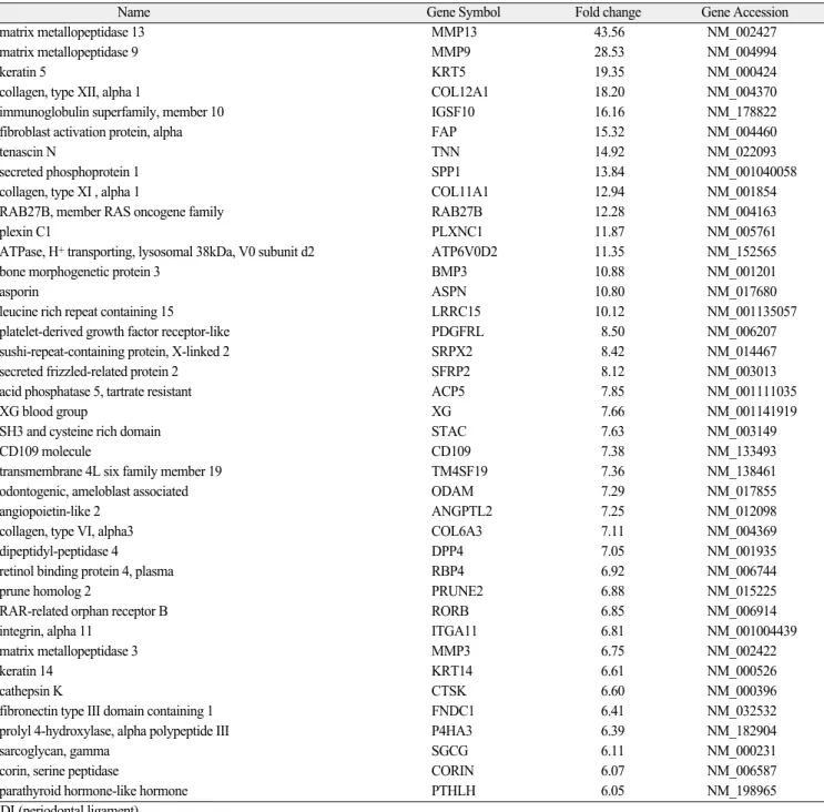
Table 1. Up-regulated genes in the PDL tissue of permanent teeth
Name Gene Symbol Fold change Gene Accession
matrix metallopeptidase 13 MMP13 43.56 NM_002427
matrix metallopeptidase 9 MMP9 28.53 NM_004994
keratin 5 KRT5 19.35 NM_000424
collagen, type XII, alpha 1 COL12A1 18.20 NM_004370
immunoglobulin superfamily, member 10 IGSF10 16.16 NM_178822
fibroblast activation protein, alpha FAP 15.32 NM_004460
tenascin N TNN 14.92 NM_022093
secreted phosphoprotein 1 SPP1 13.84 NM_001040058
collagen, type XI , alpha 1 COL11A1 12.94 NM_001854
RAB27B, member RAS oncogene family RAB27B 12.28NM_004163
plexin C1 PLXNC1 11.87 NM_005761
ATPase, H+transporting, lysosomal 38kDa, V0 subunit d2 ATP6V0D2 11.35 NM_152565
bone morphogenetic protein 3 BMP3 10.88 NM_001201
asporin ASPN 10.80 NM_017680
leucine rich repeat containing 15 LRRC15 10.12 NM_001135057
platelet-derived growth factor receptor-like PDGFRL 8.50 NM_006207
sushi-repeat-containing protein, X-linked 2 SRPX2 8.42 NM_014467
secreted frizzled-related protein 2 SFRP2 8.12 NM_003013
acid phosphatase 5, tartrate resistant ACP5 7.85 NM_001111035
XG blood group XG 7.66 NM_001141919
SH3 and cysteine rich domain STAC 7.63 NM_003149
CD109 molecule CD109 7.38NM_133493
transmembrane 4L six family member 19 TM4SF19 7.36 NM_138461
odontogenic, ameloblast associated ODAM 7.29 NM_017855
angiopoietin-like 2 ANGPTL2 7.25 NM_012098
collagen, type VI, alpha3 COL6A3 7.11 NM_004369
dipeptidyl-peptidase 4 DPP4 7.05 NM_001935
retinol binding protein 4, plasma RBP4 6.92 NM_006744
prune homolog 2 PRUNE2 6.88 NM_015225
RAR-related orphan receptor B RORB 6.85 NM_006914
integrin, alpha 11 ITGA11 6.81 NM_001004439
matrix metallopeptidase 3 MMP3 6.75 NM_002422
keratin 14 KRT14 6.61 NM_000526
cathepsin K CTSK 6.60 NM_000396
fibronectin type III domain containing 1 FNDC1 6.41 NM_032532
prolyl 4-hydroxylase, alpha polypeptide III P4HA3 6.39 NM_182904
sarcoglycan, gamma SGCG 6.11 NM_000231
corin, serine peptidase CORIN 6.07 NM_006587
parathyroid hormone-like hormone PTHLH 6.05 NM_198965
PDL(periodontal ligament)
were classified based on information regarding gene function in gene ontology from the KEGG Pathway data- base. GO classes with an F-statistic p value of < 0.05 following analysis on the basis of their biologic processes and molecular functions are shown in Figure 1 and 2, respectively. In broad outlines, the GO classes of the dental pulp tissue are relatively more counted those of the PDL tissue. Especially, there were notable differ- ences of biologic processes in regulation of cell adhesion, neurological system process, signal transduction, and ion transport, and of molecular functions in nucleotide bind- ing, ATP binding, and ion binding.
3. Quantitative PCR
Quantitative PCR (qPCR) was performed to verify the different gene-expression levels obtained through cDNA microarray analysis. The following six genes were select- ed for this verification procedure: COL12, MMP9, SPP1, CALB1, SPOCK3, and DSPP. The remaining six were compared between PDL and pulp in the form of gene-ex- pression ratios (Table 3). The pulp: PDL ratios for CALB1, SPOCK3, and DSPP were high, at 4,600, 6,800, and 15,000, respectively, while those for COL12, MMP9, and SPP1 were low, at 44, 8, and 9, respectively.
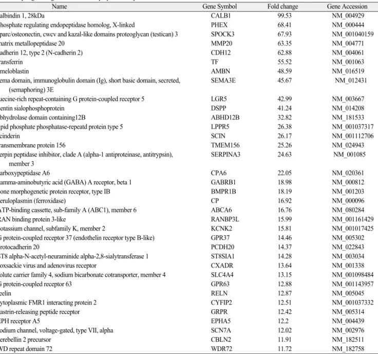
Table 2. Up-regulated genes in the dental pulp tissue of permanent teeth
Name Gene Symbol Fold change Gene Accession
calbindin 1, 28kDa CALB1 99.53 NM_004929
phosphate regulating endopeptidase homolog, X-linked PHEX 68.41 NM_000444
sparc/osteonectin, cwcv and kazal-like domains proteoglycan (testican) 3 SPOCK3 67.93 NM_001040159
matrix metallopeptidase 20 MMP20 63.35 NM_004771
cadherin 12, type 2 (N-cadherin 2) CDH12 62.88 NM_004061
transferrin TF 55.52 NM_001063
ameloblastin AMBN 48.59 NM_016519
sema domain, immunoglobulin domain (Ig), short basic domain, secreted, SEMA3E 45.67 NM_012431 (semaphoring) 3E
luecine-rich repeat-containing G protein-coupled receptor 5 LGR5 42.99 NM_003667
dentin sialophosphoprotein DSPP 41.24 NM_014208
abhydrolase domain containing12B ABHD12B 32.82 NM_181533
lipid phosphate phosphatase-repeatd protein type 5 LPPR5 26.38NM_001037317
scinderin SCIN 26.17 NM_001112706
transmembrane protein 156 TMEM156 25.26 NM_024943
serpin peptidase inhibitor, clade A (alpha-1 antiproteinase, antitrypsin), SERPINA3 24.63 NM_001085 member 3
carboxypeptidase A6 CPA6 22.05 NM_020361
gamma-aminobutyric acid (GABA) A receptor, beta 1 GABRB1 18.98 NM_000812
bone morphogenetic protein receptor, type IB BMPR1B 18.19 NM_001203
ceruloplasmin (ferroxidase) CP 16.92 NM_000096
ATP-binding cassette, sub-family A (ABC1), member 6 ABCA6 16.76 NM_080284
RAN binding protein 3-like RANBP3L 15.99 NM_001161429
potassium channel, subfamily K, member 2 KCNK2 15.81 NM_001017425
G protein-coupled receptor 37 (endothelin receptor type B-like) GPR37 14.46 NM_005302
protocadherin 20 PCDH20 14.37 NM_022843
ST8 alpha-N-acetyl-neuraminide alpha-2,8-sialytransferase 1 ST8SIA1 14.28 NM_003034
coxsackie virus and adenovirus receptor CXADR 13.64 NM_001338
solute carrier family 4, sodium bicarbonate cotransporter, member 4 SLC4A4 13.15 NM_001098484
G protein-coupled receptor 63 GPR63 12.88 NM_001143957
reelin RELN 12.87 NM_005045
cytoplasmic FMR1 interacting protein 2 CYFIP2 12.51 NM_001037332
gastrin-releasing peptide receptor GRPR 12.42 NM_005314
EPH receptor A5 EPHA5 12.2 NM_004439
sodium channel, voltage-gated, type VII, alpha SCN7A 12.02 NM_002976
cerebellin 2 precursor CBLN2 11.91 NM_182511
WD repeat domain 72 WDR72 11.72 NM_182758
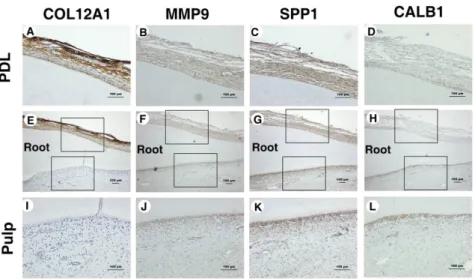
4. IHC staining
The following four proteins were the targets of the IHC study: COL12A1, MMP9, SPP1, and CALB1 (Fig. 3).
COL12A1 was stained strongly in PDL tissues but was not stained in pulp tissues, while MMP9 and SPP1 were stained more strongly in all PDL tissues than in all pulp tissues, and staining was found only in the odontoblast layer of the pulp tissues. CALB1 was located mainly in the odontoblast layer of the pulp tissue.
Fig. 1. Main categories of genes expressed in the PDL and dental pulp of human permanent teeth relative to their biologic processes (F-statistic: p <
0.05).
Fig. 2. Main categories of genes expressed in the PDL and dental pulp of human permanent teeth relative to their molecular processes (F-statistic: p
< 0.05).
Table 3. The relative difference in gene mRNA expression in periodontal ligament tissue and dental pulp
Gene Relative Gene Expression (Mean ± SD) Periodontal Ligament Tissues Dental Pulp
COL12 44 ± 6 1
MMP9 18 ± 8 1
SPP1 9 ± 2 1
CALB1 1 4,600 ± 270
SPOCK3 1 6,800 ± 270
DSPP 1 15,000 ± 2,500
Fig. 3. Immunohistochemical staining of PDL and pulp samples (A-L). IHC staining for COL12A1 in (A,E) PDL and (E,I) pulp; for MMP9 in (B,F) PDL and (F,J) pulp; for SPP1 in (C,G) PDL and (G,K) pulp; and for CALB1 in (D,H) PDL and (H,L) pulp. The micrographs in A-D and I-L are higher-magnifi- cation views of the areas outlined by the upper and lower squares, respectively, in E-H. Scale bars: all 100 ㎛.
Ⅳ. Discussion
Among the 28,869 genes that were analyzed using cDNA microarrays, 347 (1.20%) were expressed differ- entially by a factor of fourfold or more between PDL and pulp. According to the cDNA microarray and GO analy- ses, the genes that were up-regulated in the two tissue types were intimately related to their respective func- tions. PDL exhibited strong expression of genes related to collagen synthesis (FAP), collagen degradation (MMP3, MMP9, and MMP13), and bone development and remodeling (SSP1, BMP3, ACP5, CTSK, and PTHLH). In contrast, pulp exhibited strong expression of genes associated with calcium ions (CALB1, SCIN, and CDH12) and the mineralization and formation of enamel and dentin (SPARC/SPOCK3, PHEX, AMBN, and DSPP).
FAP alpha plays a major role in the production and turnover of extracellular matrix components, a process that is critical for wound healing and tissue remodeling.
Research into FAP in dentistry is rare because of its lack of expression in normal tissues and induced expression in areas of tissue remodeling and tumor stroma
19). MMP3, MMPR9, and MMP13 are members of the MMP family, which comprise proteolytic enzymes that mediate the degradation of extracellular matrix macromolecules and contribute not only to extracellular matrix home- ostasis, but also to pathologic or therapeutic situa- tions
20,21).
SPP1, widely known as osteopontin, has been impli- cated as an important factor in bone remodeling
22). BMP3 has been extensively investigated with regard to bone formation, and reportedly down-regulates bone mineralization and density, and acts as an antagonist of osteogenic BMP
23,24). ACP5, also known as tartrate-re- sistant acid phosphatase, is a glycosylated monomeric metalloenzyme that is expressed in mammals
25). CTSK is expressed predominantly in osteoclasts; high levels of CTSK are detected in patients with bone-destructive dis- ease. PTHLH is a member of the parathyroid hormone family that represses bone sialoprotein, osteocalcin, and mineralization in PDL cells and, potentially, in cemento- blasts
26).
In the dental pulp tissues, CALB1 is an intracellular, soluble, vitamin-D-dependent calcium-binding protein and a member of the troponin C superfamily
27). SCIN is an actin-severing protein and that is found in abundance in secretory tissues. Very little research into the gene
that encodes this protein (SCIN) has been conducted in the field of dentistry, and work is needed to determine the function of SCIN in dental pulp function. CDH12 mediates calcium-dependent cell-cell adhesion. This par- ticular cadherin appears during a critical period of neu- ronal development, and perhaps specifically during synaptogenesis
28). It is thought that CDH12 up-regula- tion is related to the nerve innervation of dental pulp.
As expected, genes related to the mineralization and formation of enamel or dentin were strongly expressed in pulp. SPARC/SPOCK3 is a phosphorylated glycoprotein that is associated with development, tissue remodeling, and repair
29). Phosphate-regulating endopeptidase ho- molog, X-linked (encoded by PHEX) is expressed by hu- man odontoblasts aligned at the margin of the dental pulp but not in the dental pulp cells, and its expression is up-regulated during odontoblast development
30).
AMBN is found in tooth enamel, and is produced by ameloblasts during the early secretory to late maturation stages of amelogenesis. However, it has recently been shown that reparative dentin formation is associated with the sequential expression of dentin-specific factors in wounded porcine pulp
31). DSPP is a human gene that encodes two principal proteins of the dentin extracellular matrix of the tooth.
The qPCR findings were indeed consistent with those of the cDNA microarray analysis and the IHC findings supported the cDNA microarray data and revealed the locations of the expressed genes. These data also support previous research that has shown that MMP9 may play an important role in the pathogenesis of pulpal inflam- mation
32), and that the SPP1 synthesized by odonto- blasts is associated with the initial sites of calcification within mantle dentin
33).
Most microarray studies were about comparing the gene expression of bone marrow stem cells and stem cells derived from dental tissues. Another microarray re- searches were about confirming the gene expression pat- terns of cells under certain conditions. Although there were studies about comparative gene expression analysis of the PDL tissues in deciduous and permanent teeth and of the PDL and dental follicle tissues in humans, there is no study about comparing the relative gene ex- pression of PDL and dental pulp tissues in humans. So gene expression analysis might help to explain the func- tional difference between PDL and dental pulp.
In conclusion, comparison of the PDL and the dental
pulp tissues revealed genetic differences of actively for-
mationing gene. The gene-expression profiles presented here identify candidate genes that may distinguish spe- cific functions of PDL and dental pulp. The critical point of this research is that RNA obtained from fresh func- tioning dental pulp and PDL tissues of permanent teeth, showed different specific genetic activities. And in tissue regenerative therapy, it might help to find the key re- generative factors in PDL and pulp regeneration if fur- ther studies are performed about genes with different expression pattern in two types of tissues.
Ⅴ. Conclusion
This study was conducted to identify the gene-expres- sion profiles and their molecular biological differences of PDL and dental pulp tissues from the human permanent teeth using cDNA microarray analysis, qPCR, and im- munohistochemical stain. Genes associated with collagen degradation such as MMP3, MMP9, and MMP13 and with collagen synthesis such as FAP, and with bone de- velopment and remodeling such as SPP1, BMP3, ACP5, CTSK, and PTHLH were more strongly expressed in PDL tissues of permanent teeth. They are clinically re- lated to PDL’ s functions of external force absorption and tooth supporting. In dental pulp tissues of permanent teeth, genes associated with calcium ion such as CALB1, CDH12, and SCIN and with mineralization and forma- tion of enamel or dentin such as SPOCK3, PHEX, AMBN, and DSPP were more strongly expressed. They are clinically related to dental pulp’ s functions of sec- ondary and tertiary dentin formation. The qPCR analy- sis and immunohistochemical staining analysis was also coincided with the cDNA microarray assay data.
References
1. Demarco FF, Conde MC, Cavalcanti BN, et al. : Dental pulp tissue engineering. Braz Dent J, 22:3- 13, 2011.
2. Caton J, Bostanci N, Remboutsika E, et al. : Future dentistry: cell therapy meets tooth and periodontal repair and regeneration. J Cell Mol Med, 15:1054- 1065, 2011.
3. Danesh-Meyer MJ : Tissue engineering in periodon- tics and implantology using rhBMP-2. Ann R Australas Coll Dent Surg, 15:144-149, 2000.
4. Song JS, Hwang DH, Kim SO, et al. : Comparative gene expression analysis of the human periodontal
ligament in deciduous and permanent teeth. PLoS One, 8:e61231, 2013.
5. Xie YF, Shu R, Jiang SY, et al. : Comparison of microRNA profiles of human periodontal diseased and healthy gingival tissues. Int J Oral Sci, 3:125- 134, 2011.
6. Lee YH, Na HS, Jeong SY, et al. : Comparison of inflammatory microRNA expression in healthy and periodontitis tissues. Biocell, 35:43-49, 2011.
7. Han X, Amar S : Identification of genes differentially expressed in cultured human periodontal ligament fibroblasts vs. human gingival fibroblasts by DNA microarray analysis. J Dent Res, 81:399-405, 2002.
8. de Araujo RM, Oba Y, Moriyama K : Identification of genes related to mechanical stress in human peri- odontal ligament cells using microarray analysis. J Periodontal Res, 42:15-22, 2007.
9. Paakkonen V, Ohlmeier S, Bergmann U, et al. : Analysis of gene and protein expression in healthy and carious tooth pulp with cDNA microarray and two-dimensional gel electrophoresis. Eur J Oral Sci, 113:369-379, 2005.
10. McLachlan JL, Smith AJ, Bujalska IJ, Cooper PR : Gene expression profiling of pulpal tissue reveals the molecular complexity of dental caries. Biochim Biophys Acta, 1741:271-281, 2005.
11. Paakkonen V, Vuoristo JT, Salo T, Tjaderhane L : Comparative gene expression profile analysis between native human odontoblasts and pulp tissue.
Int Endod J, 41:117-127, 2008.
12. Heikinheimo K, Jee KJ, Niini T, et al. : Gene expression profiling of ameloblastoma and human tooth germ by means of a cDNA microarray. J Dent Res, 81:525-530, 2002.
13. Lee HS, Lee J, Kim SO, et al. : Comparative gene- expression analysis of the dental follicle and peri- odontal ligament in humans. PLoS One, 8:e84201, 2013.
14. Gottlow J, Nyman S, Karring T, Lindhe J : New attachment formation as the result of controlled tis- sue regeneration. J Clin Periodontol, 11:494-503, 1984.
15. Mitchell DL, West JD : Attempted orthodontic movement in the presence of suspected ankylosis.
Am J Orthod, 68:404-411, 1975.
16. Lekic P, McCulloch CA: Periodontal ligament cell
population: the central role of fibroblasts in creating
a unique tissue. Anat Rec, 245:327-341, 1996.
17. Abrahao IJ, Martins MD, Katayama E, et al. : Collagen analysis in human tooth germ papillae.
Braz Dent J, 17:208-212, 2006.
18. Gronthos S, Mankani M, Brahim J, et al. : Postnatal human dental pulp stem cells (DPSCs) in vitro and in vivo. Proc Natl Acad Sci U S A, 97:13625-13630, 2000.
19. Shi M, Yu DH, Chen Y, et al. : Expression of fibrob- last activation protein in human pancreatic adeno- carcinoma and its clinicopathological significance.
World J Gastroenterol, 18:840-846, 2012.
20. Boushell LW, Kaku M, Mochida Y, Yamauchi M : Distribution and relative activity of matrix metallo- proteinase-2 in human coronal dentin. Int J Oral Sci, 3:192-199, 2011.
21. Hakki SS, Hakki EE, Nohutcu RM : Regulation of matrix metalloproteinases and tissue inhibitors of matrix metalloproteinases by basic fibroblast growth factor and dexamethasone in periodontal ligament cells. J Periodontal Res, 44:794-802, 2009.
22. Choi ST, Kim JH, Kang EJ, et al. : Osteopontin might be involved in bone remodelling rather than in inflammation in ankylosing spondylitis. Rheumatology (Oxford), 47:1775-1779, 2008.
23. Daluiski A, Engstrand T, Bahamonde ME, et al. : Bone morphogenetic protein-3 is a negative regula- tor of bone density. Nat Genet, 27:84-88, 2001.
24. Choi S, Cho TJ, Kwon SK, et al. : Chondrogenesis of periodontal ligament stem cells by transforming growth factor-beta3 and bone morphogenetic pro- tein-6 in a normal healthy impacted third molar. Int J Oral Sci, 5:7-13, 2013.
25. Baumbach GA, Saunders PT, Ketcham CM, et al. :
Uteroferrin contains complex and high mannose-type oligosaccharides when synthesized in vitro. Mol Cell Biochem, 105:107-117, 1991.
26. Ouyang H, McCauley LK, Berry JE, et al. : Parathyroid hormone-related protein regulates extracellular matrix gene expression in cemento- blasts and inhibits cementoblast-mediated mineral- ization in vitro. J Bone Miner Res, 15:2140-2153, 2000.
27. Wasserman RH, Taylor AN : Vitamin d3-induced calcium-binding protein in chick intestinal mucosa.
Science, 152:791-793, 1966.
28. Selig S, Bruno S, Scharf JM, et al. : Expressed cad- herin pseudogenes are localized to the critical region of the spinal muscular atrophy gene. Proc Natl Acad Sci U S A, 92:3702-3706, 1995.
29. Yan Q, Sage EH : SPARC, a matricellular glycopro- tein with important biological functions. J Histochem Cytochem, 47:1495-1506, 1999.
30. Buchaille R, Couble ML, Magloire H, Bleicher F : A substractive PCR-based cDNA library from human odontoblast cells: identification of novel genes expressed in tooth forming cells. Matrix Biol, 19:
421-430, 2000.
31. Nakamura Y, Hammarstrom L, Matsumoto K, Lyngstadaas SP : The induction of reparative dentine by enamel proteins. Int Endod J, 35:407-417, 2002.
32. Gusman H, Santana RB, Zehnder M : Matrix met- alloproteinase levels and gelatinolytic activity in clinically healthy and inflamed human dental pulps.
Eur J Oral Sci, 110:353-357, 2002.
33. Trowbridge HO : Pulp biology: progress during the
past 25 years. Aust Endod J, 29:5-12, 2003.
주요어: 유전자 발현, 치주인대, 치수, 사람 영구치
사람 영구치에서 치주인대 및 치수 조직의 유전자 발현에 대한 비교 연구
이석우
1∙전미정
1∙이효설
2∙송제선
1∙손흥규
1∙최형준
1∙정한성
3∙문석준
3∙박원서
4∙김성오
11
연세대학교 치과대학 소아치과학교실
2
경희대학교 치과대학 소아치과학교실
3
연세대학교 치과대학 구강생물학교실
4