Effects of Mineral Trioxide Aggregate on the Proliferation and Differentiation of Human Dental Pulp Stromal Cells from Permanent and Deciduous Teeth
Seunghye Kim1, Mijeong Jeon1, Dong Min Shin2, Jae Ho Lee1, Je Seon Song1
1Department of Pediatric Dentistry, College of Dentistry and Oral Science Research Center, Yonsei University, Seoul, Korea
2Department of Oral Science, College of Dentistry, Yonsei University, Seoul, Korea
Mineral trioxide aggregate (MTA) has recently been used as a pulpotomy medicament for primary molars. The aim of this study was to evaluate and compare the proliferation and differentiation potential of dental pulp stro- mal cells of permanent teeth and deciduous teeth cultured on MTA-coated surface. Human dental pulp stromal cells were obtained from human permanent premolars and deciduous teeth and cultured on MTA-coated culture plates. The cells were subjected to proliferation assay and cell cycle analysis. Their differentiation potential was evaluated by analysing changes in the mRNA expressions of runt-related transcriptional factor 2 (Runx2) and alkaline phosphatase (ALP). Morphological changes of cells in direct contact with MTA were observed using scanning electron microscopy (SEM). The proliferation rates, distribution of cell cycles and mRNA expression patterns of Runx2 and ALP were similar in both types of pulpal cells. SEM observations revealed that both types changed into more dendrite-like cells. On the surface of MTA, human dental pulp stromal cells from de- ciduous and permanent teeth were able to both proliferate and differentiate into cells that induce mineralization.
MTA is suitable as a biocompatible pulpotomy medicament for primary teeth.
Key words :Mineral Trioxide Aggregate (MTA), Human dental pulp stromal cells, Deciduous teeth, Permanent teeth, Proliferation, Differentiation
Abstract
Ⅰ. Introduction
Several pulp medicaments are commonly used for pulpotomy in primary molars, including formocresol and ferric sulphate. However, the use of formocresol is now decreasing in many countries as a result of its carcino- genic properties in human1,2) and ferric sulphate is known to irritate the pulp tissue and provoke an inflam- matory reaction in some cases3-6).
Mineral trioxide aggregate (MTA) has recently been applied clinically with successful outcomes as an alterna-
tive pulp medicament for pulpotomy in primary molars7,8). Since it was introduced by Torabinejad et al.
in 19939), MTA has been widely applied in various en- dodontic procedures, such as pulp capping, pulpotomy, apical barrier formation in immature permanent teeth with necrotic pulp, repair of root perforation, root-end filling and root canal filling. MTA is a biocompatible ma- terial, non-toxic to the host, and has been reported to induce dentin bridge formation and hard-tissue forma- tion on exposed pulp tissue without pulpal necrosis in animal studies6,10).
Corresponding author : Je Seon Song
Department of Pediatric Dentistry, College of Dentistry, Yonsei University, 50 Yonsei-ro, Seodaemun-gu, Seoul, 120-752, Korea Tel : +82-2-2228-3176 / Fax : +82-2-392-7420 / E-mail: [email protected]
Received November 30, 2012 / Revised June 4, 2013 / Accepted June 11, 2013
※This study was supported by a grant of the Korea Healthcare Technology R&D Project, Ministry for Health, Welfare and Family Affairs, Republic of Korea (no. A100517) and Basic Science Research Program through the National Research Foundation of Korea (NRF) funded by the Ministry of Education, Science and Technology (2011-
Previous in vitro research has demonstrated the bio- compatibility of MTA using human dental pulp stromal cells from permanent teeth11-13). However, few in vitro studies have investigated the pulpal reaction to MTA in deciduous teeth14). The aim of this study was to compare the viability and differentiation potential of pulpal cells from permanent and deciduous teeth in direct contact with MTA.
Ⅱ. Materials and Methods 1. Preparation of MTA
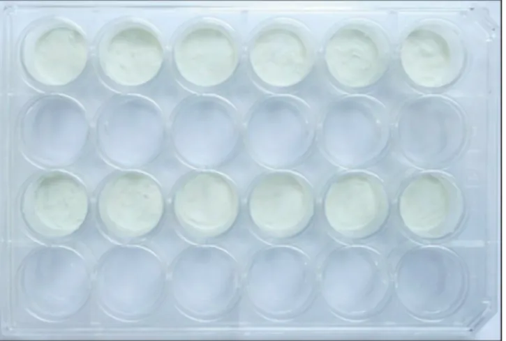
White MTA (ProRoot MTA, Dentsply Tulsa Dental, Tulsa, OK, USA) was used in all experiments. The ma- terial was mixed according to the manufacturer’s in- structions and then plated in culture plates (BD Falcon, Franklin Lakes, NJ, USA) or on round glass slides (8 mm in diameter), as shown in Fig. 1. The MTA was left to set completely for 3~4 days at 37℃ in a humidified 5%-CO2 incubator. After complete setting, the MTA- coated plates and glass slides were sterilized under eth- ylene oxide gas.
2. Cell cultures
The experimental protocol was approved by the Institutional Review Board of the Dental Hospital, Yonsei University, and informed consent to participate was obtained from all of the subjects and their parents (#2-2010-0012). Human dental pulp tissue was ob- tained from healthy permanent premolars (n = 3; pa-
tients aged 13~18 years) that had been extracted for orthodontic purposes, and from deciduous teeth (n = 4;
patients aged 2~7 years) from which mechanically ex- posed pulp tissue was extirpated during routine treat- ment for caries (Table 1). The extracted teeth and extir- pated pulp tissue were stored in Dulbecco’s Modified Eagle’s Medium (Invitrogen, Carlsbad, CA, USA). The pulp tissue, corresponding to the middle one-third of the root, was separated and subjected to primary culture by outgrowth method. Briefly, the separated pulp tissue was minced into about 1.0 mm3size and pressed gently unto 60 mm culture dish (BD Falcon, Franklin Lakes, NJ, USA) using cover glass (Superior, Lauda- Ko¨nigshofen, Germany). The explants were cultured in alpha minimum essential medium (α-MEM; Invitrogen) supplemented with 10% fetal bovine serum (FBS;
Invitrogen), 100 U/ml penicillin and 100 ug/ml strepto- mycin (Invitrogen) at 37℃ in 5% CO2. The isolated cells from 3 samples of premolars were blended at the first passage, and the third to fifth passages were used in the subsequent experiments. The same procedure was per- formed for the pulp stromal cells obtained from four samples of deciduous teeth.
3. Proliferation assay
The proliferation rate of human dental pulp cells from permanent teeth and deciduous teeth in direct contact with MTA was measured using Cell Counting Kit-8 (Dojindo, Kumamoto, Japan), according to the manufac- turer’s instructions. Human dental pulp cells were seed- ed at 5×10³ cells/well in 24-well culture plates that were either uncoated or coated with MTA. After 1, 3, 5 and 7 days of culture period, the optical density of the yellow-coloured formazan formed by vital cells was mea- sured at 450 nm using a Benchmark Plus Microplate spectrophotometer (Bio-Rad Laboratories, Hercules, CA, USA).
Table 1. Information about the source of extirpated pulp tissue regarding the tooth number, age, and gender of the subjects
Source Tooth Number Age Gender
Permanent Teeth #14 18Y F
#14 15Y M
#24 13Y 7M M
Primary Teeth #54 7Y 4M M
#53 5Y 3M F
#64 4Y 5M F
#74 2Y 2M M
Fig. 1. White MTA coated onto 24-well culture plates. The first and third rows are coated with white MTA while the second and fourth rows are left uncoated for the control.
4. Cell cycle analysis
Cells were seeded at 5×10⁵ cells/well onto MTA-coat- ed or uncoated 60-mm culture plates, cultured for 3 days, and then harvested by trypsinization and fixed in cold 70% ethanol for 1 hour at 4℃. After washing twice with phosphate-buffered saline (Invitrogen), samples were incubated in 0.2 mg/ml RNase A (LaboPass, Sapporo, Japan) for 1 hour at 37℃ and then stained with propidium iodide (40 μg/ml; #P-4170, Sigma) at 4℃
for 30 min. Flow cytometric analysis was performed us- ing a FACS Calibur flow cytometer (BD Biosciences, San Jose, CA, USA), and cell cycle analysis was conducted using FCS Express V3 software (De Novo Software, Los Angeles, CA, USA).
5. Gene expression analysis using the quantitative reverse-transcription polymerase chain reaction (RT-PCR)
Human dental pulp cells from permanent and decidu- ous teeth were seeded at a concentration of 1×10⁵ cells/well in either MTA-coated or uncoated 12-well cul- ture plates. They were harvested from the culture plates at 1, 2 and 4 days after seeding by trypsinization, and total cell RNA was extracted with the RNeasy Micro Kit (Qiagen, Valencia, CA, USA), according to the manufac- turer’s instructions. The concentration of RNA was cal- culated using a spectrophotometer (Nanodrop ND-1000, Thermo Scientific, Waltham, MA, USA). A 1-μg aliquot of each RNA sample was reverse transcribed into cDNA using an Maxime reverse transcription premix kit (Intron Biotechnology, Seoul, Korea). According to the manufacturer’s instructions, the total RNA was reverse transcribed using an oligo d(T)15 primer at 45℃ for 1 hour, and the reaction was stopped by incubation at 95℃
for 5 min.
The complemented DNA was subjected to quantitative RT-PCR analysis using SYBR premix Ex Taq (Takara Bio, Otsu, Japan) and the Thermal Cycler Dice real- time system (Takara Bio), according to the manufactur- er’s instructions. Each PCR assay was performed with.
The program was set at 95℃ for 10 s for the initial de- naturing step, followed by 45 cycles for denaturation at 95℃ for 5 s, annealing at 60℃ for 15 s and amplification at 72℃ for 10 s. Amplification specificity was confirmed by visualizing the PCR products on 1.5% agarose gels and by melting-curve analysis (from 60℃ to 95℃) after
the completion of 45 cycles. The values for each gene were normalized to the expression levels of glyceralde- hyde-3-phosphate dehydrogenase (GAPDH), and the relative quantities of the studied genes were calculated.
The specific primers used in this study included Runx2 (NM_004348.3: forward primer, 5’-CACTGGCGCTG- CAACAAGA-3’; reverse primer, 5’-CATTCCGGAGC- TCAGCAGAATAA-3’), alkaline phosphatase (ALP;
NM_000478.4: forward primer, 5’-GGACCATTCC- CACGTCTTCAC-3’; reverse primer, 5’-CCTTGTAGCC- AGGCCCATTG-3’) and GAPDH (NM_002046.3: forward primer, 5’-TCCTGCACCACCAACTGCTT-3’; reverse primer, 5’-TGGCAGTGATGGCATGGAC-3’).
6. Scanning electron microscopy (SEM) analysis
Human dental pulp cells from permanent and decidu- ous teeth were seeded at a concentration of 1×10⁵ cells/well in four-well plates, whereby MTA-coated round glass slides were placed with the MTA facing up- ward. After 5 days, the pulp cells on the MTA-coated slides were incubated in fixative solution containing 2%
paraformaldehyde, 2% glutaraldehyde and 0.5% CaCl2, followed by post-fixation using 1% OsO4(all these chem- icals were purchased from Sigma). After dehydration and drying, the samples were coated with a 100-nm lay- er of platinum and then visualized using a Hitachi S- 3000N scanning electron microscope (Hitachi, Tokyo, Japan) at an accelerating voltage of 20.0 kV and a mag- nification of ×1500.
7. Statistical analysis
All of the experiments were repeated under at least three independent conditions. All data are presented as mean and standard deviation values. Any significant dif- ferences between human dental pulp stromal cells from permanent teeth and deciduous teeth were determined using Student’s t-test with SPSS software (version 17.0, SPSS, Chicago, IL, USA). The level of statistical signifi- cance was set at p < 0.05.
Ⅲ. Results
1. Proliferation assay and cell cycle analysis
In the proliferation assay, there was no significant dif- ference between pulpal cells from permanent teeth and
deciduous teeth when cultured on either an uncoated culture plate or on the MTA surface. However, the pro- liferation rate was significantly higher on the culture plate compared to the MTA-coated surface for both cell types (Fig. 2). The cell cycle analysis revealed similar percentages of cells in the G1 phase in all samples (63~64%; Fig. 3).
2. Quantitative RT-PCR
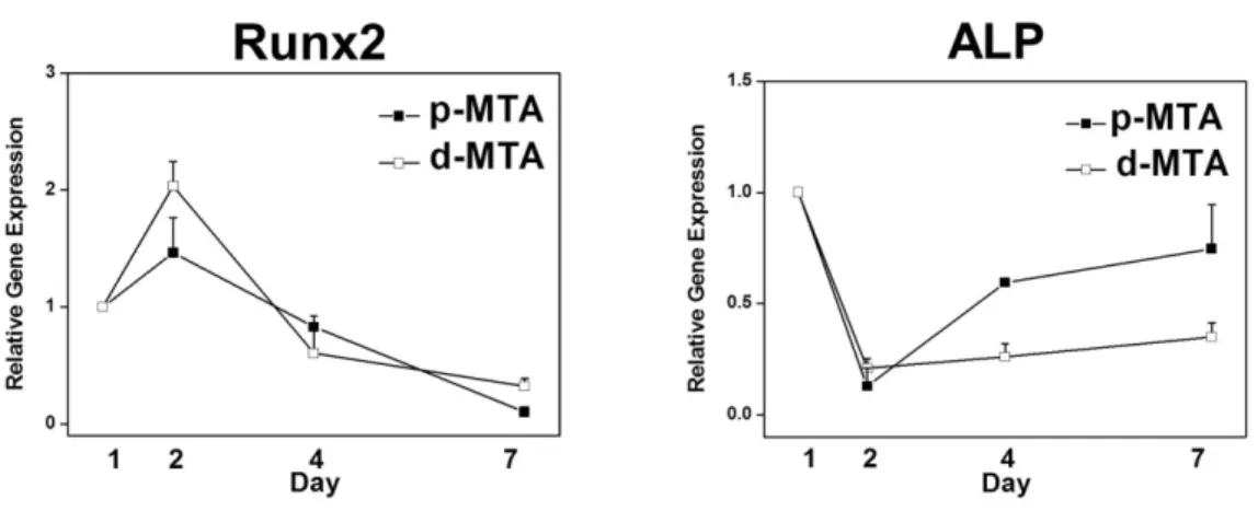
The expression of Runx2 increased at day 2, and then gradually decreased thereafter, while that of ALP con- tinued to gradually increase over the 7-day experimental period. However, these expression levels did not show significant difference between pulpal cells from perma- nent teeth and deciduous teeth cultured on an MTA- coated surface (Fig. 4).
Fig. 2. Proliferation of pulpal cells from permanent and deciduous teeth in direct contact with MTA. The Y-axis indicates the optical density of yel- low-coloured formazan converted by the cells. Abbreviations: p-Cont, pulpal cells from permanent teeth on a plate not coated with white MTA;
d-Cont, pulpal cells from deciduous teeth on a plate not coated with white MTA; p-MTA, pulpal cells from permanent teeth on a plate coated with white MTA; d-MTA, pulpal cells from deciduous teeth on a plate not coated with white MTA.
Fig. 3. Cell cycle distributions of pulpal cells on direct contact to MTA.
Fig. 4. Chronological changes in the gene expressions of Runx2 and ALP of pulpal cells in direct contact with white MTA.
Gene expression levels at 1 day after seeding were normalized to 1.
3. SEM analysis
Human pulpal cells from permanent teeth and decidu- ous teeth were able to attach to the MTA coating, pre- senting a more dendrite-like morphology with several branches, rather than a spindle shape (Fig. 5).
Ⅳ. Discussion
In previous studies, investigators have reported that dental pulp cells obtained through primary culture of dental-pulp tissue from permanent and shedding decidu- ous teeth present different features15,16). The pulp stromal cells from permanent teeth are known to be capable of differentiating into odontoblasts and form a dentin-pulp- like complex following in vivo transplantation17). In con- trast, those from exfoliated deciduous teeth fail to recon- stitute a dentin-pulp-like complex in vivo, but do induce recipient cell-mediated bone formation in vivo15). In addi- tion, the pulp cells from deciduous teeth exhibit a higher proliferation rate and increased cell-population dou- bling15,18). Such differences found in previous studies sug-
gest that the pulpal reactions to MTA may differ be- tween permanent and deciduous teeth.
In this study, unlike previous studies, the proliferation rates measured were similar in pulpal cells from perma- nent teeth and deciduous teeth, regardless of the pres- ence of an MTA surface coating. While previous studies used dental pulp from exfoliated deciduous teeth, we used dental pulp extirpated from deciduous teeth with a sound root. Given that deciduous teeth go through vari- ous developmental stages from eruption through to shed- ding, the stage at which pulp tissue is obtained may be an important determinant of the pulpal characteristics, because pulp components and gene expression patterns may differ according to the stage.
The biocompatibility of MTA has been confirmed through many cell-culture studies, and it is known to be one of the least cytotoxic dental materials19,20). In the present study, the proliferation rate of both cell types was reduced on MTA-coated culture plate, but the pro- portions of cells in the G1 phase were similar, regardless of the presence of the MTA coating. This indicates that the pulpal cells from permanent and deciduous teeth ex- Fig. 5. SEM images of pulpal cells cultured on an MTA-coated surface from permanent (A) and deciduous
(B) teeth. Arrows indicates dendritic process resembling odontoblast process. Scale bars: 30 ㎛.
A p-MTA
B d-MTA
hibit similar viabilities on MTA, and confirms that MTA is a biocompatible material. The reduced proliferation rate on the MTA-coated surface can be explained by the surface roughness of set MTA. Previous studies that found an increased proliferation of pulp cells on MTA blocks used an insert-well system to prevent direct con- tact between the cells and the tested material11,21,22). Direct contact between the test materials and the cells may introduce unfavourable factors that could obscure or inhibit the actual effect of the materials on cell survival and growth. In the present study, the MTA coating on the plate increased the roughness and irregularity of its surface, which may have inhibited the initial attachment of the cells, consequently leading to a reduction in actual numbers of seeded cells. However, the use of the insert- well system is limited in that it can only be used to mea- sure the effects of the diffusible components of the mate- rial, while our system allowed the full effect of MTA in direct contact with the cells to be investigated.
It has been reported that MTA induces mineralization under both in vitro and in vivo conditions23-27). We have shown herein that the expression levels of Runx2 and ALP of pulpal cells were up-regulated when they were cultured on an MTA-coated surface. Runx2 is a tran- scriptional activator of osteogenesis-associated genes and is essential for osteoblast differentiation and bone forma- tion28). Runx2 also regulates the proliferation and differ- entiation of odontoblasts and increases the expression of dentin sialophosphoprotein (DSPP) which is the main non-collagenous component of the odontoblastic extracel- lular matrix-in immature odontoblasts29,30). ALP is an- other important regulator during the mineralization of bone and reparative dentin31). ALP expression gradually increases during development of collagen matrix, which precedes calcium accumulation32). In this study, short expression of Runx2 with its peak at Day 4 coincides with its character as the early marker initiating os- teogenic or odontogenic differentiation. Gradual increase of ALP expression through 8-day period indicates devel- opment of collagen matrix. The SEM images showed that the deciduous and permanent pulp cells grown on an MTA block had dendritic process that resembled ma- ture odontoblastic process. These findings indicate that human pulpal stromal cells from permanent and decidu- ous teeth may differentiate to induce hard-tissue forma- tion when in direct contact with MTA. However, odonto- genic differentiation was not confirmed in this study be- cause we did not investigate either extracellular matrix
mineralization or the expression patterns of other miner- alization-related genes, such as DSPP, osteocalcin, and osteopontin, which are expressed later in the mineraliza- tion process.
In conclusion, the results of the described experiments indicate that MTA is a biocompatible material for human pulpal stromal cells in both permanent and deciduous teeth. However, neither the differentiation pattern nor the mechanism of hard-tissue formation was clarified.
Further studies are required to distinguish any differ- ence in the mechanism of hard-tissue formation and composition of the final mineral product between perma- nent and deciduous teeth.
Ⅴ. Conclusions
The present study investigated and compared the pul- pal reactions of permanent and deciduous teeth when the pulp is in direct contact with set MTA in vitro. The proliferation of these cells was evaluated by (1) deter- mining the relative numbers of vital cells and (2) cell cycle analysis. The differentiation of pulpal cells on MTA was evaluated using quantitative RT-PCR and by obser- vation under SEM.
MTA did not interfere with the proliferation of human pulpal stromal cells from deciduous teeth as in perma- nent teeth. MTA induced the differentiation of human pulpal stromal cells from deciduous teeth into cells that form hard tissue, as it did in permanent teeth. This in vitro study confirmed MTA as a biocompatible pulp medicament for deciduous teeth, thereby providing bio- logical support for its clinical use in deciduous teeth.
References
1. Judd PL, Kenny DJ: Formocresol concerns. A review. J Can Dent Assoc, 53:401-404, 1987.
2. International Agency for Research on Cancer: IARC classifies formaldehyde as carcinogenic to humans.
Press release no. 153: Lyon: World Health Organization International Agency for research on Cancer, 2004.
3. Deery C: Formocresol and ferric sulfate have similar success rates in primary molar pulpotomy. In carious primary molars does a pulpotomy performed with ferric sulphate, compared with formocresol, result in greater clinical/radiographic success? Evid Based Dent, 6:70, 2005.
4. Fei AL, Udin RD, Johnson R: A clinical study of fer-
ric sulfate as a pulpotomy agent in primary teeth.
Pediatr Dent, 13:327-332, 1991.
5. Loh A, O'Hoy P, Tran X, et al.: Evidence-based assessment: evaluation of the formocresol versus ferric sulfate primary molar pulpotomy. Pediatr Dent, 26:401-409, 2004.
6. Shayegan A, Petein M, Abbeele AV: Beta-tricalcium phosphate, white mineral trioxide aggregate, white Portland cement, ferric sulfate, and formocresol used as pulpotomy agents in primary pig teeth. Oral Surg Oral Med Oral Pathol Oral Radiol Endod, 105:536- 542, 2008.
7. Peng L, Ye L, Tan H, Zhou X: Evaluation of the formocresol versus mineral trioxide aggregate prima- ry molar pulpotomy: a meta-analysis. Oral Surg Oral Med Oral Pathol Oral Radiol Endod, 102:e40- 44, 2006.
8. Noorollahian H: Comparison of mineral trioxide aggregate and formocresol as pulp medicaments for pulpotomies in primary molars. Br Dent J, 204:E20, 2008.
9. Torabinejad M, Watson TF, Pitt Ford TR: Sealing ability of a mineral trioxide aggregate when used as a root end filling material. J Endod, 19:591-595, 1993.
10. Salako N, Joseph B, Ritwik P, et al.: Comparison of bioactive glass, mineral trioxide aggregate, ferric sul- fate, and formocresol as pulpotomy agents in rat molar. Dent Traumatol, 19:314-320, 2003.
11. Min KS, Yang SH, Kim EC: The combined effect of mineral trioxide aggregate and enamel matrix deriv- ative on odontoblastic differentiation in human den- tal pulp cells. J Endod, 35:847-851, 2009.
12. Paranjpe A, Cacalano NA, Hume WR, Jewett A: N- acetyl cysteine mediates protection from 2-hydrox- yethyl methacrylate induced apoptosis via nuclear factor kappa B-dependent and independent path- ways: potential involvement of JNK. Toxicol Sci, 108:356-366, 2009.
13. Takita T, Hayashi M, Takeichi O, et al.: Effect of mineral trioxide aggregate on proliferation of cul- tured human dental pulp cells. Int Endod J, 39:415- 422, 2006.
14. Wang MY, Liu H, Li SL, Qin M: [Effects of mineral trioxide aggregate and calcium hydroxide on the pro- liferation and differentiation capacity of pulp cells of primary teeth]. Zhonghua Kou Qiang Yi Xue Za Zhi, 43:524-527, 2008.
15. Miura M, Gronthos S, Zhao M, et al.: SHED: stem cells from human exfoliated deciduous teeth. Proc Natl Acad Sci U S A, 100:5807-5812, 2003.
16. Gronthos S, Brahim J, Li W, et al.: Stem cell prop- erties of human dental pulp stem cells. J Dent Res, 81:531-535, 2002.
17. Gronthos S, Mankani M, Brahim J, et al.: Postnatal human dental pulp stem cells (DPSCs) in vitro and in vivo. Proc Natl Acad Sci U S A, 97:13625-13630, 2000.
18. Nakamura S, Yamada Y, Katagiri W, et al.: Stem cell proliferation pathways comparison between human exfoliated deciduous teeth and dental pulp stem cells by gene expression profile from promising dental pulp. J Endod, 35:1536-1542, 2009.
19. Torabinejad M, Parirokh M: Mineral trioxide aggre- gate: a comprehensive literature review--part II:
leakage and biocompatibility investigations. J Endod, 36:190-202, 2010.
20. Camilleri J, Pitt Ford TR: Mineral trioxide aggre- gate: a review of the constituents and biological properties of the material. Int Endod J, 39:747-754, 2006.
21. Choi Y-S, Lee N-Y, Lee S-H: Gene expression of exposure to mineral trioxide aggregate (MTA) on dental pulp cells. J Korean Acad Pediatr Dent, 35:
30-37, 2008.
22. Moghaddame-Jafari S, Mantellini MG, Botero TM, et al.: Effect of ProRoot MTA on pulp cell apoptosis and proliferation in vitro. J Endod, 31:387-391, 2005.
23. Ford TR, Torabinejad M, Abedi HR, et al.: Using mineral trioxide aggregate as a pulp-capping materi- al. J Am Dent Assoc, 127:1491-1494, 1996.
24. Faraco IM, Jr., Holland R: Response of the pulp of dogs to capping with mineral trioxide aggregate or a calcium hydroxide cement. Dent Traumatol, 17:163- 166, 2001.
25. Accorinte ML, Loguercio AD, Reis A, et al.:
Response of human dental pulp capped with MTA and calcium hydroxide powder. Oper Dent, 33:488- 495, 2008.
26. Moretti AB, Sakai VT, Oliveira TM, et al.: The effectiveness of mineral trioxide aggregate, calcium hydroxide and formocresol for pulpotomies in prima- ry teeth. Int Endod J, 41:547-555, 2008.
27. Yasuda Y, Ogawa M, Arakawa T, et al.: The effect of mineral trioxide aggregate on the mineralization
ability of rat dental pulp cells: an in vitro study. J Endod, 34:1057-1060, 2008.
28. Ducy P, Starbuck M, Priemel M, et al.: A Cbfa1- dependent genetic pathway controls bone formation beyond embryonic development. Genes Dev, 13:
1025-1036, 1999.
29. Chen S, Rani S, Wu Y, et al.: Differential regulation of dentin sialophosphoprotein expression by Runx2 during odontoblast cytodifferentiation. J Biol Chem, 280:29717-29727, 2005.
30. Mizuno M, Imai T, Fujisawa R, et al.: Bone sialo-
protein (BSP) is a crucial factor for the expression of osteoblastic phenotypes of bone marrow cells cul- tured on type I collagen matrix. Calcif Tissue Int, 66:388-396, 2000.
31. Butler WT: Dentin matrix proteins and dentinogen- esis. Connect Tissue Res, 33:59-65, 1995.
32. Lian JB, Stein GS: Concepts of osteoblast growth and differentiation: basis for modulation of bone cell development and tissue formation. Crit Rev Oral Biol Med, 3:269-305, 1992.
주요어: MTA, 치수기질세포, 유치, 영구치, 증식, 분화
Mineral trioxide aggregate가 유치 및 영구치의 치수기질세포 증식 및 분화에 미치는 영향
김승혜1∙전미정1∙신동민2∙이제호1∙송제선1
연세대학교 치과대학1소아치과학교실, 구강과학연구소, 2구강생물학교실
최근 유구치의 치수절단술 약제로 MTA의 임상 적용이 문헌들에서 보고된 바 있으나 MTA 표면에서 일어나는 유치 치수 세포의 반응에 대한 시험관내 연구는 많이 보고되지 않았다. 이번 연구의 목적은 유치 및 영구치에서 유래한 치수기질세포가 경화된 MTA 표면에서 나타내는 증식 및 분화 능력을 비교 평가하는 것이었다. 사람 영구치와 유치 치수 조직에서 분리된 치 수기질세포를 경화된 MTA 표면에서 배양 후 세포증식율과 세포주기를 검사하였으며, 정량적 역전사 중합효소 연쇄반응 (RT-PCR)을 사용하여 분화양상을 분석하였다. Runt-related transcription factor 2 (Runx2)와 alkaline phosphatase (ALP)가 정량적 RT-PCR의 표지자로 사용되었고, MTA 표면에서 증식된 치수기질세포의 형태학적 변화를 주사전자현미 경 하에서 관찰하였다. 영구치와 유치의 치수기질세포군은 세포증식률, 세포주기 분포 및 mRNA 발현 양상에 있어서 차이 를 보이지 않았으며, 주사전자현미경 상에서 두 군 모두 수지상 형태를 나타내었다. MTA 상에서 관찰된 유치와 영구치의 치 수기질세포의 비슷한 증식력 및 광화를 유도하는 세포로의 분화능은 유치의 치수절단술 제재로 MTA가 생체친화적으로 적 합함을 보여준다.
국문초록