Korean J Vet Res(2018) 58(4) : 201~209 https://doi.org/10.14405/kjvr.2018.58.4.201
201
<원례보저>
개 REM134 유선종양세포의 sphere 형성을 통한 암 줄기세포 특성 분석
변정수·이지현*·정다운·구나연·조인수·차상호 농림축산검역본부 바이러스질병과
Tumorsphere formation and cancer stem cell characterization of REM134 canine mammary carcinoma cells
Jeong Su Byeon, Jienny Lee*, Da-Un Jeong, Na-Yeon Gu, In-Soo Cho, Sang-Ho Cha
Viral Disease Research Division, Animal and Plant Quarantine Agency, Gimcheon 39660, Korea(Received: July 3, 2018; Revised: September 20, 2018; Accepted: September 27, 2018)
Abstract: Canine mammary tumors are among the most frequently observed cutaneous tumors in female dogs. Cancer stem cells (CSCs), referred to as tumor-initiating cells, are thought to have properties similar to normal stem cells such as the ability to self-renewal and to differentiate into various cell types. Biological understanding of CSCs and the critical pathways involved in their maintenance are important in research and therapy for mammary tumors. We conducted the present study on sphere formation from REM134 cells by using methylcellulose to produce tumorspheres on a large scale and compared the specific markers of the spheres-formed and plating-cultured REM134 cells. The results revealed that the tumorspheres cultured in methylcellulose had higher seeding density and improved morphology compared to those produced in normal sphere formation medium. Expression levels of stemness markers and CSC- related markers were higher in tumorsphere-forming cells than in plating-cultured cells. Subsequently, we transplanted the tumorsphere-forming and plating-cultured cells into female nude mice to examine their tumorigenic potential. Tumor volume increased rapidly in mice transplanted with tumorsphere-derived cells compared to plating-cultured cells. We observed a novel sphere-forming condition for REM134 cells and showed that REM134 cell tumorspheres can exhibit improved CSC properties.
Keywords: animal mammary neoplasms, carcinogenesis, neoplastic stem cells
서 론
오늘날 반려동물은 1인 가구, 노인가구, 핵가족화 등에 의 해 가족의 일원으로 자리매김하고 있고 반려동물 관련 시장 도 가파르게 성장하고 있다. 반려동물의 주요 사망원인인 암 은 개나 고양이에서 흔히 발생한다. 2,000여 건의 동물 부검 결과에서 10살 이상 개의 약 45%가 암으로 사망했고, 약 23%는 나이와 상관없이 암으로 사망했다고 보고된 바 있다 [18]. 대표적인 반려동물인 개는 사람보다 종양 발생률이 2 배 정도 높고 빠른 속도로 진행된다 [14, 18].
특히 악성종양 중 발생률이 높고 치료가 어렵다고 알려진 유선종양은 모든 포유류 군에서 발생하는데 [25], 주로 암컷 개에서 발생하며 10만 마리의 암컷 개에서 매년 약 200~250건의 유선종양이 진단되고 [9], 이 중 약 50%는 악 성종양으로 확인되고 있다 [30]. 개의 유선종양은 여성의 유
방암과 유사한 역학적, 임상학적, 형태학적 특징이 있어서 사 람 유방암의 근본적인 분자 메커니즘을 이해하는 데에 적합 한 비교 모델로도 고려되고 있다 [18].
최근 종양의 시작, 성장 및 전이를 담당하는 종양세포 내 주요한 모집단에 대한 연구가 보고되고 있고 이러한 세포들 을 암 줄기세포라고 명명하기 시작하였다 [26]. 암 줄기세포 는 자가재생능을 통해 정상 줄기세포와 달리 종양을 개시하 고 유지할 수 있는 능력이 있으며, 악성종양 덩어리를 구성 하는 이질 계통의 암세포를 생산한다고 알려져 있다 [23].
암 줄기세포에서는 ATP 결합 카세트 전달체의 높은 발현과 특정 DNA 복구 및 정지 능력에 의한 약물 내성 증가가 확 인된다 [6]. 이러한 암 줄기세포의 생물학적 특성과 종양의 유지와 관련된 중요한 경로를 파악하는 것은 암 치료 기전 을 이해하는 데에 있어서 중요한 발전으로 이어질 수 있다.
현재 화학요법 및 방사선요법을 포함한 다양한 암 치료법에
*Corresponding author
Tel: +82-54-912-0809, Fax: +82-54-912-0812
E-mail: roska@korea.kr
는 내성이 알려져있다 [2, 8]. 종양세포를 대량으로 죽이는 기존의 암 치료법은 암 줄기세포 집단을 완전히 제거하지 못 하며 살아남은 암 줄기세포는 또다시 종양을 재생시킬 가능 성이 있으므로 암 줄기세포의 특성과 기전에 대한 이해는 효 과적인 항암요법 개발에 필수적이다.
사람 암 줄기세포는 1997년 급성골수성백혈병에서 처음 발 견되었으며 [5], 이후 유방암 [1], 난소암 [3], 대장암 [27], 흑색종 [29] 등 다양한 암에서도 확인되었다. 개과 동물에서 암 줄기세포는 개 골육종 세포에서 처음 발견되었고 [35] 신 경교종, 혈관육종, 편평상피암과 같은 고형암에서도 암 줄기 세포가 확인되었다 [24].
이러한 암 줄기세포의 특성과 기전에 대한 올바른 이해는 종양 연구 및 치료에 있어서 매우 중요하다. 그러나 in vitro 에서 종양세포로부터 암 줄기세포를 분리하고 증식하는 데 에 어려움이 있으므로 이를 이용한 약물 스크리닝 및 치료 제 개발에 한계가 있다. 암 줄기세포를 연구하기 위해서는 성장인자를 포함한 무혈청 배지를 사용하는데, 비 접착성 배 양시스템을 통한 sphere 형성은 in vitro에서 암 줄기세포의 특성을 더욱 명확하게 하는 데 이용되고 있으며 [34], 이러 한 sphere를 형성한 tumorsphere는 자가재생 및 종양 형성 능력을 갖춘 암 줄기세포의 특성을 가지게 된다 [12].
현재까지 개 유선종양으로부터 유래된 다양한 세포주들이 보고되었으며, 대표적으로 CHMp-5b, CHMp-13a [21], CF41.Mg [33], REM134 [11] 등이 개 유선종양 연구를 위 한 세포 모델로 활용되고 있다. 이 중 REM134는 1974년 Hampe & Misdorp [11]에 의해 확립되고 특성 연구가 시작 되었다. 본 연구는 기존에 보고된 다양한 연구 결과들을 바 탕으로 in vitro에서 REM134 세포로부터 tumorsphere를 더 효율적으로 형성하는 조건을 연구하고 REM134 세포의 adherent 세포와 tumorsphere 형성 세포의 특성을 비교함으 로써 암 줄기세포의 특성을 확인하여 종양의 핵심 기전 연 구에 도움이 되고자 하였다.
재료 및 방법
세포 배양
REM134 mammary carcinoma 세포주(ECACC, 12122002) 는 10% fetal bovine serum (Gibco, USA), 100 U/mL penicillin, 100 µg/mL streptomycin, 1× glutamax supple- ment (Gibco)가 포함된 Dulbecco’s modified Eagle’s medium (DMEM; Gibco)을 사용하여 37oC, 5% CO2조건의 세포배 양기에서 배양하였다. 2일 간격으로 배지를 교체하고 세포가 전체적으로 80% 정도 밀집하면 0.25% Trypsin-EDTA (Gibco)를 처리하여 세포를 탈착한 뒤 계대배양 하였다.
세포 배가시간 분석(Doubling time)
6-well plate (n = 3)에 5 × 10⁴개의 세포를 분주한 다음 24시간마다 세포 수를 측정하여 세포 배가시간을 계산하였다.
Tumorsphere 형성능 확인
Tumorsphere를 형성하고자 Ultra-low Attachment Surface 6-well plate (Corning, USA)에 2 × 10⁴개의 REM134 세포 를 분주하였다. REM134 세포의 최적의 tumorsphere 형성조 건을 확인하기 위하여 다음의 3가지 조건으로 세포를 배양하 였다. (조건 1) Human recombinant basic fibroblast growth factor (bFGF) (10 ng/mL; Invitrogen, USA), mouse recombinant EGF (10 ng/mL; Invitrogen), insulin (20 µg/
mL; Sigma, USA), progesterone (20 nM; Sigma), putrescine chloride (100 µM; Sigma), sodium selenite (30 nM; Sigma), transferrin (25 µg/mL; Sigma), 0.5% methylcellulose (R&D, USA)가 포함된 serum free DMEM/F12 medium (Gibco) (basal medium). (조건 2) Basal medium + 1× B-27 supple- ment (Invitrogen). (조건 3) Basal medium + 1× N-2 supple- ment (Invitrogen).
Growth factor가 포함된 세포 배양액을 2일 간격으로 첨가 하여 7일간 배양하였으며, tumorsphere 형성능을 비교하기 위하여 inverted microscope를 이용하여 100배율로 세포를 촬영하였다. 다음으로 tumorsphere의 수를 확인하기 위해 40 µm cell strainer (BD Falcon, USA)를 사용하여 일정 크기 이상의 sphere만을 수집한 뒤, 0.25% Trypsin-EDTA를 5분간 처리하고 25G 주사기로 5회 통과시켜 single cell을 만든 다음 tumorsphere 유래 세포 수를 측정하여 이후의 분 석에 활용하였다.
유세포 분석을 통한 세포 표면 특이단백질 발현 검사 REM134 세포의 adherent 세포와 tumorsphere 형성 세포 의 세포 표면 특이단백질의 발현을 비교하기 위하여 FACS- Calibur flow cytometer (BD science, USA)를 이용하였다.
5 × 10⁵개의 세포에 형광물질이 결합된 CD24 (12-0242;
Thermo Fisher, USA), CD44 (17-0441; Thermo Fisher), CD133 (11-1331; Thermo Fisher) 항체를 넣고 30분간 반 응한 다음 Dulbecco’s phosphate-buffered saline (DPBS)로 2회 세척한 후 Cell Quest Pro (BD science) 프로그램을 사용하여 결과를 분석하였다.
약물 저항성 검사(Drug resistance assay)
REM134 세포의 adherent 세포와 tumorsphere 형성 세포 의 약물 저항성을 비교하고자 REM134 세포를 5 × 10³ cells/well로 준비한 다음 96-well culture plate에 분주하여 세 포를 배양하였다. 24시간 경과 후, doxorubicin을 250, 500, 1,000 ng/mL의 농도로 처리하였다. 48시간 경과 후, 20/100 μL의 CellTiter 96 AQueous MTS Reagent Powder (Pro- mega, USA)를 처리하고 3시간 동안 인큐베이터에서 반응시 킨 다음 microplate reader (Thermo Fisher)를 이용하여 490 nm에서 흡광도를 측정하였다. 아무것도 처리하지 않은 세 포의 생존율을 대조군으로 계산하였다.
Quantitative real-time reverse transcriptase polymerase chain reaction (qRT-PCR)을 이용한 유전자 발현 검사
REM134 세포의 adherent 세포와 tumorsphere 형성 세포 의 암 줄기세포 관련 유전자 발현을 확인하고자 miRNeasy Mini Kit (Qiagen, Germany)를 이용하여 total RNA를 추 출하였으며, 추출된 total RNA (1 µg)의 cDNA 합성을 위 해 GoScript Reverse Transcriptase (Promega)를 사용하였다.
LightCycler 480 SYBR Green I Master kit (Roche Diagnostics, Switzerland)를 이용하여 qRT-PCR을 수행하였 다. 95oC에서 10분간 predenaturation, 60oC에서 10초 annealing, 그리고 72oC에서 10초 elongation, 이 과정을 1 cycle로 지정하여 총 45 cycle 반복하였다. Melting curve 분 석은 2−ΔCt 계산법을 사용하였으며 [17], β-actin 발현을 reference로 이용하여 qRT-PCR 결과를 분석하였다. Table 1 에 실험에 사용한 유전자 primer 정보를 제시하였다.
면역 부전 마우스에서 종양 형성
생후 5주령의 암컷 BALB/c-nude 면역 부전 마우스(Charles
River Laboratories, USA)를 1주간 특정 병원체 부재 무균 사육실에서 온도(22 ± 3oC), 습도(50 ± 5%) 및 명암주기(12시 간 낮, 12시간 밤)가 조절되는 표준사육 환경에서 적응과정을 거친 다음 동물실험에 사용하였다. 실험 기간 사료와 물의 양 은 충분히 공급하였다. 동물실험은 농림축산검역본부 동물실 험윤리위원회의 승인을 받아 수행하였다(승인번호 2018-410).
실험군은 3가지 그룹으로 나누어 그룹당 3마리씩 설정하 였다. 각 실험군에는 REM134 세포의 adherent 세포와 tumorsphere 형성 세포를 각각 1 × 10⁵개로 준비하여 60 µL DPBS에 부유한 다음 growth factor reduced matrigel (Corning)과 1:1로 섞은 뒤 21G 주사기를 이용하여 면역 부 전 마우스의 왼쪽 mammary fat pad(유방 지방체) 부위에 피하로 투여하였다. 종양 형성 유도 실험군의 대조군에는 60 µL DPBS와 동량의 matrigel만을 섞어 투여하였다. 종양 크기는 caliper를 이용하여 1주일 간격으로 측정하였으며 종 양 부피(V)는 다음 식을 사용하여 계산하였다.
V = a × b2× 1/2 (mm3) Table 1. Primer sequences
Gene Primer sequence (5'-3') PCR product
size (bp)
Annealing temperature (
oC)
GenBank accession number CD34 F- TGT CGA GTG TGT GAG TGC AA
193 60 NM_174368
R- AGA CTC CAA GGC AGG TCT GA CD44 F- TAT GCG GAA ACC TCA AAT CC
123 60 NM_001197022
R- AGC TTT TTC TTC TGC CCA CA
CD90 F- CCA CGA GAA TGC TAC CAC CT
193 60 XM_844606
R- TGT GTA TGT CCC CTC GTC CT
Oct4 F- CGA GTG AGA GGC AAC CTG GAG A
114 60 XM_538830
R- CCA CAC TCG GAC CAC ATC CTT C Sox2 F- CAG ACC TAC ATG AAC GGC TCG C
147 60 XM_005639752
R- CCT GGA GTG GGA GGA GGA GGT A Nanog F- GGT ACC TGC TGA ACC CTT CT
195 60 XM_014108418
R- GCA GCG ATT CCT CTT CAC AG CD133 F- AGC CCT GTT GAA CGT GAA CA
141 60 KJ654317
R- GTT GTA GCC ACT GGA GGG AC Bmi-1 F- CAC TGT GAA TAA TGA CTT CTT GCA T
100 60 NM_001287063
R- AAG TTT ACT TTC CTT TGA TCG GTT T
EGFR F- AAG ATC AAG GTG CTG GGC TC
124 60 AY527212
R- CTT TGG GAG ACG TGG CTT CT HER-2 F- ATC AAC TGC ACC CAC TCC TG
89 60 NM_001003217
R- GCG GCA ATG ATG GAT GTC AC
PR F- TAT TGC CGC ACA GTT ACC CA
272 60 NM_001003074
R- TGG GCA CAC AGA AAC ACC TT Vimentin F- GAC CTT GAG CGG AAA GTG GA
242 60 NM_001287023
R- GAG AGG TCG GCA AAC TTG GA β-actin F- GCT ACG TCG CCC TGG ACT TC
86 60 NM_001003349
R- GCC CGT CGG GTA GTT CGT AG
PCR, polymerase chain reaction; F, forward; R, reverse; EGFR, epidermal growth factor receptor; HER-2, human epidermal growth factor 2;
PR, progesterone receptor.
a, b는 종양의 길이(mm)와 너비(mm)를 나타낸다.
총 6주간의 실험 종료 후 마우스는 CO2 가스를 이용하여 안락사하고 멸균된 가위와 핀셋을 이용하여 실험동물의 종 양 조직을 절제하여 종양의 무게를 측정하였다.
통계학적 분석
모든 실험 결과는 평균과 표준편차(mean ± SD)로 나타내 었으며, 각 실험군의 평균값 비교는 one-way ANOVA (analysis of variance)와 Student’s t-test (JMP 6.0; SAS Institute, USA)에 의해 검정하였다.
결 과
REM134 세포의 모양 및 세포 배가시간 분석
REM134 세포 1 × 10⁶개를 T75 플라스크에 3일간 배양할 경우, 약 80% confluency로 되는 것이 관찰되었고 상피세포 형태로 부착하여 잘 자랐으며, 41.0 ± 0.8 시간의 세포 배가 시간을 확인할 수 있었다(Fig. 1).
REM134 세포의 tumorsphere 형성 확인
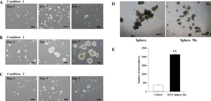
REM134 세포의 tumorsphere 형성을 통한 악성종양 특성 을 극대화하고자 서로 다른 3가지 실험조건에서 세포를 배 양하였다. Tumorsphere 형성 유도 7일 경과 후 inverted microscope를 이용하여 세포 모양을 확인한 결과, basal medium에 B27 supplement를 첨가한 조건 2에서 tumorsphere
형성 수와 크기가 증가함을 확인하였다. 형성된 tumorsphere 를 단일 세포로 분리하여 정량적으로 확인한 결과, 각각 조 건 1에서 7.9 × 10⁴개, 조건 2에서 470 × 104개, 조건 3에서 7.22 × 10⁴개로 나타났으며, 조건 2에서 형성된 tumorsphere 로부터 분리된 단일 세포수가 조건 1과 조건 3보다 약 60배 가량 증가하였음을 확인하였다. 이 결과를 바탕으로 차후의 실험에는 조건 2를 적용하여 진행하였다.
다음으로 methylcellulose의 유무에 따른 tumorsphere 형 성능을 비교하였다. 그 결과 methylcellulose를 첨가하지 않 은 실험조건에서는 tumorsphere 형성 시 크기가 일정치 않 고 370 ± 43.43개가 형성되었으며, 형성과정 중 자연적으로 뭉쳐 자라는 현상이 관찰되었다. 반면에 0.5% methylcellulose 를 첨가한 실험조건에서는 tumorsphere 형성 시 크기는 작 지만 비교적 일정한 크기를 나타내었으며 2,140 ± 56.57개로
Fig. 1. The morphology of REM134 cells. REM134 cells show an epithelial-like phenotype at the following magnifi- cations (A) 100× and (B) 200×. Scale bars = 100 µm.
Fig. 2. Sphere-forming capacity of REM134 cells. Spheres formed from REM134 cells plated at a density of 2 × 10⁴ cells/well in six well ultra-low attachment plates for different tumorsphere formation mediums: (A) condition 1, (B) condition 2, and (C) condition 3 for 7 days. (D) Representative images and (E) histogram showing the sphere forming capacity of spheres from REM134 cells in condition 2 medium with methylcellulose (Sphere Me) or without methylcellulose (Sphere). Values are mean± SD of three independent experiments (**p < 0.001). Scale bars = 100 µm (A–D).
methylcellulose를 첨가하지 않았을 때 보다 많은 수의 tumorsphere가 형성됨을 확인할 수 있었다(Fig. 2).
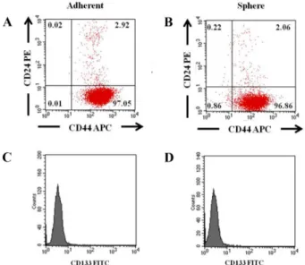
Fig. 3. Flow cytometry analysis of the expression of CD24, CD44, and CD133 in REM134 cells. (A) Expression of the CD44+/CD24− phenotype in plating-cultured and (B) sphere- forming cells of REM134 cells. (C) Expression of CD133 in plating-cultured and (D) sphere-forming cells of REM134 cells compared to an isotype control. Values are mean± SD of three independent experiments.
Fig. 4. Drug resistance assay of plating-cultured and sphere- forming cells from REM134 cells. Cell viability assays in the presence of doxorubicin. Sphere-forming (white dots) and plating-cultured cells (black dots) were treated with different concentrations of doxorubicin for 48 h. Values are means± SD of three individual experiments. **p < 0.001, compared with the plating-cultured cells.
Fig. 5. Expression of stem cell-related markers. (A) Expression of surface molecules (CD34, CD44, and CD90), (B) pluripotency markers (Oct4, Sox2, and Nanog), and (C) cancer stem cell markers (CD133, Bmi-1, EGFR, HER-2, PR, and vimentin) in plating- cultured and sphere-forming cells from REM134 cells with methylcellulose (Sphere Me) or without methylcellulose (Sphere) by qRT-PCR analysis. β-actin was used as a housekeeping gene. Values are means ± SD of three individual experiments. *p < 0.05,
**p < 0.001, compared with the plating-cultured cells.
유세포 분석을 통한 세포 표면 특이단백질 발현 조사 REM134 세포의 adherent 세포와 tumorsphere 형성 세포 의 세포 표면 특이단백질 발현을 비교하고자 유세포 분석방 법을 이용하여 CD24, CD44 및 CD133 발현 여부를 조사 하였다. 그 결과, 두 세포 모두 CD24−/CD44+인 것으로 확 인되었으며, 유세포 분석으로는 암 줄기세포의 특이 마커로 알려진 CD133의 발현은 확인되지 않았다(Fig. 3).
Tumorsphere 형성 세포의 약물 저항성 조사
암 화학치료 약물인 doxorubicin을 이용하여 REM134 세 포의 adherent 세포와 tumorsphere 형성 세포의 약물 저항 성을 비교하였다. 종양세포의 우선 생존능을 비교하고자 adherent 세포와 tumorsphere 형성 세포로부터 분리한 단일 세포에 doxorubicin을 처리한 다음 세포 생존율을 조사하였 다. 그 결과 adherent 세포는 doxorubicin을 250, 500, 1,000 ng/mL로 처리하였을 때 각각 59.19 ± 2.80%, 51.07 ± 1.07%, 37.56± 0.44%의 세포 생존율을 나타내었고, tumors-
phere 형성 세포로부터 유래한 세포는 각각 82.95 ± 3.45%, 68.21± 1.98%, 58.47 ± 2.73% 세포 생존율을 나타내어 tumorsphere 형성이 adherent 세포보다 doxorubicin에 대한 세포 독성에 높은 저항성을 가지는 것을 확인할 수 있었다 (Fig. 4).
qRT-PCR을 이용한 악성종양 특이유전자 발현 조사 Methylcellulose 유무에 따른 REM134 세포의 tumorsphere 형성 시, 악성종양 특이유전자 발현을 확인하고자 qRT-PCR 을 수행하였다. 그 결과 adherent 세포보다 tumorsphere 형 성 세포에서 CD34, CD90, Oct4, Sox2, Nanog, CD133, Bmi-1, human epidermal growth factor 2 (HER-2), vimentin 등의 발현이 증가하였고 progesterone receptor (PR)의 발현 은 감소하였다. 즉, tumorsphere 형성 시 악성종양 특이적 유전자의 발현이 증가함을 확인하였다(Fig. 5).
면역 부전 마우스에서 종양 형성 및 관찰
REM134 세포의 adherent 세포와 tumorsphere 형성 세포 로부터 분리한 단일 세포를 암컷 면역 부전 마우스의 유방 지방체에 투여한 다음 종양 형성과정을 관찰하였다. 2주 후 두 가지 실험군에서 모두 종양이 형성되는 것을 확인할 수 있었으며, adherent 세포를 투여하였을 때보다 tumorsphere 형성 세포를 투여한 실험군에서 종양의 크기가 증가하였음 을 확인하였다. 종양 형성 6주 차에 종양의 크기를 측정한 결과, adherent 세포를 투여한 마우스에서 57.67 ± 50.65 mm3, tumorsphere 형성 세포를 투여한 마우스에서 69.50 ± 23.06 mm3를 나타내었다(Fig. 6).
고 찰
반려동물 종양 치료는 외과적 절제가 대부분이고 아직까 지 명확한 치료법은 없다. 종양 치료 시 종양의 핵심이 되는 암 줄기세포를 완전히 제거하지 못할 경우 살아남은 암 줄 기세포는 다시 종양을 재생시키므로 종양의 진행, 재발 및 전이에 중요한 역할을 한다. Tumorsphere 형성은 암 줄기세 포의 특성을 더욱 분명하게 하는 중요한 방법으로 항암 기 초연구 및 약물검사에 널리 이용되고 있으며, 기존의 배양시 스템은 sphere 간의 융합을 초래하고 이로 인한 암 줄기세포 의 자발적인 분화, 구의 크기 및 모양의 불안정, 큰 구의 형 성 등 여러 가지 단점을 보유하고 있다. 이러한 sphere 간의 융합을 방지하고자 배양액에 methylcellulose와 같은 증점제 를 첨가하여 3D matrix 구조를 형성하거나 매질의 점도를 증가시키기도 한다 [16].
사람 암세포는 무혈청 배지에 bFGF, EGF, insulin, heparin 및 B27을 첨가한 저부착 세포배양 조건에서 세포를 배양할 경우 tumorsphere가 형성된다고 알려져 있으며 [10], 위의 방법으로 Cocola 등 [7]도 개 유선암 줄기세포의 분리 를 보고한 바 있다. 그러나 REM134 세포로부터 tumors- phere를 형성하기 위해서는 기본적인 성장인자 외 추가적인 Fig. 6. Tumorigenic capacity of REM134 cells. (A) 1 × 10⁵
cells of singly suspended REM134 cells from plating-cultured or sphere-forming cells into injected nude mice. Tumor formation was observed for six weeks after injection. Signifi- cant differences were detected within four weeks in sphere- forming cells injected mice compared to that in plating- cultured cells. Values are means± SD of three mice. *p < 0.05, compared with the adherent cells. (B) Macroscopic appearances of dissected tumors from plating-cultured cells on the left and sphere-forming cells on the right at six weeks. Scale bars = 12 mm (left), 15 mm (right).
요소가 필요하다. Blacking 등 [4]도 REM134 세포에 bFGF, EGF, N2 및 methylcellulose (0.8%)를 포함한 배양 액을 사용하여 tumorsphere를 형성한 것을 확인하였으며, Pang 등 [24]도 REM134 세포에 bFGF, EGF, insulin, progesterone, putrescine, sodium selenite, transferrin 및 추 가적인 성장인자를 첨가하여 tumorsphere 형성을 확인한 바 있다. Zhu 등 [37]은 다양한 사람 암세포에서 methyl- cellulose와 gellan gum을 이용하여 tumorsphere 형성과 그 특성을 비교하였다. 그 결과 3D 배양시스템에서 형성된 tumorsphere의 높은 세포밀도와 형태학적 특성이 개선되고 암 줄기세포의 특성을 유지함으로써 0.3% methylcellulose의 첨가는 종양의 융합을 조절하기 위한 적절한 농도라고 보고 한 바 있다 [37].
본 연구에서는 REM134 세포로부터 암 줄기세포의 중요 한 특성 중 하나인 tumorsphere를 효율적으로 형성할 수 있 는 배양 조건을 확립하고 기존의 연구 결과들을 바탕으로 tumorsphere 형성에 필요한 성장인자들을 다양하게 조합하여 배양액에 첨가한 다음 tumorsphere 형성능과 그 특성을 비 교하고자 하였다. 기존에 보고된 연구 결과들을 바탕으로 여 러 가지 조건으로 tumorsphere 형성을 유도한 결과, 최종적 으로 bFGF, EGF, insulin, progesterone, putrescine chloride, transferrin, sodium selenite, B27 supplement 및 0.5%
methylcellulose를 포함한 배양액에서 tumorsphere가 가장 잘 형성되고 균일한 크기의 tumorsphere가 대량으로 형성되는 것을 확인하였다.
다양한 종양에서 CD34, CD44, CD90 및 CD133 등이 암 줄기세포의 마커로 보고되었다 [1, 22, 36]. Michishita 등 [20]은 개 유선종양 유래 CHMp 세포주에서 형성한 tumorsphere 형성 세포에서 CD44와 CD133의 발현 증가 및 암 줄기세포의 자가재생능과 관련된 Oct4, Sox2, Nanog 와 종양 마커인 Bmi-1의 발현 증가를 보고하였다 [31].
Pang 등 [24]도 REM134 세포에 존재하는 소량의 암 줄기 세포가 이들 세포의 주요한 특징인 tumorsphere 형성능을 증가시킨다고 보고한 바 있다. 유선종양에서 표현형의 존재 는 악성종양을 확인하는 데에 있어 상당히 중요한 부분이다.
개 유선종양 연구에서 CD44+/CD24− 표현형의 존재는 높은 등급의 악성종양과 관련된다고 보고되었다 [19]. 이러한 연 구 결과를 바탕으로 유세포 분석을 통해 REM134 세포의 adherent 세포와 tumorsphere 형성 세포에서 각각 CD44+/
CD24− 표현형의 존재를 확인하였으나, 유세포 분석 결과에 서는 암 줄기세포의 대표적인 표현형인 CD133은 확인되지 않았다.
DNA 손상과 세포 증식을 억제하는 항암제 doxorubicin을 이용한 약물 저항성 실험은 암 줄기세포의 약물 저항 효과 를 확인하고자 널리 이용되는 방법이다 [24]. REM134 세포 의 adherent 세포와 tumorsphere 형성 세포의 약물 저항성 을 비교한 결과 tumorsphere 형성 세포가 adherent 세포보 다 약물 저항성이 크다는 것을 확인할 수 있었으며, 이는 곧 항암치료에 대한 내성이 크다는 것을 의미한다. 또한,
tumorsphere 형성 세포에서 methylcellulose 첨가와 상관없이 악성종양 특이적 마커로 알려진 유전자의 발현량이 증가하 는 것으로 나타났다. CD34, CD90, Oct4, Sox2, Nanog, CD133, Bmi-1, HER-2, vimentin의 발현이 tumorsphere 형 성 세포에서 높았으나 PR의 발현은 낮았다.
사람 유방암은 암 줄기세포를 확인한 최초의 종양이며, Al-Hajj 등 [1]은 종양 개체군 내의 기능적 이질성 존재에 대해 언급한 바 있다. 이러한 암 줄기세포는 종양 발달 및 전이를 무한으로 유지하기 위한 다분화능과 자가재생능을 가 진다 [23]. 사람 유방암 치료에서 저항성은 치료를 위한 주 요한 문제 중 하나이다. 저항성 관련 주요 인자로 epidermal growth factor receptor (EGFR)와 HER-2의 과발현, estrogen receptor와 PR의 발현 감소 등이 논의된다 [32]. 개 유선종 양 연구에 대한 많은 연구가 수행되었는데, 이 중 자발적인 악성 유선종양에서 스테로이드 호르몬 의존성의 감소가 관 찰되고, PR 음성인 악성종양이 양성종양보다 더 빠른 속도 로 증식한다고 보고되었다 [13]. EGFR은 ErbB 계열의 tyrosine kinase 수용체로 사람 유방암 치료의 표적인자로 연 구되고 있다. 개 유선종양에서 EGFR의 역할을 연구한 보고 는 많지 않다. Rutteman 등 [28]은 EGFR의 과발현이 있는 종양에서 무병 기간과 전체적인 생존율은 감소하였으나 통 계적 유의성은 없었다고 보고한 바 있다.
Korsching 등 [15]은 전이성을 가진 사람 유방암에서의 vimentin 발현을 보고하였다. 또한 이러한 vimentin의 발현 은 일반적으로 상피-중간엽 전이(epithelial-mesenchymal transition) 또는 근상피 조직 형성(myoepithelial histogenesis) 에 의한 것으로 추정되며 높은 종양 침윤 및 내 화학성과도 관련이 있다고 보고하였다. Pang 등 [24]은 Boyden chamber를 이용한 invasion 분석 결과에서 REM134 세포의 adherent 세포보다 tumorsphere 형성 세포가 침윤 가능성이 높고 tumorsphere 형성 세포의 단백질 발현량을 조사한 결 과에서 vimentin의 발현 증가를 보고하였다. 본 연구에서도 REM134 세포가 sphere를 형성하였을 때 CD133, Bmi-1, HER-2, vimentin 등 다양한 악성종양 마커가 증가하는 것을 확인하였고 tumorsphere 형성 세포의 마우스 투여 시 adherent 세포보다 더 빠른 종양 형성능을 확인할 수 있었다.
Else 등 [11]도 CBA Nu/Nu 암컷 마우스에 107개의 REM134 adherent 세포를 투여한 경우, 5일 차에 0.4 cm 크기의 고형 종양 확인과 3주 차에 2 cm 크기에 달하는 종 양을 확인하였으며, 1 × 10⁵개 이하의 세포를 투여한 경우의 마우스에서는 종양이 형성되지 않았다고 보고하였다. 본 연 구에서는 1 × 10⁵개의 REM134 세포 유래 adherent 세포와 tumorsphere 형성 세포를 암컷 면역 부전 마우스에 투여한 결과, 2주 차에 종양 형성이 확인되었고 6주 차까지 관찰한 결과, tumorsphere 형성 세포를 투여한 실험군에서 종양의 크기가 현저하게 증가함을 확인하였다. 또한, 4주 차부터는 외부 괴사가 시작되어 REM134 세포 유래 종양 형성과정에 는 악성종양의 분자적 특성뿐만 아니라 염증 및 괴사 관련 인자도 함께 관여하는 것으로 추정된다.
본 연구는 개 유선종양에서 유래한 REM134 세포의 tumorsphere 형성을 통해 악성종양 특이적 마커의 발현 변화 와 종양의 진행 속도가 더욱 촉진되는 것을 확인함으로써 유 선종양을 포함한 향후 반려동물 종양 치료를 위한 항암기전 연구 및 항 종양물질 개발 연구에 도움이 되고자 하였다.
감사의 글
본 연구는 농림축산검역본부 연구사업(B-1543083-2018-20- 01)의 지원으로 수행되었으며, 이에 감사드립니다.