Anti-oxidant and Anti-inflammatory Activities of Barley Sprout Extract
Cheong-Su Eun†, Eun-Young Hwang†, Syng-Ook Lee, Seun-Ah Yang and Mi-Hee Yu*
Department of Food Science and Technology, Keimyung University, Daegu 704-701, Korea Received January 11, 2016 /Revised March 11, 2016 /Accepted April 1, 2016
Barley (Hardeum vulgare L.) sprout has received much attention in recent years as a functional food in many countries, especially in Korea and Japan. It has been reported that barley sprouts are com- prised of 52.6% polysaccharides, 34.1% proteins, and 4.97% fats, along with a variety of vitamins, min- erals, and polyphenols. The purpose of this study was to assess the anti-oxidant and anti-in- flammatory activities of the ethanol extracts of barley sprouts. We examined the inhibitory effect of barley sprout extracts (BSE) on inducible nitric oxide synthase (iNOS) and cyclooxygenase-2 (COX-2) expression, and nitric oxide (NO), prostaglandin E2 (PGE2) and cytokine production in lip- opolysaccharide (LPS)-stimulated RAW 264.7 mouse macrophage cells. BSE contains high amounts of phenolics and flavonoids and exhibits potent anti-oxidative activity, as depicted by the DPPH radi- cal-scavenging experiment. The concentration of total phenols was 17.55 μg/ml, and flavonoids, 13.98 μg/ml. We also investigated the anti-inflammatory activities of BSE in LPS-stimulated RAW 264.7 cells. Tumor necrosis factor-alpha and PGE2 production, which had increased as a result of treatment with LPS, were significantly inhibited by BSE in a dose-dependent manner. BSE also significantly sup- pressed LPS-induced production of NO, and this was accompanied by a decrease in the expression of the iNOS and COX-2 proteins. These results indicate that barley sprouts may be a highly valuable natural product owing to its high-quality functional components as well as its anti-oxidant and an- ti-inflammatory activities.
Key words : Anti-inflammatory, anti-oxidant, barley sprout, macrophage, Raw 264.7
†Authors contributed equally.
*Corresponding author
*Tel : +82-53-580-5538, Fax : +82-53-580-5538
*E-mail : [email protected]
This is an Open-Access article distributed under the terms of the Creative Commons Attribution Non-Commercial License (http://creativecommons.org/licenses/by-nc/3.0) which permits unrestricted non-commercial use, distribution, and reproduction in any medium, provided the original work is properly cited.
Journal of Life Science 2016 Vol. 26. No. 5. 537~544 DOI : http://dx.doi.org/10.5352/JLS.2016.26.5.537
서 론
생체의 대사과정에서 발생되는 활성 산소류(reactive oxy- gen species, ROS)는 지질, 단백질, 그리고 핵산과 같은 체내 주요 물질의 비가역적 손상을 야기시키며, 세포가 지속적으로 과도한 산화적 스트레스에 노출되면 세포사멸뿐만 아니라 퇴 행성 질환을 일으키는 특정세포의 유전자 발현이 증가되어 만성적 염증 반응이 유도된다[13, 29]. 염증반응은 외부의 자극 에 의한 생체조직의 국소적인 방어보호 반응으로, 대식세포인 macrophage 등이 일산화질소(nitric oxide, NO)와 프로스타 글란딘(prostaglandin, PG), 그리고 tumor necrotic factor-α (TNF-α)와 같은 pro-inflammatory cytokine 등과 같은 염증매 개물질의 생성에 관여하고 이를 조절한다[22, 24]. NO는 염증 반응에서 중요한 역할을 하는 염증매개조절 인자로, nitric ox- ide synthases (NOS)에 의해 L-arginine으로부터 생성되며, iNOS (inducible nitric oxide synthase)는 염증반응에 관여하
는 대식세포에서 NO를 생성하여 염증반응을 촉진하는 역할 을 한다. 또한 Cyclooxygenase-2 (COX-2)는 arachidonic acid 를 prostaglandin E2 (PGE2)로 전환시키는 효소로 prosta- glandin (PG)류의 다양한 염증반응 매개체들을 생성한다[19, 23].
흡연이나 음주, 잘못된 생활 습관, 각종 환경오염물질 및 약물 등에 의해 활성 산소종이 세포 내에 과도하게 발생하면 체내 산화적 스트레스가 증가하여 만성질환을 일으키는 원인 이 된다[39]. 이러한 산화적 스트레스를 경감시킬 수 있는 항산 화제에 대한 현대인들의 관심이 높아짐에 따라 국내외적으로 항산화 및 항염증에 대한 연구가 활발히 진행되고 있다[1, 41].
특히, 최근에는 만성질환의 예방과 치료를 위한 방법으로 채 소류나 곡류와 같은 식물성 식품 섭취에 대한 관심이 증가하 고 있으며[28, 37], 그 중 새싹 채소는 건강식품으로 각광 받고 있어 이에 따라 소비량과 기호도가 높아지고 있다[21, 26].
보리(Hordeum vulgare L.)에는 lutonarin (isoorientin-7-O- glucoside), saponarin (flavone-C-glycosides) 등 다양한 폴리 페놀과 플라보노이드가 함유되어 있으며, 수용성 식이섬유인 β-glycan, 아미노산, 탄수화물이 함유되어 있다. 또한, 보리 잎 중 풍부하게 들어있는 superoxide dismutase (SOD)와 보리 잎에서 분리된 glycoprotein인 P4-D1, D1-G1은 강한 항염증 작용에 대해 알려져 있다[26]. 새싹 보리의 경우 외부 공격으로 부터 자신을 보호하고 싹을 틔우기 위한 다양한 생리활성물질
들을 함유하고 있으며[18, 20, 23], 백색광 등 저 에너지의 광을 받으면 C-glycosylflavone 생성이 증가되며[8, 10], 특히, 6-c- glycosyl-7-o-glucosylapigenin, 2-C-glycosylisovitexin은 보리 잎에서 발견된 대표적인 flavone계 항산화 물질로 알려져 있 다[31]. 플라보노이드는 이 외에도 항암작용, 항박테리아작용, 항바이러스작용, 항염작용, 효소활성 억제능 및 면역계에 대 한 작용 등 여러 가지 생물학적 활성을 지니고 있다[30, 31, 34].
이에 본 연구에서는 보리싹 추출물을 이용하여 DPPH radi- cal 소거활성 및 총 폴리페놀, 플라보노이드 함량을 조사하여 항산화 효과를 확인하고, LPS로 염증반응이 유도된 RAW 264.7 cell을 통해 NO, PGE2의 활성과 함께 iNOS, COX-2의 단백질 발현에 미치는 보리싹의 항염증 효과를 확인하여 보리 싹을 이용한 기능성 바이오 소재 개발을 위한 기초자료로 제 공 및 응용하고자 한다.
재료 및 방법
시료제조
본 실험에서 사용한 보리싹은 (주)에이팜(Daegu, Korea)에 서 제공받아 사용하였다. 시료는 불순물을 제거하기 위해 수 세한 후 건조하여 사용하였고, 무게의 10배량(w/v)의 70% 에 탄올을 가하여 24시간 동안 정치하여 총 3회 반복 추출 하였 다. 추출액은 여과지(Whatman NO. 3, Whatman Internatio- nal Ltd., Maidstone, England)로 여과한 다음 rotary evapo- rator (UT-1000, EYELA, Tokyo, Japan)로 55℃에서 농축한 후 동결 건조하여 에탄올 추출물로 사용하였다.
세포주 배양
대식세포 계열(murine macrophage cell line)인 RAW 264.7 세포주는 한국 세포주 은행(KCLB, Seoul, Korea)으로부터 분 양 받았으며, 10% FBS (fetal bovine serum)와 1% antibiotics (penicillin/streptomycin)를 첨가한 Dulbeco’s modified Eagle’s medium high glucose (DMEM) (Welgene, Daegu, Korea)배지를 이용하여 5% CO2가 존재하는 37℃ incubator에 서 1주일에 2~3회 계대 배양하였다. 세포는 D60 culture dish (1×10⁶ cells/dish) 및 6 well plate (5×10⁵ cells/dish)에 주입하 여 부착시키고, 시료를 농도별로 처리하여 24시간 배양한 후, lipopolysaccharide (LPS) 100 ng/ml를 첨가하여 24시간 배양 시킨 후 실험에 이용하였다. 이때, LPS를 처리하지 않은 실험 군을 negative control로 하였으며, LPS를 처리한 것을 pos- itive control로 하였다.
α-α-diphenyl-β-picrylhydrazyl (DPPH) radical 소거 활성
DPPH radical에 대한 각 시료의 환원력을 측정하기 위해
99% 메탄올에 각 시료를 녹여 농도별로 희석한 희석액 800 μl와 메탄올에 녹인 0.15 mM DPPH용액 200 μl를 가하여 실온 에 30분 방치한 후 biotekmicroplate spectrophotometer (Bio- tek instruments)를 이용하여 517 nm에서 흡광도를 측정하였 다. 각 시료 추출물의 유리 라디칼 소거활성은 시료를 첨가하 지 않은 대조구의 흡광도를 1/2로 환원시키는데 필요한 시료 의 농도인 RC50 값으로 나타내었다. 이때 활성비교를 위하여 합성항산화제인 butylated hydroxyl anisole (BHA)를 사용하 여 항산화 효과를 측정하였다.
총 폴리페놀 함량
총 폴리페놀 함럄은 Folin-Denis법[3]을 응용하여 측정하였 다. 즉 각 메탄올 추출물 시료 1 mg을 증류수 1 ml에 녹이고 10배 희석한 희석액 2 ml에 2배 희석한 Folin 시약 2 ml을 첨가하고 잘 혼합한 후 3분간 방치한 후 10% Na2CO3 2 ml을 넣고 1시간 반응 시킨 후 biotekmicroplate spectrophotometer (Biotek instruments)를 이용하여 700 nm에서 흡광도를 측정 하여 작성한 표준곡선으로부터 함량을 구하였다. 이때 tannic acid를 이용한 표준곡선은 5, 10, 25, 50, 75, 100 μg/ml가 되도 록 하여 위와 같은 방법으로 700 nm에서 흡광도를 측정하였 다.
총 플라보노이드 함량
시료 추출물의 총 플라보노이드 함량은 Nieva Moreno법 [33]을 이용하여 측정하였다. 각 시료 추출물 0.1 ml에 80%
ethyl alcohol 0.9 ml, 10% aluminum nitrate 0.1 ml, 1 M po- tassium acetate 0.1 ml 및 80% ethyl alcohol 4.3 ml를 혼합하 여 실온에서 30분간 반응시킨 후 biotekmicroplate spectro- photometer (Biotek instruments)를 이용하여 415 nm에서 흡 광도를 측정하였다. 표준물질로 quercetin을 사용하였으며, 표 준물질의 검량선과 비교하여 함량을 구하였다.
세포독성 측정
시료의 세포증식과 독성을 측정하기 위해 Green 등의 방법 [4]에 따라 3-(3,4-dimethyl- thiazolyl-2)-2,5-diphenyl tetrazo- lium bromide (MTT) assay를 실시하였다. MTT assay는 미토 콘드리아의 탈 수소 효소작용에 의하여 노란색의 수용성 기질 인 MTT가 불용성의 보라색 formazan으로 환원되는 원리를 이용한 방법으로, 생성된 formazan의 흡광도는 살아있고 대 사가 왕성한 세포의 농도를 반영한다. RAW 264.7 cell이 1×10⁵ cells/well의 농도가 되게 96 well plate에 분주하고, 37℃, 5%
CO2 incubator에서 24시간 동안 배양하였다. 배양한 RAW 264.7 세포는 serum free 배지로 교체한 후 LPS (100 ng/ml)와 시료를 처리하여 24시간 배양하여 5 mg/ml의 MTT 시약 (Amresco, Solon Ind., Columbus, Ohio, USA)을 5 mg/ml 농 도로 10 μl를 각 well에 처리하고 incubator에서 4시간 동안
배양하였다. 배양 종료 후 상등액을 제거하고 각 well에 100 μl의 DMSO를 첨가하여 생성된 formazan 결정을 용해시켜 biotekmicroplate spectrophotometer로 550 nm에서 흡광도를 측정하였고, 세포독성은 시료의 흡광도를 대조군의 흡광도에 대한 백분율로 나타내었다.
Nitric oxide (NO) 생성량 측정
RAW 264.7 세포로부터 생성되는 활성질소종인 nitric ox- ide (NO)의 양은 Green 등[5]의 방법을 이용하여 세포 배양액 중 존재하는 NO2-
형태를 Griess Reagent와 반응시켜 측정하 였다. RAW 264.7 cell을 DMEM 배지를 이용하여 1×10⁵ cells/
ml 농도로 96 well plate에 분주한 후 시료를 농도별로 처리하 여 24시간 배양하였으며 Lipopolysaccharide(LPS) 100 ng/ml 를 첨가하여 다시 24시간 배양시켰다. 세포배양 상등액 100 μl와 Griess시약(1% sulfanilamide, 0.1% naphthylethylendi- amine in 25% phosphoric acid) 100 μl를 혼합하여 96 well plates에서 10분간 반응시킨 후 biotekmicroplate spectropho- tometer를 이용하여 540 nm에서 흡광도를 측정하였다. NO2-
표준곡선은 NaNO2를 농도별로 조제하여 사용하였다.
Prostaglandin E2 (PGE2) 생성량 측정
활성화된 RAW 264.7 세포로부터 생성되는 염증 매개 물질 인 PGE2의 양을 측정하기 위해 Commercial competitive en- zyme immunoassay kit (Cayman Chemicals, San Diego, CA, USA)를 구입하여 실험하였다. Raw 264.7세포를 1×10⁶ cells/
ml 농도로 6 well plate에 분주하고 24시간 배양한 후, 농도별 로 시료와 LPS (100 ng/ml)를 처리하고 다시 24시간 배양시켜 세포배양 상등액을 이용하여 실험하였다. 즉, Goat polyclonal anti-mouse IgG가 coating된 96 well plate에 세포 배양 상등액 50 μl와 prostaglandinE2 acetylcholinesterase 50 μl와 anti- body를 4℃에서 18시간 배양한 후에 wash buffer (0.2%, Tween 20)를 이용하여 수세하였다. Ellman's reagent (5,5'-di- thio-bis-(2-nitrobenzoic acid, acetylthiocholine)를 200 μl 첨가 하여 60~90분 동안 반응시킨 후에 420 nm에서 흡광도를 측정 하였다.
Tumor necrosis factor-alpha (TNF-α) 생성량 측정 Raw 264.7 cell을 DMEM배지를 이용하여 1×10⁶ cells/ml 농도로 6 well plate에 분주하여 24시간 배양한 후, 시료를 농 도별로 처리하고 LPS (100 ng/ml)를 첨가하여 다시 24시간 배양시켰다. TNF-α의 생성량은 Commercial competitive en- zyme immunoassay kit (PEPROTECH, USA)을 이용하여 측 정하였다. TNF-α antibody가 coating된 96well plate에 세포배 양 상등액 100 μl를 첨가하여 2시간 동안 실온에서 반응시킨 후 wash buffer를 이용하여 수세한 후 Biotin conjugate (15 mM sodium azide) 100 μl를 첨가하여 다시 1시간 동안 실온에
서 반응시킨 다음 수세한다. Streptavidin-HRP (3.3 mM thy- mol) 100 μl와 살균된 chromogen 역시 같은 방법으로 30분 반응과 수세를 반복한 후 stop solution (0.18 M phosphoric acid)으로 반응을 정지 시키고 450 nm에서 흡광도를 측정하였 다.
Western bolt analysis
보리싹 추출물이 염증반응에서 발현하는 인자들의 발현에 미치는 영향을 조사하기 위해 Western bolt을 실시하였다.
RAW 264.7 cell이 1×10⁶ cells/well의 농도가 되게 6 well plate 에 분주하고, 37℃, 5% CO2 incubator에서 24시간 동안 배양하 였다. 배양한 RAW 264.7 세포는 serum free 배지로 교체한 후 LPS (100 ng/ml)와 시료를 처리하여 24시간 배양하였다.
샘플 처리가 완료된 세포를 2-3회 PBS로 세척한 후 1 ml의 lysis buffer를 첨가, 30분에서 1시간 동안 용해시킨 후 13,000 rpm에서 10분간 원심 분리하여 세포막 성분 등을 제거하였다.
단백질 농도는 bovine serum albumin (BSA)를 표준화 한 Bio-Rad protein assay kit (Hercules, CA, USA)를 사용하여 정량하였다. 4℃에서 13,000 rpm으로 10분간 원심 분리한 상 등액은 단백질을 정량한 후 10% running gel과 4.5% stacking gel을 이용하여 125 V에서 SDS-polyacrylamide gel 전기영동 (SE260, Amersham Biosciences, Uppsala, Sweden)을 실시하 였다. 전기영동으로 분리한 단백질은 immobilon-P transfer membrane (PVDF transfer membrane, Port Washington, NY, USA)과 transfer buffer (20% methanol, 25 mM Tris-HCl, 192 mM glycine)를 사용하여 350 mA에서 120분간 transfer하였 다. 단백질이 이동된 membrane은 fast green solution으로 transfer의 유무를 확인한 후, 5% non-fat skim milk solution 으로 blocking하였다. 4℃에서 일차 항체의 발현 정도를 검토 하기 위하여 TST (Tris-Buffered Saline and Tween 20) 용액에 1:1,000으로 희석하여 24시간 반응시킨 후 TST로 3 회 세척하 였다. 계속하여 이차항체를 2시간 TST 용액에 1:2000으로 희 석하여 반응시키고 다시 TST로 3회 세척하였다. 증류수로 한 번 더 세척하고 membrane에 ECL detection kit (WSE-7120s, ATTO Co., Tokyo, Japan)의 발색시약 Ⅰ과 Ⅱ를 1:1로 섞은 후에 혼합액을 도포하고, X-ray film (CP-G plus, Agfa Health- care Ltd, New Orleans, LA, USA)에 노출하여 현상한 후 film 상에서 iNOS, COX-2의 발현정도를 관찰하였다.
통계처리
모든 결과는 평균±표준편차로 나타내었으며, 유의성 검사 는 SPSS TM version 21 (SPSS Inc., Chicago, USA)을 이용하여 one-way ANOVA 를 실시하였고, 대조구인 분화 Control에 대한 시료 처리구의 통계적 유의성은 Duncan’s multiple range test로 검증하였다. p<0.05 이상일 때만 통계적 유의성이 있는 것으로 판단하였다.
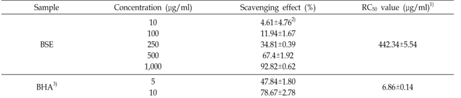
Table 1. DPPH radical scavenging activity of BSE
Sample Concentration (μg/ml) Scavenging effect (%) RC50 value (μg/ml)1)
BSE
10 100 250 500 1,000
4.61±4.762) 11.94±1.67 34.81±0.39 67.4±1.92 92.82±0.62
442.34±5.54
BHA3) 5
10
47.84±1.80
78.67±2.78 6.86±0.14
1)Concentration required for 50% reduction of DPPH at 30 min after starting the reaction
2)Each value is expressed as mean±SD in triplicate experiments.
3)t-Butylatedhydroxyanisole (BHA) were used as positive references.
Table 2. Total polyphenols and flavonoids contents of BSE
Sample Total polyphenols (μg/mg)1)
Total flavonoids (μg/mg)2)
BSE 17.55±0.533) 13.98±6.91
1)Micrograms of total polyphenol content/mg of plants based on tannic acid as standard.
2)Micrograms of total flavonoid content/mg of plants based on quercetin acid as standard.
3)Each value is expressed as mean±SD in triplicate experiments.
결과 및 고찰
DPPH radicals 소거활성
DPPH는 산화된 형태에서 free radical이 cysteine, gluta- thion, aromatic amine, BHA (butylated hydroxyl anisole) 등 에 의해 전자를 얻고 환원되어 짙은 자색의 DPPH가 diphe- nylpicrylhydrazine으로 탈색되는데 이것은 다양한 천연소재 로부터 항산화 물질을 검색 하는데 널리 이용되고 있다[12, 35]. 보리싹 추출물을 대상으로 DPPH 라디칼 소거 활성을 알 아보기 위해 합성 항산화제로 알려진 BHA와 비교하여 조사 하였다. 보리싹 추출물의 DPPH radical 소거능은 Table 1에 나타내었고, DPPH radical을 50% 저해하는 시료의 농도를 RC50값으로 나타내었다. 보리싹 추출물은 1,000 μg/ml 농도에 서 92.82±0.62%의 DPPH radical 소거활성을 보였으며, RC50
값은 442.34±5.54 μg/ml로 나타났다. Cho 등[32]의 연구 논문 에 따르면 콜라비 새싹과 무순 추출물 1,000 μg/ml의 농도에 서 각각 40%, 35%의 DPPH radical을 소거하였으며, Han 등 [6]의 보리순 추출물의 항산화 활성 보고에서 RC50 값이 1.226 mg/ml로 나타났던 것과 비교하면 본 논문의 보리싹이 더욱 우수한 DPPH radical 소거능을 보였다. 이는 재배 지역에 따 라 또는 추출 조건에 따라 보리싹 추출물의 항산화능에 차이 가 있는 것으로 사료된다.
총 폴리페놀, 총 플라보노이드 함량
폴리페놀계 물질들은 다양한 구조와 분자량을 가지며, 한 분자 내에 2개 이상의 phenolic hydroxyl (OH) 기를 가진 ben- zene 화합물을 가리킨다. 이는 색소 화합물인 플라보노이드와 탄닌이 주성분으로 항산화, 항균, 항암, 충치예방 등의 생리 기능을 가지는 것으로 알려져 있다[2]. 본 실험에서는 보리싹 추출물에 존재하는 폴리페놀 및 플라보노이드 함량을 각각 tannic acid, quercetin을 기준 물질로 하여 측정하였다(Table 2). 그 결과, 보리싹 추출물의 총 폴리페놀 함량과 플라보노이 드 함량은 각각 17.55±0.53, 13.98±6.91 μg/mg으로 나타났다.
Kim [17] 등이 연구한 발아 벼 부위별 에탄올 추출물의 폴리페
놀 함량과 비교하였을 때 발아 벼 11.23 mg/g, 발아 전곡, 왕겨 및 현미가 각각 2.10, 3.76 및 1.93 mg/g의 함량을 보여 보리싹 추출물의 높은 폴리페놀 함량을 확인할 수 있었다. 또한, Park [37] 등의 보고에 의하면, 5종 보리품종의 총 폴리페놀 함량은 0.58 μg/mg에서 많게는 1.63 μg/mg으로 나타났고, 총 플라보 노이드 함량은 0.24 μg/mg에서 0.40 μg/mg으로 나타났다.
5종 보리와 비교해본 결과 본 논문에서 연구한 보리싹 추출물 의 폴리페놀 및 플라보노이드 함량(폴리페놀: 17.55 μg/mg, 플라보노이드: 13.98 μg/mg)이 더 높은 편임을 알 수 있었다.
보리싹 추출물의 nitric oxide 소거활성
Nitric Oxide (NO)는 활성 질소종(reactive nitrogen spe- cies)의 하나이며, 일반적으로 종양을 제거하는 면역반응에 관 여하지만 과량 존재할 경우 인체에 염증 반응을 조절하여 다 양한 병리적인 반응을 유도한다[43]. NO 생성 저해에 대한 보리싹 추출물의 효과를 알아보기 위해 lipopolysaccharide (LPS)로 NO의 생성을 유도한 뒤 50, 100, 250, 500 μg/ml 농도 의 보리싹 추출물을 처리하고 생성된 NO의 양을 측정하였다 (Fig. 1A). LPS 처리군은 LPS 무첨가 군에 비하여 NO 생성량 이 증가하는 것을 확인하였고, 보리싹 추출물을 농도별로 처 리한 결과 모두 농도 유의적으로 NO 생성량이 감소하는 것을 확인 하였다. Karki [14] 등이 연구한 메밀순 추출물의 NO 생성량은 50, 100 μg/ml 농도에서 약 52, 45 μM이였으며, 보리 싹 추출물은 26, 22 μM이었다. 이러한 NO 생성 저해가 세포독
A
B
Fig. 1. Effect of BSE on NO production and cell viability in LPS- induced RAW 264.7 cells. RAW 264.7 cells were treated with Barley sprout ethanol extract and LPS (100 ng/ml) for 24 hr. NO production was determined in culture su- pernatant by Griess reagent. Cell viability was calculated as a percentage of MTT metabolism in controls. Results represent the mean±SD of three independent experi- ments. *p<0.05, **p<0.01, ***p<0.001 compared to the group treated with LPS.
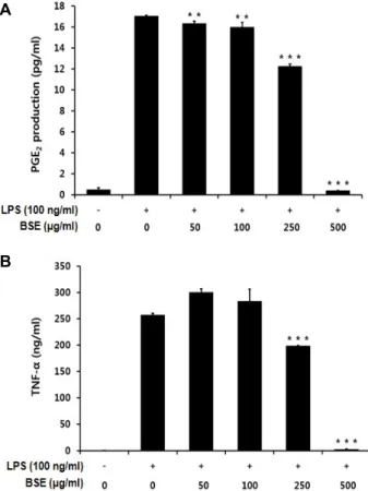
A
B
Fig. 2. Effect of BSE on PGE2 and TNF-αlevels in LPS-induced RAW 264.7 cells. RAW 264.7 cells were treated with Barley sprout ethanol extract and LPS (100 ng/ml) for 24 hr. Results represent the mean±SD of three independ- ent experiments. *p<0.05, **p<0.01, ***p<0.001 compared to the group treated with LPS.
성에 기인하는 것인지를 확인하기 위해 MTT assay를 이용하 여 세포독성을 확인하였다(Fig. 1B). 그 결과 보리싹 추출물의 50 μg/ml 에서 500 μg/ml의 전 농도 범위에서 100% 이상의 생존율을 보여 세포 생장에 영향을 미치지 않는 것을 확인하 였다.
보리싹 추출물이 염증성 사이토카인 PGE2, TNF-α 생성과 iNOS, COX-2에 미치는 영향
외부 자극으로 인해 염증이 발생하면 염증 세포에서 TNF- α, IL-1β와 IL-6 및 염증 매개 물질인 nitric oxide와 prosta- glandin E2가 분비된다[9]. PGE2는 prostaglandin endoper- oxide synthase 효소인 COX-2의해 합성되는 염증 매개 물질 로서 다양한 염증 질환을 야기시킨다. COX-2는 염증과 같은 병적인 환경에서 대식세포, 단핵구, 조골세포 등에서 분비되 며 조직이 손상되면 염증을 매개하는 물질들이 다량 생성되어 통증이 유발되게 된다[42]. 보리싹 추출물이 염증 매개물질을
억제할 수 있는지 알아보기 위해 LPS로 자극된 RAW 264.7 세포를 대상으로 PGE2 및 TNF-α 생성량을 조사하였다(Fig.
2B). 먼저 PGE2의 경우에는 Raw 264.7 세포에 100 ng/ml의 LPS를 처리했을 때, 처리 하지 않은 군에 비해 PGE2의 생성량 이 급격히 증가되었으며, 보리싹 추출물을 250, 500 μg/ml의 농도로 처리 했을 때 250 μg/ml에서는 미미하였지만, 500 μ g/ml에서 PGE2의 생성량이 현저하게 감소하였다(Fig. 2A).
TNF-α 생성은 LPS 무처리군에 비하여 LPS 처리구에서 200배 이상 크게 증가하였으며, 보리싹 추출물을 250, 500 μg/ml 농 도로 처리하였을 때 TNF-α의 생성이 유의적으로 감소되는 것 으로 나타났다(Fig. 2B).
iNOS는 평소에는 세포 내에 존재하지 않으나 LPS에 의해 유도되면 장시간 동안 다량의 NO를 생성하며, 염증상태에서 iNOS에 의해 생성된 NO는 혈관투과성, 부종 등의 염증반응 을 촉진시킬 뿐만 아니라 염증매개체의 생합성을 촉진한다.
이는 macrophage에서 LPS나 cytokine에 의해 염증성 매개물 질들이 과잉 생산되는 중요한 mechanism이 된다[11]. 또한, 염증 억제 약물들의 작용기전은 arachidonic acid를 PGE2로
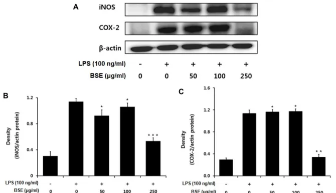
A
B C
Fig. 3. Inhibition effects of BSE on the expression of iNOS and COX-2 in LPS-stimulated RAW 264.7 cells. The RAW 264.7 cells (1×10⁶ cells/ml) were incubated 24 hr and then pretreated with and then pretreated with Barley sprout ethanol extract in the presence or absence of LPS (100 ng/ml) for 24 hr. Equal amounts of total protein were resolved by SDS-PAGE. Results represent the mean±SD of three independent experiments.*p<0.05, **p<0.01, ***p<0.001 compared to the group treated with LPS.
전환시키는 효소인 COX-2의 활성저해와 관계가 깊다[38, 40].
따라서 보리싹 추출물이 iNOS 및 COX-2 단백질의 발현에 미 치는 영향을 알아보기 위하여 세포질 내에서의 iNOS와 COX- 2 단백질의 발현양을 조사하였다. 음성 대조군은 LPS (100 ng/ml) 처리로 인해 iNOS 단백질의 발현이 증가한 것을 확인 하였으며, 보리싹 추출물을 각각 농도 별로 처리 하였을 때 250 μg/ml 에서부터 농도 의존적으로 iNOS의 발현이 억제되 는 것을 확인할 수 있었다(Fig. 3). 또한, COX-2 단백질 역시 LPS 처리로 인해 그 발현량이 증가하였으며, 보리싹 추출물 50, 100 μg/ml 에서는 COX-2의 발현의 변화가 거의 없었지만 250 μg/ml에서 COX-2의 발현이 크게 억제되는 것을 확인 할 수 있었다. Karki [14] 등과 Kim [22] 등은 NO 저해 활성이 높게 나타난 메밀순과 왕주똥나무잎 추출물에서 iNOS 단백질 발현을 저해한다고 보고하였으며, 보리싹 추출물의 NO생성 과 iNOS 단백질 발현 저해와 유사한 경향을 확인하였다. 또한, Park [36] 등과 Huang [7] 등의 연구에서는 철쭉나무와 자소잎 추출물이 COX-2 단백질의 발현 저해를 통해 염증성 사이토카 인(PGE2, TNF-a)이 감소됨을 확인하였다. 향후 보리싹 추출물 의 항염증 활성을 중심으로 기전 연구와 활성성분 규명에 대 한 연구가 추가적으로 이루어져야 할 것이다.
감사의 글
이 논문은 2014년도 교육부의 재원으로 한국연구재단의 지 원을 받아 수행된 기초연구사업임(NRF-2014R1A1A2056033).
References
1. Bak, M. J., Jeong, J. H., Kang, H. S., Jin, K. S., Seon, O. K.
and Jeong, W. S. 2009. Cedrela sinensis leaves suppress oxida- tive stress and expressions of iNOS and COX-2 via MAPK signaling pathways in RAW 264.7 cells. J. Food Sci. Nutr.
14, 269-276.
2. Chio, J. H., Lee, S. H., Park, Y. H., Lee, S. G., Jung, T. E., Lee, I. S., Park, J. H. and Kim, H. J. 2013. Antioixdant and alcohol degradation activities of extracts from Acertegmen- tosum Maxim. J. Kor. Soc. Food Sci. Nutr. 42, 378-383.
3. Folin, O. and Denis, W. 1912. On phosphotungstic-phospho- mo-lybdic compounds as color reagents. J. Biol. Chem. 12, 239-249.
4. Green, L. C., Reade, J. L. and Ware, C. F. 1984. Rapid colori- metric assay for cell viability: application to the quantitation of cytotoxic and growth inhibitory lymphokines. J. Immunol.
Methods 70, 257-268.
5. Green, L. C., Wagner, D. A. and Glogowski, J. 1982. Analysis of nitrate, and [15N]nitrate in biological fluids. Anal. Biochem.
126, 131-138.
6. Han, S. I. 2011. Antioxidative effects of the extract from bar- ley sprout and application of the extracts to an edible oil and fat. Th.M. dissertation, Chonbuk National University, Chonbuk, Korea.
7. Huang, B. P., Lin, C. H., Chen, Y. C. and Kao, S. H. 2014.
Anti-inflammatory effects of Perilla frutescens leaf extract on lipopolysaccharide-stimulated RAW264.7 cells. Mol. Med.
Rep. 10, 1077-1083.
8. James, A. S. and Jerry, W. M. 1976. The occurrence and pho- toregulation of flavonoid in barley plastids. Phytochemistry 15, 805-807.
9. Jeong, S. I., Kim, H. S., Jeon, I. W., Kang, H. J., Mok, J.
Y., Cheon, C. J., Yu, H. H. and Jang, S. I. 2014. Antioxidant and anti-inflammatory effects of ethanol extracts from Perilla fruescens. Kor. J. Food Sci. Technol. 46, 87-93.
10. Jerry, W. M. and Wilson, K. G. 1970. Photocontrol of C-gly- cosylflavone in barley seedlings. Phytochemistry 9, 763-773.
11. Jung, Y. S., Eun, C. S., Jung, Y. T., Kim, H. J. and Yu, M.
H. 2013. Anti-inflammatory effects of Picrasma Quassidides (D.DON) BENN leaves extract. J. Life Sci. 23, 629-636.
12. Jung, Y. T., Lee, I. S., Whang, K. and Yu, M. H. 2012.
Antioxidant effect of Picrasma quassioides and Chamaecyparis obtuasa (S. et Z.) ENDL extracts. J. Life Sci. 22, 354-359.
13. Kang, H. W. 2012. Antioxidant and anti-inflammatory effect of extracts from Flammulina velutipes (Curtis) Singer. J. Kor.
Soc. Food Sci. Nutr. 41, 1072-1078.
14. Karki, R., Park, C. H. and Kim, D. W. 2013. Extract of buck- wheat sprouts scavenges oxidation and inhibits pro-in- flammatory mediators in lipopolysaccharide-stimulated macrophages (RAW264.7). J. Integr. Med. 11, 246-252.
15. Kazuhiko, K., Yutaka, M. and Kirohumi, S. 1983. Isolation of protent anti-inflammatory protein from 29 barley leaf. J.
Inflammation 3, 238-245.
16. Kim, D. H., Park, S. R., Debnath, T., Hasnat, M. A., Pervin, M. and Lim, B. O. 2013. Evaluation of the antioxidant activ- ity and anti-inflammatory effect of Hericium erinaceus water extracts. Kor. J. Med. Crop Sci. 21, 112-117.
17. Kim, H. Y, Hwang, I. G., Kim, T. M., Park, D. S., Kim, J.
H., Kim, D. J., Lee, J. S. and Jeong, H. S. 2011. Antioxidant and angiotensin converting enzyme I inhibitory activity on different parts of germinated rough rice. J. Kor. Soc. Food Sci. Nutr. 40, 775-780.
18. Kim, I. S., Han, S. H. and Han, K. W. 1997. Study on the chemical change of amino acid and vitamin of rapeseed dur- ing germination. J. Kor. Soc. Food Sci. Nutr. 26, 1058.
19. Kim, K. A., Yi, H. S., Yun, H. J. and Park, S. D. 2009. Anti-ox- idative and anti-inflammatory effect of fractionated extracts of Cynomorium songaricum. Kor. J. Ori. Physiol. Pathol. 23, 1320-1331.
20. Kim, K. T., Kim, S. S. and Lee, S. H. 2003. The functionality of barley leaves and its application on functional foods. Food Science and Industry 36, 45-49.
21. Kim, Y. J., Park, H. T. and Han, H. S. 2006. A study on the production and marketing of sprouts and leaf vegetables.
Korea Rural Economic Institute 6-15.
22. Kim, Y. S., Lee, S. J., Hwang, J. W., Kim, E. H., Park, P.
J. and Jeong, J. H. 2012. Anti-inflammatory effects of extracts from Ligustrum ovalifolium H. leaves on RAW264.7 macro- phages. J. Kor. Soc. Food Sci. Nutr. 41, 1205-1210.
23. Lee, C. H., Shin, S. L., Kim, N. R., Yoon, S. E., Kim, S. I., Baek, S. H. and Hwang, J. H. 2009. Changes of antioxidant effect according to greening period of Astrangalus mem- branaceus var. membranaeus, Senna occidentalis, Dianthus long- icalyx, and Plantago asiatica sprout vegetables. Kor. J. Plant Res. 22, 349-358.
24. Lee, D. S., Kim, K. S., Li, B. Choi, H. G., Keo, S., Jun, K.
Y., Park, J. H. and Kim, Y. C. 2012. Anti-inflammatory effect of the Cirsium japonicum var. ussuriense 70% ethanolic extract in RAW 264.7 cells by heme oxygenase-1 expression. Kor.
J. Pharmachogn. 43, 39-45.
25. Lee, J. Y., Kang, M. C., Lee, J. A., Ko, K. H., Kim, B. S., Han, J. H., Kim, S. J. and Kim, G. O. 2009. Antioxidant and anti-inflammatory activity of ethanol extract of Malus micro- malus Makino in Jeju isiand. KSBB J. 24, 327-333.
26. Lee, K. S. and Park, G. S. 2014. Studies in the consumption and preference for sprout vegetable. J. East Asian Soc. Dietary Life 24, 896-905.
27. Leem, H. H., Kim, E. O., Seo, M. J. and Choi, S. W. 2011.
Antioxidant and anti-inflammatory activities of eugenol and its derivatives from Clove. J. Kor. Soc. Food Sci. Nutr. 40, 1361-1370.
28. Lee, M. Y., Shin, S. R., Chang, Y. D. and Lee, C. H. 2009.
Environment factors for germination, growing and storage of sprout vegetables of Coreopsis tincoria Nutt., Saussurea pul- chella (Fisch.) Fisch. and Matricaria recurica L. Kor. J. Plant Res. 22, 136-144.
29. Lee, S. G., Jeong, H. J., Lee, B. J., Kim, J. B. and Choi, S.
W. 2011. Antioxidant and anti-inflammatory activities of ethanol extracts from medicinal herb mixture. Kor. J. Food Sci. Technol. 43, 200-205.
30. Lee, S. H., Lee, Y. M., Lee, H. S. and Kim, D. K. 2009.
Anti-oxidative and anti-hyperglycemia effects of Triticum ae- sivum wheat sprout water extracts on the streptozotocin-in- duced diavetic mice. Kor. J. Pharmacogn. 40, 408-414.
31. Lee, Y. C., Son, J. Y., Kim, T. J. and Kim, S. S. 1994. Antiox- idant activity of solvent extract isolated from barley leaf.
Kor. J. Food Nutr. 7, 332-337.
32. Lee, Y. J., Kim, J. H., Oh, J. W., Shin, G. H., Lee, J. S., Cho, J. H., Park, J. J., Lim, J. H. and Lee, O. H. 2014. Antioxidant and anti-adipogenic effects of kohlrabi and radish sprout extracts. Kor. J. Food Sci. Technol. 5, 531-537.
33. Nivea, M., Sampieto, A. and Vattuone, M. 2000. Comparison of the free radical scavenging activity of propolis from sev- eral relisions of Argentia. J. Ethnopharmacol. 71, 109-114.
34. Ohtake, H., Nonaka, S., Sawada, Y., Hagiwara, Y., Hagi- wara, H. and Kubota, K. 1985. Studies on the constituents of green juice from young barley leaves. J. Pharm. Soc. Japan 105, 1052-1057.
35. Park, E. J., Kim, S. H., Kim, K. S. and Oh, H. J. 2013. Effect of administration method on blood Garcinia cambogia con- centration and antioxidative activity. J. Kor. Soc. Food Sci.
Nutr. 42, 856-860.
초록:보리싹 추출물의 항산화 및 항염증 활성
은청수†․황은영†․이승욱․양선아․유미희*
(계명대학교 식품가공학 전공)
본 연구에서는 보리싹을 이용하여 항산화 활성 및 RAW 264.7 세포에서의 항염증 활성에 미치는 영향을 확인 하였다. 보리싹 에탄올 추출물의 DPPH라디칼 소거활성은 1,000 μg/ml 농도에서 92.82±0.62%로 나타났으며, RC50
값은 442.34±5.54 μg/ml이었다. 보리싹 추출물의 총 폴리페놀과 플라보노이드 함량은 각각 17.55 μg/ml, 13.98 μg/ml로 나타났다. 또한 LPS로 염증을 유도한 RAW 264.7 세포에서 TNF-α와 PGE2 생성량은 LPS처리에 따라 증가하였으며, 보리싹 추출물 250, 500 μg/ml의 농도에서 유의적인 감소를 보였다. LPS에 의해 증가된 iNOS, COX-2 단백질 발현에 대해 보리싹 추출물 250 μg/ml의 농도에서 현저히 감소됨을 확인하였다. 이러한 결과들을 통해 보리싹은 항산화 및 항염증 효과를 가진 천연물 소재로 활용 가능할 것으로 생각된다.
36. Park, J. W., Kwon, O. K., Kim, J. H., Oh, S. R., Kim, J. H., Paik, J. H., Marwoto, B., Widjhati, R., Juniarti, F., Irawan, D. and Ahn, K. S. 2015. Rhododendron album Blume inhibits iNOS and COX-2 expression in LPS-stimulated RAW264.7 cells through the downregulation of NF-κB signaling. Int.
J. Mol. Med. 35, 987-994.
37. Park, S. M., Choi, Y. M., Kim, Y. W., Ham, H. M., Jeong, H. S. and Lee, J. S. 2011. Antioxidant content and activity in methanolic extracts from colored barley. J. Kor. Soc. Food Sci. Nutr. 40, 1043-1047.
38. Rocca, B. and FitzGerald, G. A. 2002. Cyclooxygenases and prostaglandins: shaping up the immune response. Int.
Immunopharmacol. 2, 603-630.
39. Ryu, J. H., Ahn, H., Kim, J. Y. and Kim, Y. K. 2003.
Inhibitory activity of plant extracts on nitric oxide synthesis
in LPS-activated macrophage. Phytother. Res. 17, 485-489.
40. Turini, M. E. and DuBois, R. N. 2002. Cyclooxygenase-2:
a therapeutic target. Annu. Rev. Med. 53, 35-57.
41. Uttara, B., Singh, A. V., Zamboni, P. and Mahajan, R. T.
2009. Oxidative stress and neurodegenerative diseases: a re- view of upstream and downstream antioxidant therapeutic options. Curr. Neuropharmacol. 7, 65-74.
42. Yang, H., Oh, K. H. and Yoo, Y. C. 2015. Anti-inflammatory effect of hot water extract on Aronia fruits in LPS-stimulated RAW 264.7 macrophages. J. Kor. Soc. Food Sci. Nutr. 44, 7-13.
43. Yoon, J. H., Park, S. G., Lee, M. J., Park, J. Y., Seo, K. S., Woo, K. C. and Lee, C. E. 2013. Antioxidant and anti-in- flammatory effects of Bletilla striata Reichenbach fil. Fraction as cosmetic. J. Life Sci. 23, 1073-1078.