Identification and Molecular Characterization of Superoxide Dismutase Genes in Pseudomonas rhodesiae KK1 Capable of Polycyclic Aromatic Hydrocarbon Degradation
Dong-Heon Lee
1, Kye-Heon Oh
2, Seung Il Kim
3and Hyung-Yeel Kahng
1*
1Department of Environmental Education, Sunchon National University, 255 Jung-Ang Ro, Suncheon 540-742, Korea
2Department of Life Science, Soonchunhyang University, P.O. Box 97, Asan, Chung-Nam 336-600, Korea
3Proteomics Team, Korea Basic Science Institute, Daejeon 305-333, Korea
Received August 28, 2015 /Revised November 3, 2015 /Accepted November 3, 2015
Pseudomonas rhodesiae KK1 has been reported to degrade polycyclic aromatic hydrocarbons (PAHs), such as anthracene, naphthalene, and phenanthrene, which are considered major environmental contaminants. Interestingly, antioxidant genes, including superoxide dismutase, are known to be ex- pressed at different levels in response to environmental contaminants. This study was performed to identify the superoxide dismutase gene in strain KK1, which may be indirectly involved with degra- dation of PAHs, as well as to investigate the expression pattern of the superoxide dismutase gene in cells grown on different PAHs. Two types of superoxide dismutase genes responsible for the anti- oxidant defense mechanism, Mn-superoxide dismutase (sodA) and Fe-superoxide dismutase (sodB), were identified in P. rhodesiae KK1. The sodA gene in strain KK1 shared 95% similarity, based on 141 amino acids, with the Mn-sod of P. fluorescens Pf-5. The sodB strain, based on 135 amino acids, shared 99% similarity with the Fe-sod of P. fluorescens Pf-5. Southern hybridization using the sod gene frag- ment as a probe showed that at least two copies of superoxide dismutase genes exist in strain KK1.
RT-PCR analysis revealed that the sodA and sodB genes were more strongly expressed in response to naphthalene and phenanthrene than to anthracene. Interestingly, sodA and sodB activities were re- vealed to be maintained in cells grown on all of the tested substrates, including glucose.
Key words :
Antioxidant enzyme, PAHs, Pseudomonas rhodesiae KK1, RT-PCR, SOD
*Corresponding author
*Tel : +82-61-750-3385, Fax : +82-61-750-3308
*E-mail : [email protected]
This is an Open-Access article distributed under the terms of the Creative Commons Attribution Non-Commercial License (http://creativecommons.org/licenses/by-nc/3.0) which permits unrestricted non-commercial use, distribution, and reproduction in any medium, provided the original work is properly cited.
Journal of Life Science 2016 Vol. 26. No. 1. 75~82 DOI : http://dx.doi.org/10.5352/JLS.2016.26.1.75
Introduction
Recently, studies on the roles of antioxidant enzymes such as catalase and superoxide dismutase for degradation of pol- lutants have been attracted by environmental micro- giologists [5, 10, 17, 21]. Polycylcic aromatic hydrocarbons (PAHs) are representative ones which have been reported cytotoxic, mutagenic, and potentially carcinogenic [2]. These chemicals also might play a role of environmental stressors to microorganisms. Reactive oxygen species (ROS) such as H
2O
2, OH
-and O
2-are strong oxidants and often produced in microorganisms during metabolism of PAHs.
Mechanisms for production and the removal of ROS in mi- croorganisms have been studied for ages by many micro-
biologists, resulting in elucidation of the gene structures and functions of catalases and superoxide dismutases which are involved with removal of ROS [3, 11, 12, 16, 20]. Strong oxi- dative stress caused by high concentration of ROS might be lethal to most organisms, because many antioxidant en- zymes including catalases and superoxide dismutases so far identified are not able to function at such high concentration.
It has been reported that sodA activities are inducible un- der oxidative stress in Pseudomonas strains [15, 20]. The su- peroxide dismutase (SOD) is among the microbial defense systems against oxidative stress from ROS. ROS may not only be harmful or damaging to microbial cells, but it may decrease the survival rate of microorganisms in environment.
Antioxidant enzymes including SOD have been known to
play critical roles of scavenging ROS. Microorganisms fre-
quently face oxidative stresses caused by the pollutants
themselves or intermediates generated during biodegrada-
tion processes even though they can utilize a pollutant as
a substrate. Methyl-tert butyl ether was found to induce the
expression of two types of superoxide dismutase (SodM and
SodF) in Pseudomonas putida KT2440 [13]. The overexpression
of these antioxidant enzymes may be effective in scavenging the ROS generated during naphthalene degradation in P. pu- tida KT2440 [10]. Pseudomonas rhodesiae KK1 has been re- ported to be able to utilize PAHs such as anthracene, naph- thalene and phenanthrene [9]. This study focuses on the identification of SOD as well as on the analysis of relative transcriptional expression of antioxidant enzymes respond- ing to PAHs in P. rhodesiae KK1.
Material and Methods
Cell growth and PAHs-induced stress
Pseudomonas rhodesiae KK1 cells were pre-grown in LB me- dium at 30°C for 18 hr, and 1 ml of the culture was trans- ferred to a set of flasks containing 100 ml of the same me- dium, and further grown at 30°C until the optical density reached 0.5-0.6 at 600 nm. The grown cells were recovered by centrifugation at 4
oC, 4,000x g for 10 min, washed two times with BM buffer (pH 6.8) containing 0.1 g CaCl
2·H
2O, 0.1 g FeCl
3, 0.1 g MgSO
4·7H
2O, 0.1 g NH
4NO
3, 0.2 g KH
2PO
4, 0.8 g K
2HPO
4in 1 liter distilled water, and suspended with BM buffer. In order to obtain cells stressed by PAH the same amount of the suspended cells was transferred to the same buffer which contains either glucose, anthracene, naph- thalene, or phenanthrene, and incubated at 30
oC, 180 rpm for 6-hr. Besides, cells were exposed to one of the three oxi- dative agents of 0.5 mM H
2O
2, 0.5 mM tert-butyl hydro- peroxide, 0.2 mM menadione and 0.2 mM paraquat in order to induce oxidative chemical stress in KK1 cells. After the addition of the oxidative stressors, cell growth continued at 30°C for 15 min or 30 min, and the cells were then collected by centrifugation for 20 min at 4,000 xg for the test of super- oxide dismutase activity. The effect of the three PAHs on the activity of superoxide dismutase was investigated by the ferric cyanide stain method on 7.5% non-denaturing poly- acrylamide slab gel [14].
Superoxide dismutase activity test
For the test of enzyme activities, cells of strain KK1 were incubated in 100 ml LB medium at 30
oC until cell growth reached to early-stationary phase. Cells were harvested by centrifugation at 4
oC, 10,000× g for 30 min, washed three times with 50 mM phosphate buffer (pH 7.0) and disrupted with Mini-Bead Beater
TMCell Disruptor (Biospec products Co., Bartlesville, OK, U.S.A.). Preparation of crude cell ex- tract was performed according to the method previously
published [16]. Superoxide dismutase activity was measured according to the method mentioned previously [1]. A re- action mixture containing 3 ml of 100 mM K-phosphate buf- fer (pH 7.8), 0.1 mM EDTA, 12 mM L-methionine, 75 μM nitroblue tetrazolium chloride (NBT), 2 μM riboflavin, and 0-50 μl of enzymatic extract was exposed to illumination from a 30-W fluorescent lamp for 15 min at 15, 20, 30, 40, and 50˚C to start the photochemical reduction of NBT to blue formazan, which was measured as the increase in absorb- ance at 540 nm using an ELISA microplate reader. One SOD unit was defined as the amount of enzyme required to in- hibit 50% of the NBT photo reduction in comparison with tubes without the tissue extract that were kept in the dark.
All the activity tests were performed in three times.
Cloning and identification of superoxide dismutase genes
DNA was extracted by using the Wizard genomic DNA purification kit (Promega Co., Madison, WI, U.S.A.) and used for the amplification of superoxide dismutase and 16S rRNA from strain KK1. The superoxide dismutase gene frag- ments were amplified through the PCR using a set of degen- erate primers, sod-F (5‘-AAR CAY CAY CAR ACN TAY GT-3’)–sod-R (5’-TAR TAN SHR TGY TCC CA-3‘) designed in this study. The amplified gene sequences were compared and analyzed with relevant gene sequences available from GenBank database (http://www.ncbi.nlm.nih.gov/blast/) to draw the phylogenetic affiliation using CLUSTAL W soft- ware as mentioned previously [16].
RNA extraction and transcriptional expression analysis by RT-PCR
Total RNA was extracted from KK1 cells grown on PAH
such as anthracene, naphthalene and/or phenanthrene using
RNeasy Mini kit (Qiagen, Valencia, CA, U.S.A.) with RNase-
free DNase, and quantified at 260 nm by spectrophotometer
for synthesis of cDNA. KK1 cells grown on glucose were
also used for RNA extraction as the positive control. cDNA
was constructed using
TMReverse Transcription System
(Promega Co., Madison, WI, U.S.A.) according to the method
provided by the manufacturer. One microgram of total RNA
and 0.5 μg/μl of random primer were mixed in a microfuge
tube, and the mixed solution was adjusted to 5 μl by nucle-
ase-free water. It was heated at 70℃ for 5 min, cooled on
ice for 5 min, and the solution was added to 15 μl of the
prepared reaction solution [Nuclease-Free Water, imProm-
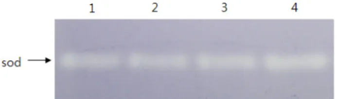
Fig. 1. Activity of superoxide dismutase in P. rhodesiae KK1.
Cells were pre-grown on LB medium and collected.
Cells grown to 0.5-0.6 at 600 nm were transferred to BM medium containing PAHs or oxidative stressors and in- cubated at 160 rpm at 30℃. Glucose was used as control.
Total protein (10ug) isolated from cells following 6h in- cubation was used activity staining. A, PAH-induced.
Lanes 1, glucose; 2, anthracene; 3, naphthalene; 4, phe- nanthrene.
II
TM5X reaction buffer, 10 mM dNTP (final concentration 0.5 mM), recombinant RNasin
Rribonuclease inhibitor, im- Prom-II
TMreverse transcriptase]. The final reaction solution was incubated at 25℃ for 5 min, followed by sequential in- cubation at 42℃ for 60 min for cDNA synthesis. The reaction was stopped by heating at 70℃ for 15 min. PCR was per- formed using 1 μl cDNA as template and the reaction sol- ution [10X buffer 2.5 μl, 25 mM MgCl
22 μl, 10 mM dNTP 0.5 μl, 10 pmol primer set 2.5 μl, Taq polymerase (5 U/μl) 0.25 μl], and the final volume was adjusted to 25 μl with nuclease-free water. RT-PCR was performed using a set of following primers: sodAF (5‘-GGT GGG CAT GCC AAC CAT TCG-3’) - sodAR (5’-GTA GGT ARG TTC CCA CAC ATC-3‘) for superoxide dismutase A, and sodBF (5‘-GCT CAG GTC TGG AAC CAC ACC-3’) – sodBR (5’-GTA TGC GTG TTC CCA GAC GTC-3‘) for superoxide dismutase B.
And, KK1-16F (5‘-CAG ACT CCT ACG GGA GGC A-3’) - KK1-16R (5’-CGT GGA CTA CCA GGG TAT C-3‘) for 16S rRNA gene were also amplified as the positive control in the RT-PCR for the analysis of transcriptional expression ac- cording to the method published previously [14].
Results and Discussion
Enzyme activity of superoxide dismutase in Pseu- domonas rhodesiae KK1
P. rhodesiae KK1 has the degradation ability for PAHs such as anthracene, naphthalene and phenanthrene [9]. Negative stain-based analysis of superoxide dismutase (SOD) in cell extracts of strain KK1 grown on BM medium containing glu- cose, anthracene, naphthalene, and/or phenanthrene re- vealed the existence and expression of Sod (Fig. 1). SOD
activity was observed in the similar level in all the cells grown with glucose and PAHs, even though there was a little difference in SOD activity on PAHs-induced cells. This result suggested that a sod gene is constitutively expressed.
In order to further analyze the expression pattern of sod gene under different conditions, sod genes in strain KK1 were investigated using molecular techniques
Polycyclic aromatic ring-hydroxylating dioxygenase gene in Pseudomonas rhodesiae KK1
A 300-bp aromatic ring-hydroxylating gene for α-subunit of dioxygenase responsible for degradation of polycyclic ar- omatic hydrocarbons in strain KK1 was obtained by PCR amplification using a set of degenerate primers, DioF and DioR [9]. Sequence analysis of the gene revealed naph- thalene 1,2-dioxygenase composed of 94 amino acids, which shared 98.9% similarity with nahAc of Pseudomonas fluo- rescens PC20 [7], 96.8% with nahAc of Pseudomonas putida G7 [8], 95.7% with ndoC2 of Pseudomonas fluorescens ATTC 17483 [4], 93.6% with pahAc of Pseudomonas putida OUS82 [24], and 86.2% with nahAc of Pseudomonas balearica SP1402 [4] and Pseudomonas stutzeri AN11 [4] (Fig. 2). A part of the amplified PAH-ring hydroylating gene sequence was used for the analysis of the transcriptional gene expression using RT-PCR as well as for the comparative analysis of transcriptional gene expression of PAH-ring hydroxylating dioxygenase and sod genes.
Identification and analysis of SOD genes in Pseudomonas rhodesiae KK1
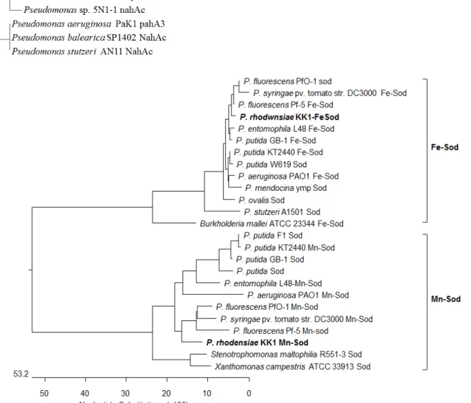
The approximately 420-bp putative superoxide dismutase gene fragment amplified by PCR was found to contain two types of superoxide dismutase in P. rhodesiae KK1. One of them was Mn-superoxide dismutase (sodA) composed of 423-bp and 141 amino acids, and the other is Fe-superoxide dismutase (sodB) composed of 405-bp and 135 amino acids (Fig. 3, Fig. 4).
Both Mn- and Fe-SOD have been found in many prokary- otic bacteria including Pseudomonas species [6, 13, 15, 19, 20].
Multialignment analysis based on 141 amino acids showed
that sodA in strain KK1 shared 95% similarity with Mn-sod
of P. fluorescens Pf-5 [19], 92% with Mn/Fe-Sod of P. fluo-
rescens PfO-1 [22], 89% with of Mn-sod of P. syringae pv. toma-
to str. DC3000 [12] and 88% with Mn-sod of P. aeruginosa
PAO1 [23]. The sodB shared 99% similarity, based on 135
amino acids, with Fe-sod of P. fluorescens Pf-5 [19] and super-
Fig. 2. Phylogenetic analysis of ring-hy- droxylating dioxygenase genes from P. rhodesiae KK1 and other bacterial strains based on multi- ple alignment of the deduced amino acid sequence.
Fig. 3. Phylogenetic analysis of two types of superoxide dismutase (FeSOD and MnSOD) genes in P. rhodesiae KK1 and other bacterial strains based on deduced amino acid sequences.
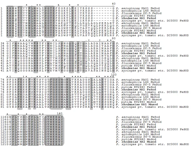
oxide dismutase of P. fluorescens PfO-1 [22], 97% with Fe-sod of P. putida KT2440 [18], and 94% with Fe-sod of P. stutzeri A1501. When twelve Fe-SOD and/or Mn-SOD homologues in Pseudomonas species are aligned, highly conserved resi- dues which have identical amino acid at the same position
were found in several regions as highlighted in grey shading
in Fig. 4. The N-terminal and C-terminal domain regions are
more densely conserved than the central domain region. It
is noticeable that the amino acid residues highly conserved
in either Fe- or Mn-SOD, but different between the two
Fig. 4. Multialignment of two types of superoxide dismutase (FeSOD and MnSOD) genes based on deduced amino acid sequences in P. rhodesiae KK1 with those found in other bacteria. Identically conserved residues in twelve MnSOD and FeSOD homologues are highlighted in grey shading. The amino acid residues identically conserved in either MnSOD or FeSOD, but different between the two genes are indicated with asterisks. Gaps are represented by dashes and were introduced to maximize the alignment. The multiple sequence alignment analysis was carried out using the Clustal method within the MEGALIGN pro- gram of Lasergene.
genes were found in forty regions as indicated with an asterisk. This result suggested that there are several more conserved regions unique to Fe- or Mn-SOD.
Southern hybridization using the PCR antioxidant gene fragment as a probe showed that at least more than two copies of superoxide dismutase genes exist in strain KK1 (Fig. 5). Restriction patterns with superoxide dismutase gene signals were found at 20 kb- and 1.8 kb-ApaI fragments, 20 kb-BamHI fragment, 8.0 kb-, 3.0 kb- and 0.5 kb-EcoRI frag- ments, 20 kb-HindIII fragment, 4.0 kb- and 2.5 kb-PstI frag- ments, and 4.0 kb- and 2.7 kb-SalI fragments. These facts suggested that at least more than two copies of superoxide dismutase exist in strain KK1. These results were consistent with the previous studies [6, 18, 19, 23], in that more than two copies of superoxide dismutase genes have been found in many Pseudomonas species such as P. putida, P. fluorescens,
P. syringae and P. aeruginosa.
Effect of PAHs on transcriptional expression of SOD genes in Pseudomonas rhodesiae KK1
The transcriptional gene expression pattern of SOD genes
in response to PAHs in P. rhodesiae KK1 cells was inves-
tigated based on RT-PCR analysis, along with that of the
ring-hydroxylation gene responsible for the cleavage of ar-
omatic ring. It was found that the ring-hydroxylating gene
expression in the transcriptional level was more stimulated
in KK1 cells grown with naphthalene and phenanthrene
than glucose and anthracene, suggesting that expression of
ring-hydroxylating gene for the degradation of PAHs in
strain KK1 might be quickly stimulated by naphthalene and
phenanthrene (Fig. 6). Interestingly, the PAH ring-hydrox-
ylating gene product is found in the basic level in the glu-
A B
Fig. 5. Southern hybridization of superoxide dismutase genes using total genomic DNA from Pseudomonas rhodesiae KK1. The DNA fragments digested with several restriction enzymes such as ApaI, BamHI, EcoRI, HindIII, PstI, and SalI were hybridized with the superoxide dismutase gene DNA frag- ment obtained from the PCR amplification. Restriction pat- terns of total genomic DNA of strain KK1 are shown in left side of the panel (A), while right side shows the signals hybridized with superoxide dismutase gene probe labeled with Dig DNA labeling kit (B). Lanes M, DNA size marker (λ- HindⅢ); 1, Genomic DNA digested with ApaI, 2, BamHI;
3, EcoRI; 4, HindⅢ; 5. PstI; 6, SalI.
Fig. 6. Transcriptional expression analysis of ring-hydroxylating enzyme (dioxygenase)- and/or superoxide dismutase isomers (sodA and sodB)-encoding genes of Pseudomonas rhodesiae KK1 using RT-PCR with total RNA from cells incubated with glucose (lane 1), anthracene (lane 2), naphthalene (lane 3) or phenanthrene(lane 4) as a substrate. M, size marker. The expression level was relatively determined based on the amount of transcriptional products.
cose grown-cells without PAHs. Relative transcriptional lev- el of the PAH ring-hydroxylating gene grown with glucose and anthracene was approximately 65% and 70%, re- spectively. Transcriptional gene expressions of Mn-SOD (sodA) and Fe-SOD (sodB) genes were commonly more
strongly stimulated in response to naphthalene and phenan-
threne than anthracene. It is notable that sodA gene is ex-
pressed in glucose-grown cells in the similar level with those
grown with naphthalene and phenanthrene. Whereas the
transcriptional expression level of sodB gene was lowest in
cells grown with glucose. Vattanaviboon et al [26] reported the constitutive expression of Fe-SOD in Vibrio harveyi, whereas Mn-SOD was expressed at the stationary phase and could be induced by a superoxide generator. However, SodA1 gene in Bacillus anthracis, which is cambialistic for magnesium and iron, was found to be constitutively ex- pressed [25]. It is remarkable that our findings suggested the possibility Mn-SOD gene might be under constitutive expression, warranting further study with other substrates such as fructose and citrate.
In conclusion, the expression pattern of sodA and sodB genes is very similar in KK1 cells exposed to PAHs, but dif- ferent in KK1 cells grown with glucose. Interestingly, sodA gene from KK1 cells grown with glucose was found to be transcriptionally expressed in the similar level with cells grown with naphthalene and phenanthrene, while sodB gene was not. These facts suggested that SODA might play a more important role in cells exposed to naphthalene and phenan- threne for the removal ROS generated in cells during oxida- tive metabolism even though both SODA and SODB are re- sponsible for the degradation of ROS.
Acknowledgments
This work was supported by Korea Research Foundation Grant (KRF-2006-351-D00018). KSI was supported by the re- search grant (T34414) from the Korea Basic Science Institute.
References
1. Beyer, W. F. and Fridovich, I. 1987. Assaying for superoxide dismutase activity: some large consequences of minor changes in conditions. Ann. Biochem. 161, 559-556.
2. Cerniglia, C. E. 1993. Biodegradation of polycyclic aromatic hydrocarbons. Curr. Opin. Biotechnol. 4, 331-338.
3. Chae, H. Z., Robison, K., Poole, L. B., Church, G., Storz, G. and Rhee, S. G. 1994. Cloning and sequencing of thi- ol-specific antioxidant from mammalian brain: alkyl hydro- peroxide reductase and thiol-specific antioxidant define a large family of antioxidant enzymes. Proc. Natl. Acad. Sci.
USA 91, 7017-7021.
4. Ferrero, M., Llobet-Brossa, E., Lalucat, J., Garcia-Valdes, E., Rossello-Mora, R. and Bosch, R. 2002. Coexistence of two distinct copies of naphthalene degradation genes in Pseudomonas strains isolated from the western Mediterranean region. Appl. Environ. Microbiol. 68, 957-962.
5. Gaupp, R., Ledala, N. and Somerville, G. A. 2012. Staphylo- coccal response to oxidative stress. Front Cell Infect.
Microbiol. 2, 1-19.
6. Guo, M., Block, A., Bryan, C. D., Becker, D. F. and Alfano,
J. R. 2012. Pseudomonas syringae catalases are collectively re- quired for plant pathogenesis. J. Bacteriol. 194, 5054-5064 7. Heinaru, E., Vedler, E., Jutkina, J., Aava, M. and Heinaru,
A. 2009. Conjugal transfer and mobilization capacity of the completely sequenced naphthalene plasmid pNAH20 from multiplasmid strain Pseudomonas fluorescens PC20. FEMS Microbiol. Ecol. 70, 563-574.
8. Herrick, J. B., Madsen, E. L., Batt, C. A. and Ghiorse, W.
C. 1993. Polymerase chain reaction amplification of naph- thalene-catabolic and 16S rRNA gene sequences from in- digenous sediment bacteria. Appl. Environ. Microbiol. 59, 687-694.
9. Kahng H. Y., Nam, K., Kukor, J. J., Yoon, B. J., Lee, D. H., Oh, D. C., Kam, S. K. and Oh, K. H. 2002. PAH utilization by Pseudomonas rhodesiae KK1 isolated from a former manu- factured-gas plant site. Appl. Microbiol. Biotechnol. 60, 475-480.
10. Kang, Y. S., Lee, Y., Jung, H., Jeon, C. O., Madsen, E. L.
and Park, W. 2007. Overexpressing antioxidant enzymes en- hances naphthalene biodegradation in Pseudomonas sp.
strain As1. Microbiology 153, 3246-3254.
11. Kim, H., Lee, J. H., Hah, Y. C. and Roe, J. H. 1994.
Characterization of the major catalase from Strepomyces coeli- color ATCC 10146. Microbiology 140, 3391-3397.
12. Klotz, M. G. and Hutcheson, S. W. 1992. Multiple peri- plasmic catalase of phytopathogenic strains of Pseudomonas syringae. Appl. Environ. Microbiol. 58, 2468-2473.
13. Krayl, M., Benndorf, D. and Loffhagen, N. 2003. Use of pro- teomics and physiological characteristics to elucidate eco- toxic effects of methyl-ter-butyl ether in Pseudomonas putida KT2440. Proteomics 3, 1544-1552.
14. Lee, D. H., Oh, K. H. and Kahng, H. Y. 2009. Molecular analysis of antioxidant genes in the extremohalophile ma- rine bacterium Exiguobacterium sp. CNU020. Biotechnol. Lett.
31, 1245-1251.
15. Lee. Y., Pena-Llopis, S., Kang, Y. S., Shin, H. D., Demple, B., Madsen, E. L., Jeon, C. O. and Park, W. 2006. Expression analysis of the fpr (ferredoxin-NADP+ reductase) gene in Pseudomonas putida KT2440. Biochem. Biophys. Res. Comm.
339, 1246-1254.
16. Lee, D. H., Oh, D. C., Oh, Y. S., .Malinverni, J. C., Kukor, J. J. and Kahng, H. Y. 2007. Cloning and characterization of monofunctional catalase from photosynthetic bacterium Rhodospirillum rubrum S1. J. Microbiol. Biotechnol. 17, 1460-1468.
17. Lü, Z., Sang, L., Li, Z. and Min, H. 2009. Catalase and super- oxide dismutase activities in a Stenotrophomonas maltophilia WZ2 resistant to herbicide pollution. Ecotoxicol. Environ.
Safety 72, 136-143.
18. Nelson, K., Paulsen, I., Weinel, C., Dodson, R., Hilbert, H., Fouts, D., Gill, S., Pop, M., Martins Dos Santos, V., Holmes, M., Brinkac, L., Beanan, M., DeBoy, R., Daugherty, S., Kolonay, J., Madupu, R., Nelson, W., White, O., Peterson, J., Khouri, H., Hance, I., Lee, P., Holtzapple, E., Scanlan, D., Tran, K., Moazzez, A., Utterback, T., Rizzo, M., Lee, K., Kosack, D., Moestl, D., Wedler, H., Lauber, J., Hoheisel, J.,
초록:PAH를 분해할 수 있는 Pseudomonas rhodesiae KK1의 SOD 유전자의 동정 및 분자학적 특성 분석
이동헌
1․오계헌
2․김승일
3․강형일
1*
(1순천대학교 환경교육과, 2순천향대학교 생명과학과, 3한국기초과학지원연구원 프로테옴팀)
Pseudomonas rhodesiae KK1은 이미 주요한 환경오염물질인 anthracene, naphthalene, phenanthrene과 같은 다 환성 방향족 화합물(PAHs)을 분해할 수 있음을 보고한 바 있다. 흥미롭게도, superoxide dismutase를 비롯한 항 산화 유전자는 환경오염물질에 반응하여 다른 수준으로 발현됨이 알려져 있다. 본 연구는 균주 KK1에서 PAHs 분해에 간접적으로 관계될 것으로 여겨지는 superoxide dismutase 유전자의 존재를 동정하고 세 가지 PAHs를 기질로 하여 생장한 세포에서 superoxide dismutase 유전자의 발현 양상을 조사하고자 수행하였다. P. rhodesiae KK1에서 항산화 기작에 관여하는 두 가지지 형의 superoxide dismutase인 Mn-superoxide dismutase (sodA)와 Fe-superoxide dismutase (sodB) 유전자를 동정하고 그 특성을 규명하였다. 균주 KK1에서 발견된 sodA 유전자는 141개의 아미노산 유전자를 기준으로 P. fluorescens Pf-5의 Mn-sod와 95%, sodB 유전자는 135개 아미노산을 기준 으로 P. fluorescens Pf-5의 Fe-sod와 99%의 가장 높은 상동성을 나타내었다. sod 유전자 단편을 탐침자로 사용한 Southern 혼성화 반응 결과 적어도 두 개 이상의 superoxide dismutase 유전자가 균주 KK1에 존재함을 규명하였 다. RT-PCR 분석을 통해 sodA 및 sodB 유전자들은 anthracene보다 naphthalene과 phenanthrene에 반응하여 더 강하게 발현함을 보여주었다. 포도당과 PAHs를 기질로 사용하여 생장한 세포에서 sodA와 sodB 유전자는 활성 상태로 존재함이 밝혀졌다.
Straetz, M., Heim, S., Kiewitz, C., Eisen, J., Timmis, K., Duesterhoft, A., Tummler, B. and Fraser, C. 2002. Complete genome sequence and comparative analysis of the metabol- ically versatile Pseudomonas putida KT2440. Environ.
Microbiol. 4, 799-808.
19. Paulsen, I. T., Press, C. M., Ravel, J., Kobayashi, D. Y., Myers, G. S., Mavrodi, D. V., DeBoy, R. T., Seshadri, R., Ren, Q., Madupu, R., Dodson, R. J., Durkin, A. S., Brinkac, L. M., Daugherty, S. C., Sullivan, S. A., Rosovitz, M. J., Gwinn, M. L., Zhou, L., Schneider, D. J., Cartinhour, S. W., Nelson, W. C., Weidman, J., Watkins, K., Tran, K., Khouri, H., Pierson, E. A., Pierson, L. S. 3rd, Thomashow, L. S. and Loper, J. E. 2005. Complete genome sequence of the plant commensal Pseudomonas fluorescens Pf-5. Nat. Biotechnol. 23, 873-878.
20. Poole, L. B. 2005. Bacterial defenses against oxidants: mecha- nistic features of cysteine-based peroxidases and their fla- voprotein reductases. Arch. Biochem. Biophys. 433, 240-254.
21. Roy, S., Genin, S., Sen, C. K. and Hänninen, O. 1996.
Monitoring of polycyclic hydrocarbons using ‘moss bags’:
bioaccumulation and responses of antioxidant enzymes in Fontinalis antipyretica hedw. Chemosphere 32, 2305-2315.
22. Silby, M. W., Cerdeno-Tarraga, A. M., Vernikos, G. S., Giddens, S. R., Jackson, R. W., Preston, G. M., Zhang, X.
X., Moon, C. D., Gehrig, S. M., Godfrey, S. A., Knight, C.
G., Malone, J. G., Robinson, Z., Spiers, A. J., Harris, S., Challis, G. L., Yaxley, A. M., Harris, D., Seeger, K., Murphy, L., Rutter, S., Squares, R., Quail, M. A., Saunders, E., Mavromatis, K., Brettin, T. S., Bentley, S. D., Hothersall, J.,
Stephens, E., Thomas, C. M., Parkhill, J., Levy, S. B., Rainey, P. B. and Thomson, N. R. 2009. Genomic and genetic analy-
ses of diversity and plant interactions of Pseudomonas fluorescens. Genome Biol. 10, R51.1-16
23. Stover, C. K., Pham, X. Q., Erwin, A. L., Mizoguchi, S. D., Warrener, P., Hickey, M. J., Brinkman, F. S., Hufnagle, W.
O., Kowalik, D. J., Lagrou, M., Garber, R. L., Goltry, L., Tolentino, E., Westbrock-Wadman, S., Yuan, Y., Brody, L.
L., Coulter, S. N., Folger, K. R., Kas, A., Larbig, K., Lim, R., Smith, K., Spencer, D., Wong, G. K., Wu, Z., Paulsen, I. T., Reizer, J., Saier, M. H., Hancock, R. E., Lory, S. and Olson, M. V. 2000. Complete genome sequence of Pseudomo- nas aeruginosa PA01, an opportunistic pathogen. Nature 406, 959-964.
24. Takizawa, N., Kaida, N., Torigoe, S., Moritani, T., Sawada, T., Satoh, S. and Kiyohara, H. 1994. Identification and char- acterization of genes encoding polycyclic aromatic hydro- carbon dioxygenase and polycyclic aromatic hydrocarbon dihydrodiol dehydrogenase in Pseudomonas putida OUS82.
J. Bacteriol. 176, 2444-2449.
25. Tu, W. Y., Pohl, S., Summpunn, P., Hering, S., Kerstan, S.
and Harwood, C. R. 2012. Comparative analysis of the re- sponses of related pathogenic and environmental bacteria to oxidative stress. Microbiology 158, 636-647.
26. Vattanaviboon, P., Panmanee, W. and Mongkolsuk, S. 2003.
Induction of peroxide and superoxide protective enzymes and physiological cross-protection against peroxide killing by a superoxide generator in Vibrio harveyi. FEMS Microbiol.
Lett. 221, 89-95.