Anti-Inflammatory Effects of Picrasma Quassioides (D.DON) BENN Leaves Extracts
Yeon Seop Jung
1, Cheong Su Eun
1, Young Tae Jung
1, Hyun Jeong Kim
2and Mi Hee Yu
1,2*
1
Department of Food Science and Technology, Keimyung University, Deagu 704-701, Korea
2
The Center for Traditional Microorganism Resurces, Keimyung University, Deagu 704-701, Korea
Received March 11, 2013 /Revised May 10, 2013 /Accepted May 27, 2013This study was performed to evaluate the anti-inflammatory and antioxidant activities of methanol ex- tract from the leaves of Picrasma quassioides BENNET (PLME). The antioxidant effects of PLME were measured based on polyphenol and flavonoid contents. PLME was found to have 367.52 μg/mg and 46.61 μg/mg high polyphenol and flavonoid contents. Cell viability was determined by MTT assay.
The production of nitric oxide (NO) and prostaglandin E
2(PGE
2) was measured by Griess assay and enzyme-linked immunosorbent assay (ELISA). In order to effectively anti-inflammatory agents, we ex- amined the inhibitory effects on the production of lipopolysaccharide (LPS)-induced NO and PGE
2in RAW 264.7 cells. PLME significantly decreased the production of NO and PGE
2in a dose-dependent manner, and also reduced the expression of iNOS, a COX-2 protein. In addition, PLME reduced the NF-kB, IκB phosphorylation in RAW 264.7 cells upon stimulation with LPS (100 ng/ml) for 24 h.
These results provide evidence for the anti-inflammatory and antioxidant effects of Picrasma quassioides leaves.
Key words : Anti-inflammation, Picrasma quassioides BENNET, nitric oxide, NF-κB, prostaglandin E
2*Corresponding author
*Tel:+82-53-580-5538, Fax:+82-53-580-5538
*E-mail : [email protected]
This is an Open-Access article distributed under the terms of the Creative Commons Attribution Non-Commercial License (http://creativecommons.org/licenses/by-nc/3.0) which permits unrestricted non-commercial use, distribution, and reproduction in any medium, provided the original work is properly cited.
Journal of Life Science 2013 Vol. 23. No. 5. 629~636 DOI : http://dx.doi.org/10.5352/JLS.2013.23.5.629
서 론
염증반응이란 체내에 외부로부터 물리적, 화학적 자극이나 세균감염에 대해 면역세포가 이를 인지하여 다양한 염증 매개 물질을 분비함으로써 손상된 조직을 수복하거나 재생하려는 기전이다[34]. 그러나 지속적으로 또는 과도하게 발생된 만성 염증반응은 조식의 손상을 유발하며 이와 관련한 활성 산소종 과 염증성 cytokine은 내독소 자극을 포함한 다양한 질병의 매개체로써 중요한 역할을 한다[4]. 대식세포는 선천면역뿐만 아니라 획득면역 등 다양한 숙주반응에 관여하여 항상성 유지 에 관여하는 것으로 알려져 있으며, 염증반응이 일어나면 이 들 대식세포와 같은 염증세포들은 nitric oxide (NO), prosta- glandin E
2(PGE
2)를 생성함으로써 통증, 부종, 열 등의 염증 반응을 유발하고, 염증 부위로 면역세포의 이동을 촉진 시킨 다[11, 15].
염증반응의 대표적인 예로, 대식세포는 염증성 매개물질 중 하나인 lipopolysaccharide (LPS)를 toll like receptor 4 (TLR4) 의 heterodimerization 형성으로 인식하며 세포내 전사요소인
nuclear factor-ĸB (NF-ĸB)의 활성화를 유도한다[22]. 핵으로 이동한 NF-ĸB는 염증성 cytokine, inducible nitric oxide syn- thase (iNOS), cyclooxygenase-2 (COX-2)의 유전자 발현을 유 도하며, 염증반응의 지표물질인 NO가 L-arginine으로부터 NO synthase (NOS)에 의해서 합성된다[29]. NOS의 경우, 유 사형태가 3가지 존재하는데 neuronal NOS (nNOS), endothe- lial NOS (eNOS), 그리고 inducible NOS (iNOS)이다. 그 중에 서 특히 iNOS에 의한 NO생성은 병리학적으로 중요한 역할을 한다[14]. nNOS와 eNOS는 항상 발현되어 있지만 iNOS의 경 우 lipopolysaccharide (LPS)와 염증성 cytokine등에 의해 발 현이 유도되고 iNOS에 의해 생성된 과다한 NO는 염증 반응 을 심화시켜 조직의 손상, 유전자 변이 및 신경 손상 등을 일으 킨다[20, 30, 33, 36]. Cyclooxygenase (COX)는 arachidonic acid를 Prostaglandin으로 변화를 촉진시키는 효소로써, 대부 분의 조직에서 정상적인 상태에 발현하여 위장관 보호, 신장 기능 조절과 같은 신체의 항상성 유지에 관여하는 COX-1과 일부 신생조직과 염증이나 기타 면역반응시 세포분열인자나 cytokine에 의해 염증의 부위에서 세포 내 발현이 증가하는 COX-2가 있다. 즉, iNOS와 COX-2의 발현과 NO, PGE
2는 면 역세포의 대표적인 염증인자이다[8].
한편, NO, HNO
2, ONOO
-와 같은 활성 질소종(reactive ni-
trogen species, RNS)은 염증반응 시 대식세포, 호중구 및 다른
면역 세포들의 면역 반응으로 다량 생성되며, 이때 활성 산소
종(reactive oxygen species, ROS)도 같이 생성된다[2, 6]. 이러
한 ROS와 RNS는 생체 내 여러 가지 세포의 작용을 조절 하는
염증반응과 깊이 관련되어 있다[1]. 페놀 화합물은 식물계에 널리 분포되어 있는 2차 대사산물 중 하나로서, 탄소 수에 따 라 페놀산, 탄닌, 플라보노이드 등의 다양한 물질로 나뉠 수 있고 최근에는 다양한 식물을 대상으로 이러한 ROS를 조절할 수 있는 항산화 활성과 기능성 소재로의 연구가 진행되고 있 다[5]. 페놀화합물의 다양한 항산화력은 그 구조적인 특징과 관련성이 높은데, 이들은 금속 킬레이트제, 환원제, 활성산소 의 소거제, 사슬전단 항산화제(chain breaking antioxidants) 등으로서의 역할에 기인하는 것으로 알려져 있다. 그리고 플 라보노이드는 페놀화합물 중에서 자연적으로 생성되는 가장 큰 그룹의 하나로서, anthocyanins, chalcones, aurones, fla- vones, flavonols 및 이들의 유도체 등으로 나눌 수 있다. 현재 까지 페놀화합물 및 플라보노이드의 구조에 따른 항산화 활성 과의 상관성에 대한 연구도 보고되었다[31].
소태나무(Picrasma quassioides (D.DON) BENN.)는 쌍떡잎 식물 쥐손이풀목 소태나무과의 소교목으로 잎과 줄기의 속껍 질에서 소의 태처럼 지독히 쓴맛이 나는 것에서 유래되었다.
건위, 조습, 살균의 효능이 있어 한방에서는 잔가지와 열매를 채취하여 소화불량, 위장염, 폐결핵, 습진, 옹종(癰腫), 개선 (疥癬) 등의 증상에 치료제로 사용하며, 민간에서는 나무 전 체를 솥에 넣고 끓인 물을 살충제로 이용한다. 소태나무의 주 요성분으로는 quasinoids, tirucallanes, ionone, alkaloids등이 알려져 있으며[21, 26], 소태나무 열매에는 arbutin, phlorin, koaburaside, syringin, citrusin B, cnidioside B, flavaprenin 7,4-diglucoside, phenyl propanoids와 phenolic compound 등[13]이 함유되어 있는 것으로 보고되어 있다. 또한 소태나 무과는 항암활성과 면역조절작용[19]을 지니고 있으며, 항고 혈압, 항산화, 소염작용, 해독 작용 등을 가지는 것으로 알려 져 있다[23].
현재 국내·외적으로 천연물로부터 기능성 성분의 소재를 발굴하기 위한 연구가 활발히 진행되고 있으나, 소태나무 잎 의 생리활성에 관한 연구는 아직까지 미진한 편이며, 따라서 본 연구에서는 소태나무 잎 추출물을 이용하여 총 폴리페놀, 플라보노이드 함량을 조사하고 LPS로 염증반응이 유도된 RAW 264.7 cell에 소태나무 잎 메탄올 추출물을 처리하여, 항 염증 활성 효과를 알아보았다.
재료 및 방법
시료 제조
본 실험에서 사용한 소태나무 잎은 대구시 약령시장에서 건조 상태의 것을 구입하여 사용하였다. 시료는 불순물을 제 거하기 위하여 수세한 후 건조하여 사용하였고, 무게의 10배 량(w/v)의 80% 메탄올을 가하여 24시간 동안 정치하여 총 3회 반복 추출 하였다. 추출액은 여과지(Whatman No. 3, Whatman International Ltd., Maidstone, England)로 여과한
다음 rotary evaporator (UT-1000, EYELA, Tokyo, Japan)로 55℃에서 농축한 후 동결 건조하여 메탄올 추출물로 사용하 였다.
세포주 배양
대식세포 계열(murine macrophage cell line)인 RAW 264.7 세포주는 한국 세포주 은행(KCLB, Seoul, Korea)으로부터 분 양 받았으며, 10% FBS (fetal bovine serum)와 1% antibiotics (penicillin/streptomycin)를 첨가한 DMEM (Gibco-BRL, Rockville, MD, USA)배지를 이용하여 5% CO
2가 존재하는 3 7℃ incubator에서 1주일에 2~3회 계대 배양하였다.
총 폴리페놀 함량
총 폴리페놀 함량은 Folin-Denis법[7]을 응용하여 측정하 였다. 즉 각 메탄올 추출물 시료 1 mg을 증류수 1 ml에 녹이 고 10배 희석한 희석액 2 ml에 2배 희석한 Folin시약 2 ml를 첨가하고 잘 혼합한 후 3분간 방치한 후 10% Na
2CO
32 ml를 넣고 1시간 반응시킨 후 UV/Visible spectrophotometer (UVIKON 922, Kontron, Italy)를 사용하여 700 nm에서 흡광 도를 측정하여 작성한 표준곡선으로부터 함량을 구하였다.
이 때 tannic acid를 이용한 표준곡선은 5, 10, 25, 50, 75, 100 μ g/ml가 되도록 하여 위와 같은 방법으로 700 nm에서 흡광 도를 측정하였다.
총 플라보노이드 함량
시료 추출물의 총 플라보노이드 함량은 Nieva Moreno법 [24]을 이용하여 측정하였다. 각 시료 추출물 0.1 ml에 80%
ethyl alcohol 0.9 ml, 10% aluminum nitrate 0.1 ml, 1 M po- tassium acetate 0.1 ml 및 80% ethyl alcohol 4.3 ml를 혼합 하여 실온에서 30분간 반응시킨 후 spectrophotometer를 이 용하여 415 nm에서 흡광도를 측정하였다. 표준물질로 quer- cetin을 사용하였으며, 표준물질의 검량선과 비교하여 함량 을 구하였다.
세포독성 측정
시료의 세포증식과 독성을 측정하기 위해 Green 등의 방법 [9]에 따라 3-(3,4-dimethyl- thiazolyl-2)-2,5-diphenyl tetrazo- lium bromide (MTT) assay를 실시하였다. MTT assay는 미토 콘드리아의 탈수소 효소작용에 의하여 노란색의 수용성 기질 인 MTT가 불용성의 보라색 formazan으로 환원되는 원리를 이용한 방법으로, 생성된 formazan의 흡광도는 살아있고 대 사가 왕성한 세포의 농도를 반영한다. RAW 264.7 cell이 1×10
5cells/well의 농도가 되게 96 well plate에 분주하고, 37℃, 5%
CO
2incubator에서 24시간 동안 배양하였다. 배양한 RAW
264.7 세포는 serum free 배지로 교체한 후 LPS (100 ng/ml)와
시료를 처리하여 24시간 배양하여 5 mg/ml의 MTT 용액 10
μ l를 각 well에 넣고 incubator에서 4시간 동안 배양하였다.
배양 종료 후 상등액을 제거하고 각 well에 100 μl의 DMSO를 첨가하여 생성된 formazan 결정을 용해시켜 microplate read- er로 550 nm에서 흡광도를 측정하였고, 세포독성은 시료의 흡광도를 대조군의 흡광도에 대한 백분율로 나타내었다.
Nitric oxide (NO) 생성량 측정
RAW 264.7 세포로부터 생성되는 활성 질소종인 nitric ox- ide (NO)의 양은 Green 등[10]의 방법을 이용하여 세포 배양 액 중 존재하는 NO
2-형태를 Griess Reagent와 반응시켜 측정 하였다. RAW 264.7 cell을 DMEM 배지를 이용하여 1×10
5cells/ml 농도로 96 well plate에 분주한 후 시료를 농도별로 처리하여 24시간 배양하였으며 LPS (100 ng/ml)를 첨가하여 다시 24시간 배양시켰다. 세포배양 상등액 100 μl와 Griess 시 약(1% sulfanilamide, 0.1% naphthylethylendiamine in 25%
phosphoric acid) 100 μl를 혼합하여 96 well plates에서 10분 간 반응시킨 후 ELISA reader를 이용하여 540 nm에서 흡광도 를 측정하였다. NO
2-표준곡선은 NaNO
2를 농도별로 조제하 여 사용하였다.
Prostaglandin E2 (PGE2) 생성량 측정
활성화된 RAW 264.7 세포로부터 생성되는 염증 매개 물질 인 PGE
2의 양을 측정하기 위해 Commercial competitive en- zyme immunoassay kit를 구입하여 실험하였다. RAW 264.7 세포를 1×10
5cells/ml 농도로 24 well plate에 분주하고 시료 를 농도별로 처리하여 24시간 배양한 후, LPS (100 ng/ml)를 첨가하여 다시 24시간 배양시켜 세포배양 상등액을 이용하여 실험하였다. 즉, Goat polyclonal anti-mouse IgG가 coating된 96 well plate에 세포 배양 상등액 50 μl와 prostaglandinE2 acetylcholinesterase 50 μl와 antibody를 4℃에서 18시간 배양 한 후에 wash buffer (0.2%, Tween 20)를 이용하여 수세하였 다. Ellman's reagent (5,5'-dithio-bis-(2-nitrobenzoic acid), acetylthiocholine)를 200 μl 첨가하여 60~90분 동안 반응시킨 후에 420 nm에서 흡광도를 측정하였다.
Western blot analysis
RAW 264.7 cell이 1×10
6cells/well의 농도가 되게 6 well plate에 분주하고, 37℃, 5% CO
2incubator에서 24시간 동안 배양하였다. 배양한 RAW 264.7 세포는 serum free 배지로 교 체한 후 LPS (100 ng/ml)와 시료를 함께 처리하여 24시간 배 양하였다. 시료 처리가 완료된 세포를 2~3회 PBS로 세척한 후 1 ml의 lysis buffer를 첨가, 30분~1시간 동안 용해시킨 후 13,000 rpm에서 10분간 원심 분리하여 세포막 성분 등을 제거 하였다. 단백질 농도는 bovine serum albumin (BSA)를 표준 화 한 Bio-Rad protein assay kit를 사용하여 정량 하였다. 4℃
에서 13,000 rpm으로 10분간 원심 분리한 상등액은 단백질을
정량 한 후 10% running gel과 4.5% stacking gel을 이용하여 125 V에서 SDS-polyacrylamide gel 전기영동을 실시하였다.
전기영동으로 분리한 단백질은 immobilon-P transfer membrane과 transfer buffer (20% methanol, 25 mM Tris- HCl, 192 mM glycine)를 사용하여 350 mA에서 120분간 transfer시켰다. 단백질이 이동된 membrane은 fast green solution으로 transfer의 유무를 확인한 후, 5% non-fat skim milk solution으로 blocking하였다. 4℃에서 일차 항체의 발 현 정도를 검토하기 위하여 TBST (Tris-Buffered Saline and Tween 20) 용액에 1:1,000으로 희석하여 24시간 반응시킨 후 TBST로 3회 세척하였다. 계속하여 이차항체를 2시간 반응시 키고 다시 TBST로 3회 세척하였다. 증류수로 세척하고 membrane에 ECL detection kit의 발색시약 Ⅰ과 Ⅱ를 1:1로 섞은 후에 혼합액을 도포하고, X-ray film에 노출하여 현상한 후 film 상에서 iNOS, COX-2 및 IκB, NF-κB의 phosphor- ylation을 관찰하였다.
통계학적 분석
모든 결과는 평균±표준편차로 나타내었으며, 유의성 검사 는 SPSS TM version 20.0 (SPSS Inc., Chicago, USA)을 이용하 여 one-way ANOVA를 실시하였고, 대조구인 분화 Control에 대한 시료 처리군의 통계적 유의성은 Duncan's multiple range test로 검증하였다. p<0.05 이상일 때만 통계적 유의성이 있는 것으로 판단하였다.
결과 및 고찰
폴리페놀 및 플라보노이드 함량
폴리페놀계 물질들은 식물체에 특수한 색깔을 부여하고 산 화 환원반응에서 기질로 작용하며, 한 분자 내에 2개 이상의 phenolic hydroxyl (OH)기를 가진 방향족 화합물을 가리키며 플라보노이드와 탄닌이 주성분으로 충치 예방, 고혈압 억제, 항에이즈, 항산화, 항암 등의 다양한 생리활성을 가진다[35].
먼저 소태나무 잎 메탄올 추출물에 존재하는 총 폴리페놀 및 플라보노이드 함량을 각각 tannic acid, quercetin을 기준 물질 로 하여 측정하였다(Table 1). 그 결과, 소태나무 잎 추출물의 총 폴리페놀 함량은 367.52 μg/mg으로 높은 폴리페놀 함량을 보였다. 총 플라보노이드 함량은 46.41 μg/mg으로 나타났다.
Lee [17] 등은 6종의 단일 생약재인 두충, 어성초, 오가피, 우슬,
홍화, 해동피 추출물과 생약 복합제인 MIX-1, MIX-2, MIX-3의
ethanol 추출물의 폴리페놀과 플라보노이드 함량을 측정 보고
하였다. 폴리페놀 함량은 어성초, 홍화, MIX-2 ethanol 추출물
이 각각 58.98, 60.79, 57.74 μg/mg으로 가장 높게 나타났으며
플라보노이드 함량은 어성초, 홍화가 각각 36.86, 19.42 μg/mg
으로 나타나 본 연구에서 나타난 소태나무 잎 추출물(폴리페
놀: 367.52 μg/mg, 플라보노이드: 46.41 μg/mg)의 폴리페놀
Table 1. Contents of total polyphenols and flavonoids in methanol extracts of
Picrasma quassioides
leavesPlant Part used Total polyphenols1) (μg/mg) Total flavonoids2)(μg/mg)
Picrasma quassioides
(D. DON) BENN Leaf 367.52±10.413) 46.41±0.321)Micrograms of total polyphenol content/mg of plants based on tannic acid as standard.
2)Micrograms of total flavonoid content/mg of plants based on quercetin acid as standard.
3)Each value is mean±S.D. (n≥3).
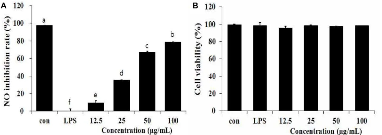
A B
Fig. 1. Effect of methanol extracts of
Picrasma quassioides
leaves on NO production and cell viability in LPS-induced RAW 264.7 cells. RAW 264.7 cells were treated withPicrasma quassioides
leaves methanol extract and LPS (100 ng/ml) for 24 hr. NO production was determined in culture supernatant by Griess reagent. Results represent the mean±S.D. of three independent experiments.및 플라보노이드 함량은 매우 높은 것으로 사료된다.
소태나무 잎 추출물의 ntric oxide 소거활성과 세포 생존율 활성 질소종(reactive nitrogen species)의 하나이며, 최근 염 증반응의 중요한 작용인자로 알려진 nitric oxide (NO) 생성 저해에 대한 소태나무 잎 추출물의 효과를 알아보기 위해 LPS 로 NO의 생성을 유도한 뒤 12.5, 25, 50, 100 μg/ml의 농도의 소태나무 추출물을 처리하였다. 그 결과, LPS에 의해 RAW 264.7 세포로부터 생성이 증가된 NO를 농도 유의적으로 저해 하는 것으로 나타났으며(Fig. 1A), 이러한 NO생성 저해가 세 포독성에 기인하는 것인지를 확인하기 위해 MTT assay를 이 용하여 세포독성을 측정하였다. 그 결과, 소태나무 잎 추출물 100 μg/ml의 농도 까지 세포의 생존율에 영향을 미치지 않았 기 때문에 RAW 264.7 세포에 독성을 나타내지 않은 것을 확인 하였다(Fig. 1B). Cheon [3] 등은 Bulnesia sarmienti 열수추출물 을 이용하여 NO 저해활성을 확인 하였다. LPS에 의해 NO의 생성이 증가한 RAW 264.7 세포에 Bulnesia sarmienti 추출물을 처리하여 100 μg/ml의 농도에서 NO의 생성을 효과적으로 억제시키는 것을 확인 하였으며 200 μg/ml의 농도까지 세포 독성이 없는 것을 확인 하였다. 본 실험에 사용한 소태나무 잎 추출물이 25 μg/ml의 농도에서부터 유의적인 NO생성 억 제 효과를 나타낸 것을 보면 유창목 추출물과 비교하여 뛰어
난 항염증 활성을 나타낸다고 사료된다.
소태나무 잎 추출물의 prostaglandin E
2(PGE
2) 생성 억제능 측정
PGE
2는 prostaglandin endoperoxide synthase 효소인
COX-2에 의해 합성되는 염증 매개 물질로서 다양한 염증 질
환에 기여한다. 즉 PGE
2의 생성은 혈관 확장을 일으키는 작용
에 의해 일어나는 염증 전 단계로써 통증과 발열을 일으킨다
[12]. 따라서 RAW 264.7 세포에 LPS와 소태나무 잎 추출물을
처리한 후, PGE
2생성량을 ELISA kit을 이용하여 측정하였다
(Fig. 2). RAW 264.7 세포에 100 ng/ml의 LPS를 처리했을 때,
PGE
2의 생성은 약 100배 증가 되었으며, 소태나무 잎 추출물
을 각각 50, 100 μg/ml로 처리했을 때 PGE
2함량은 약 837,
577 pg/ml의 농도를 보여 소태나무 잎 추출물이 각각 85%,
90% 이상의 PGE
2생성 억제를 보였다(Fig. 2). Lee [16] 등의
연구에 따르면 등골나물 ethanol 추출물이 1 mg/ml의 농도에
서 농도 유의적으로 PGE
2생성을 감소시키며 특히 10 mg/ml
의 농도에서 49%까지 감소시키는 것으로 나타났다. 소태나무
추출물의 경우 다소 낮은 50 μg/ml의 농도에서도 PGE
2생성
을 85% 감소시키는 것으로 나타나 뛰어난 PGE
2생성 억제
활성을 확인 할 수 있었다.
Fig. 3. Inhibition effects of methanol extracts of
Picrasma quassioides
leaves on the expression of iNOS and COX-2 in LPS-stimulated RAW 264.7 cells. The RAW 264.7 cells (1×10⁶cells/ml) were incubated 24 hr and then pretreated withPicrasma quassioides
leaves methanol extract in the presence or absence of LPS (100 ng/ml) for 24 hr. Equal amounts of total protein were resolved by SDS-PAGE. PLME: Methanol extracts ofPicrasma quassioides
leaves.Fig. 2. Effect of methanol extracts of
Picrasma quassioides
leaves on LPS-induced PGE2levels in RAW 264.7 cells. RAW 264.7 cells were treated withPicrasma quassioides
leaves methanol extract and LPS (100 ng/ml) for 24 hr. Results represent the mean±S.D. of three independent experiments.소태나무 잎 추출물의 iNOS, COX-2의 발현에 미치는 영향 iNOS는 평소에는 세포 내 존재하지 않으나 유도되면 장시 간 동안 다량의 NO를 생성하며, 염증상태에서 iNOS에 의해 생성된 NO는 혈관투과성, 부종 등의 염증반응을 촉진시킬 뿐 만 아니라 염증매개체의 생합성을 촉진하며, iNOS의 발현은 NF-κB 활성으로 유도되며, 이는 macrophage에서 LPS나 cy- tokine에 의해 염증성 매개물질들이 과잉 생산되는 중요한
mechanism이 된다[25]. 또한, COX는 arachidonic acid를 PGE
2로 전환시키는 효소로 COX-1과 COX-2로 분류된다.
COX-1은 체내에서 혈소판을 형성, 위벽보호, 신장 기능 유지 등 정상적인 생체기능에 작용한다. 많은 염증 억제 약물들의 작용기전은 prostaglandin 합성 억제를 나타내며, 이는 COX-2 의 생성 및 활성저해에 의한 것이다[28, 32]. 따라서 소태나무 잎 추출물의 iNOS 및 COX-2 단백질의 발현에 미치는 영향을 알아보기 위하여 Western blot 방법을 이용하여 세포질 내에 서의 iNOS와 COX-2 단백질의 발현량을 조사하였다(Fig. 3).
RAW 264.7 cell에서 LPS (100 ng/ml) 처리로 인해 iNOS 단백 질의 발현이 증가하였고 소태나무 잎 추출물을 농도 별로 처 리 하였을 때 12.5 μg/ml의 농도에서부터 유의적으로 억제되 는 것을 확인할 수 있었다(Fig. 3A). 또한, 염증반응에서 중요 한 역할을 하는 COX-2 단백질 역시 LPS 처리로 인해 그 발현 량이 증가하였으며 소태나무 잎 추출물을 농도 별로 처리하였 을 때 12.5, 25, 50 μg/ml에서는 COX-2의 발현의 변화가 거의 없었지만 100 μg/ml에서는 COX-2의 발현이 크게 억제되는 것을 확인 할 수 있었다(Fig. 3B).
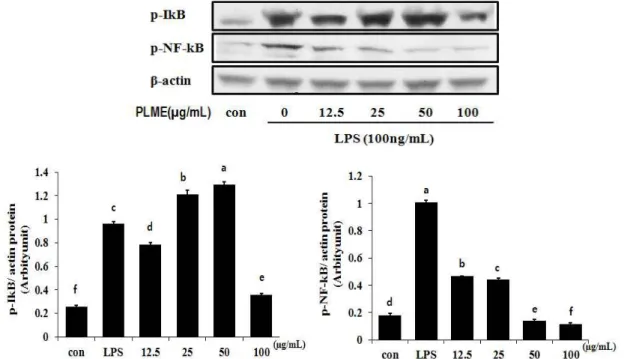
소태나무 잎 추출물의 p-NF-κB, p-Iκ-B 발현에 미치는 영향
염증반응에 있어서 NF-κB는 iNOS 및 COX-2의 발현을 조
절하는 것으로 알려져 있는데, 일반적으로 NF-κB는 Iκ-B와 결
합하여 세포질에 존재하지만 inflammatory stimuli에 의하여
Fig. 4. Inhibition effects of methanol extracts of
Picrasma quassioides
leaves on the expression of p-IkB, p-NF-kB in LPS-stimulated RAW 264.7 cells. The RAW 264.7 cells (1×10⁶cells/ml) were incubated 1 hr and then pretreated withPicrasma quassioides
leaves methanol extract in the presence or absence of LPS (100 ng/ml) for 1 hr. Equal amounts of total protein were resolved by SDS-PAGE. PLME: Methanol extracts ofPicrasma quassioides
leaves.Iκ-B kinase가 활성화 되고 Iκ-Bα의 인산화에 의한 분해가 촉 진된다. IκB가 degradation되면 자유로워진 NF-κB는 핵 내로 의 이동이 촉진되어 전사조절인자로 작용하면서 iNOS와 COX-2의 발현을 증가시켜 염증반응을 촉진시킨다[18, 27]. 따 라서 소태나무 잎 추출물이 p-NF-κB, p-Iκ-B의 발현에 어떠한 영향을 미치는지 확인하고 Fig. 4에 나타내었다. p-Iκ-B는 LPS 에 의해 증가되었으며 소태나무 잎 추출물을 농도 별로 처리 하였을 때, 100 μg/ml의 농도에서 발현을 효과적으로 억제되 는 것으로 나타나 소태나무 잎 추출물이 Iκ-B의 인산화를 감소 시키는 것을 알 수 있었다. 이와 더불어 LPS에 의해 증가하는 p-NF-κB의 발현을 소태나무 잎 추출물 처리에 의하여 농도 유의적으로 감소시켰으며 이 결과는 소태나무 잎 추출물이 대식세포에서 LPS에 의해 활성화되는 NF-κB signaling을 억 제할 수 있음을 시사한다.
감사의 글
본 연구는 지식경제부 지원 계명대학교 전통미생물자원개 발 및 산업화 연구센터에 의한 것입니다.
References
1. Azad, N., Rojanasakul, Y. and Vallyathan, V. 2008.
Inflammation and lung cancer: roles of reactive oxy-
gen/nitrogen species.
J Toxicol Env Heal B
11, 11-15.2. Brune, B., Zhou. J. and Von Knethen, A. 2003. Nitric oxide, oxidative stress, and apoptosis.
Kidney Int Suppl
84, 22-24.3. Cheon, Y. P., Mollah, M. L., Park, C. H., Hong, J. H., Lee, G. D., Song, J. C. and Kim, K. S. 2009. Bulnesia Sarmienti Aqueous Extract Inhibits Inflammation in LPS-Stimulated RAW 264.7 Cells.
J Life Sci
19, 479-485.4. Chung, E. K., Seo, E. H., Park. J. H., Shim, J. H., Kim. K.
H. and Lee, B. R. 2011. Anti-inflammaatory and anti-allergic effect of extracts from organic soybean.
Korean J Organic Agric
2, 245-253.5. Chung, Y. A. and Lee, J. K. 2003. Antioxidative properties of phonolic compounds extracted from black rice.
J Korean Soc Food Sci Nutr
32, 948-951.6. Delanty, N. and Dichter, M. A. 1998. Oxidative injury in the nervous system.
Acta Neurol Scand
117, 463-466.7. Folin, O. and Denis, W. 1912. On Phosphotungastic-phoso- ho-mdybdic compounds as color reagents.
J Biol Chem
12, 239-249.8. Golden, B. D. and Abramson, S. B. 1999. Selective cyclo- oxygenase-2 inhibitor.
Rheum Dis Clin N Am
25, 359-378.9. Green, L. C., Reade, J. L. and Ware, C. F. 1984. Rapid colori- metric assay for cell viability: application to the quantitation of cytotoxic and growth inhibitory lymphokines.
J Immunol Methods
126, 131-138.10. Green, L. C., Wagner, D. A. and Glogowski, J. 1982. Analysis of nitrate, and nitrate in biological fluids.
Anal Biochem
126, 131-138.11. Higuchi, M., Hisgahi, N., Taku, H. and Osaea, T. 1990.
Cytolytic mechanisms of activated macrophages. Tumor ne- crosis factor and L-arginine-dependent mechanisms act syn- ergistically as the major cytolytic mechanisms of activated macrophage.
J Immunol
144, 1425-1431.12. Ilorton, J, K., Williams, A. S., Smith-Philips, Z., Martin, R.
C. and O'Beirne, G. 1999. Intracellular measurement of pros- taglandin E2 : Effects of anti- inflammatory drugs on cyclo- oxygenase activity and prostanoid expression.
Anal Biochem
271, 18-28.13. Kazuko, Y., Saori, S. and Shigenobu, A. 1995. Phenylpropan- ooids and other secondary metabolites from fresh fruits of Picrasma quassioides.
Phytochemistry
40, 253-256.14. Kim, D. H., An, B. J., Kim, S. G., Park, T. S., Park, G. H.
and Son, J. H. 2011. Anti-inflammatory effect of Ligularia fischeri, Solidago virga-aurea and Aruncus dioicus complex extract in RAW 264.7 cells.
J Life Sci
21, 648-683.15. Lee, E. S., Ju, H. K., Moon, T. C., Lee, E., Jahng, Y., Lee, S. H., Son, J. K., Baek, S. H. and Chang, H. W. 2004.
Inhibition of nitric oxide and tumor necrosis factor-α (TNF- α) production by propenone compound through blockade of nuclear factor (NF)-kB activation in cultured murine macrophages.
Biol Pharm Bull
27, 617-620.16. Lee, H. N., Lim, D. Y., Lim, J. D., Kim, J. D. and Park, H.
Y. 2011. Anti-inflammatory effect of ethanol extract from Eupatorium japonicum.
Korean J Food Sci Technol
43, 65-71.17. Lee, S. G., Jeong, H. J., Lee, E. J., Kim, J. B. and Choi, S.
W. 2011. Antioxidant and anti-inflammatory activities of ethanol extracts from medicinal herb mixtures.
Korean J Food Sci Technol
43, 200-205.18. Liden, J., Rafter, I., Truss, M., Gustafsson, J. A. and Okret, S. 2000. Glucocorticoid effects on NF-kappaB binding in the transcription of the ICAM-1 gene.
Biochem Bioph Res Co
273, 1008-1014.19. Luyengi, L., Suh, N. and Fong, H. 1996. A lignan and four terpenoids from Brucea javanica that induce differentiation with cultured HL-60 promyelocytic leukemia cells.
Phytochemistry
43, 409-412.20. McCartney-Francis, N., Allen, J. B., Mizel, D. E., Albina, J.
E., Xie, Q. W., Nathan, C. F. and Wahl, S. M. 1993.
Suppression of arthritis by an inhibitor of nitric oxide synthase.
J Exp Med
178, 749-754.21. Murae, T., Tsuyuki, T., Ikeda, T., Nishihama, T. and Takahasshi, T. 1971. Bitter principles of
Picrasma ailanthoides Planchon, Nigakilactones
A, B, C, D, E, and F.Tetrahedron
27, 1545-1555.22. Miyake, K. 2004. Innate recognition of lipopolysaccharide by Toll-like receptor 4-MD-2.
Trends Microbiol
12, 186-192.23. Jung, Y. T., Lee, I. S., Whang, K., and Yu, M. H. 2012.
Antioxidant effects of
Picrasma quassioides
andChamaecyparis obtusa
(S. et Z.) ENDL extracts.J Life Sci
22, 354-359.24. Nivea, M., Sampietro, A. and Vattuone, M. 2000.
Comparison of the free radical scavenging activity of prop- olis from several relisions of Argentina.
J Ethnopharmacol
71, 109-114.25. Noh, K. H., Jang, J. H, Min, K. H., Chinzoring, R., Lee, M.
O. and Song, Y. S. 2011. Suppressive effect of green tea seed coat ethly acetate fraction on inflammation and its mecha- nism in RAW 264.7 macrophage cell.
J Korean Soc Food Sci Nutr
40, 625-634.26. Ohmoto, T., Nikaido, T., Koike, K., Kohda, K. and Sankawa, U. 1988. Inhibition of adenosine 3',5'-cyclic monophosphate phosphodiesterase by alkaloids. II.
Chem Pharm Bull Tokyo
36, 4588-4592.27. Pruett, S. B., Fan, R. and Zheng, Q. 2003. Characterization of glucocorticoid receptor translocation, cytoplasmic IkappaB, nuclear NFkappaB, and activation of NFkappaB in T lymphocytes exposed to stress-inducible concentrations of corticosterone
in vivo
.Int Immunopharmacol
3, 1-16.28. Rocca, B. and FitzGerald, G. A. 2002. Cyclooxygenases and prostaglandins: shaping up the immune response.
Int Immunopharmacol
2, 603-630.29. Shon, D. H., Choi. D. W. and Kim, M. H. 2012. Improvement of anti-inflammation activity of Gardeniae fructus extract by the tratment of β-Glucisudase.
Korean J Food Sci Technol
44, 331-336.30. Stuehr, D. J., Cho, H. J., Weon, N. S., Weise, M. F. and Nathan, C. F. 1991. Purification and characterization of the cytokine-induced macrophage nitric oxide synthase: An FAD-and FMN-containing flavoprotein.
P Natl Acad Sci USA
88, 7773-7777.31. Kim, H. K., Lee, M. Y., Hong, I. P., Choi, Y. S., Kim, N.
S., Lee, M. L. and Lee, S. C. 2010. Antioxidant and anti- microbial capacity of several monofloral honey correlation with phenolic and flavonoid contents.
Korean J Apiculture
25, 275-282.32. Turini, M. E. and DuBois, R. N. 2002. Cyclooxygenase-2:
a therapeutic target.
Annu Rev Med
53, 35-57.33. Weisz, A., Cicatiello, L. and Esumi, H. 1996. Regulation of the mouse inducible-type nitric oxid synthase gene pro- moter by interferon-γ, bacterial lipopolysaccharide, and NG-monomethyl-L-arginine.
Biochem J
316, 209-215.34. Willoughby, D. A. 1975. Human arthritis applied to animal models. Towards a better therapy.
Ann Rheum Dis
34, 471-478.35. Yoshizawa, S., Horiuchi, T., Hirota, F., Takashi, Y., Okuda, T. and Sugimura, T. 1987. Antitumor promoting activity of (-)-epigallocatechin gallate, the main constituent of tannin in green tea.
Phytother Res
1, 44-47.36. Yun, H. Y., Dawson, V. L. and Dawson, T. M. 1996.
Neurobiology of nitric oxide.