관절와 상완 관절 불안정증의 치료 방침
이화여자대학교 의학전문대학원 정형외과학교실 신 상 진
� The management of shoulder instability should be individualized based on cause, host factors, and associated pathology
� Treatment modality
1. Conservative treatment : Immobilization followed by rehabilitation and exercise 2. Surgical treatment: Arthroscope / Open
� Considerations for the choice of treatment 1. Frequency (first episode/recurrent) 2. Age (young/old)
3. Pathology (soft tissue/bone) 4. Duration (acute/chronic) 5. Force (traumatic/atraumatic)
Nonoperative treatment
1. Immobilization- Purpose: prevent abduction and external rotation (avoid the dislocation position) - Position: traditional sling (arm in internal rotation) vs. external rotation brace - Duration: 3 to 6 weeks (longer immobilization in younger patients, shorter in older) 2. Rehabilitation
- Individualizing a rehabilitation program should take into account several factors, including knowledge of the patient’s instability pattern and associated injury pathology
- Stretching exercise: restoration of range of motion using pendulum, pulley or cane - Strengthening exercise
: isometric exercise (resisted internal rotation and adduction by rubber tubing) : isotonic exercise (strengthen the stabilizer)
: scapular stabilizer
- Proprioception and neuromuscular control (balance)
3. Factors for return to sports after rehabilitation
a. normal rotator muscle strength
b. comfortable and nearly full forward elevation
c. confidence in shoulder with it in the necessary position
4. Risk factors of recurrence after nonoperative treatment in acute traumatic dislocation
- Age: 18~25 yrs (80~94% recurrent rate) - Sports: contact athletes, overhead sports
- Activity and occupation: the military, overhead worker, manual labors - Trauma: 78% of recurrent shoulder within 2~5 yrs had trauma history
Surgical treatment
1. Indications- Irreducible, open, recurrent dislocation, failed nonoperative treatment, glenoid bone loss greater than 25% and large engaging Hill-Sachs lesion
2. Relative contraindication for surgical treatment
a. Recurrent instability associated with uncontrolled epilepsy
b. Inferior subluxation in a patient with stroke or deltoid insufficiency c. Multidirectional instability with voluntary instability
3. Arthroscopic repair
- Arthroscopic stabilization for shoulder instability has grown increasingly popular and shown comparable results with the gold standard open Bankart repair.
1) Relative indications
a. Traumatic unidirectional injury b. Noncontact-sport athletes c. Thick mobile Bankart lesion
d. Little or no discernible capsular laxity
e. Glenoid bone loss less than 25% of the glenoid surface f. Non-engaging Hill-Sachs lesion
2) Relative contraindications
a. Large engaging Hill-sachs lesion
b. Humeral avulsion of glenoid humeral ligament
c. Significant glenoid bone loss (>25% of the glenoid surface) d. Contact sportsman
3) Advantages
a. Improve anatomic repair
b. Better mobility and early range of motion
c. Lower morbidity (minimize surgical dissection and less damage to surrounding tissue and scar) d. Improved cosmesis
e. Less postoperative pain f. Identify concomitant pathology
4) Disadvantages
a. Complication rate depending on technique b. Steep learning curve (technical skill) c. Undefined patient selection criteria d. Equivocal higher recurrence rate
5) Major improvement methods in arthroscopic approach a. Patients selection
b. Restoration of all anatomic abnormalities c. Arthroscopic technique and instruments d. Individualized rehabilitation programs
6) Surgical tips
a. Proper portal location: posterior, anteroinferior, anterosuperior, Port of Wilmington, transsubscapularis portals
b. Accurate diagnosis and evaluation of associated pathology
c. Adequate soft tissue mobilization of the nonanatomic labrum from anterior glenoid neck by either rasp, electric shaver, or elevator
d. Glenoid preparation by gentle decortication, trough formation (?) e. Proper anchor numbers: minimum 3 anchors (5:30, 4,3 o’clock position) f. Proper anchor placements: 2 mm medial to the edge of the articular surface g. Precise anchor position respect to insertion angle and position
� Too superficially insertion; chondral damage to the humeral head
� Too deep; sutures can be abraded by the bone
� Too medial; glenoid concavity reconstruction fail
7) Factors affecting recurrence of instability after arthroscopic repair a. Incorrect diagnosis
b. Surgical errors: technical errors, strength of repair c. Others: severe recurrent trauma, early mobilization
d. Anatomic factors: glenoid concavity defect, residual capsular laxity, anterior capsular deficiency, engaging Hill-Sachs lesion, unrecognized HAGL lesion, rotator interval defect
� Risk factors for recurrent instability:
Age, Intensity of sports activity, Type of sports, Hyperlaxity, Hill-Sachs lesion, Glenoid bone loss (Boileau, ICSS, 2007)
4. Open repair
1) Relative indication
a. Humeral avulsions of the glenohumeral ligaments b. Capsule ruptures
c. Previous failed open or arthroscopic repair d. Prior failed thermal capsulorrhaphy e. Significant glenoid or humeral bone loss f. Irreducible chronic dislocation
2) Major procedures: capsular, subscapularis, and bony manipulation a. Capsule and labrum reattachement: Bankart and Matsen procedures b. Subscapularis tightening procedures: Magnuson-Stack and Putti-Platt c. Bone block: Eden-Hybbinette procedures
d. Coracoid transfer: Bristow-Helfet and Latarjet
3) Complications after open anterior instability repair a. Surgical procedures: infection, hematoma b. Recurrence of instability: 0-30%
c. Loss of motion: especially external rotation
d. Capsulorrhaphy arthropathy: arise from excessive surgical tightening of anterior capsule causing obligate posterior translation with secondary degenerative joint disease
e. Subscapularis failure f. Hardware complications
g. Neurovascular injury (musculocutaneous, axillary nerve)
� Considerations for the choice of treatment
Bone defect
- Compression fractures of the posterior superior humeral head (Hill-Sachs lesion) can occur in 32%
to 51% of initial anterior dislocations and anteroinferior glenoid deficiency has been reported in 22%.
- 50%of patients with recurrent shoulder dislocation had bony fragments and an additional 40% had bone loss from erosion or compression.
- Burkhart and DeBeer reported a recurrence rate of 67% in patients with significant bone defects of either the glenoid or humeral head compared with 4% in those without such defects
1. Glenoid defect
- 79% of shoulders with recurrent dislocation revealed osseous glenoid lesion
1) Cartilage of glenoid lip erosion
- Loss of depth of the glenoid can be restored by repairing the labrum and capsule upon the surface of the glenoid at its lip. A labrum that is intact but not as high and stabilizing as desired can be augmented with capsulolabral plication
2) Glenoid labrum avulsion or small bony fragment (Bony Bankart lesion)
- Fossa deepening effect of the labrum can be restored by securely reattaching capsulolabral complex to the face of the glenoid with or without excision of the fragment
3) Major glenoid bone loss
- Definition of major bone loss is different according to biomechanical studies
(1) Glenoid bone loss greater than 25% of the diameter of the inferior glenoid (inverted pear shape)
= over 6 mm difference between the anteroinferior glenoid radius and the posteroinferior glenoid radius
(2) Defects of 21% of the total glenoid length (distance from supraglenoid to infraglenoid tubercle) or greater
(3) Anterior-inferior glenoid defects with a total length greater than half the maximum anterior-to- posterior diameter
- Major glenoid bone loss caused continued instability and decreased range of motion after Bankart repair and treated with a bone graft placed so that the graft restore the extent of the glenoid fossa ex) Latarjet operation or autoiliac bone graft
2. Humeral bone loss
- Engaging Hill-Sachs lesion: the long axis of the grooved humeral head defect is parallel to the anterior rim of the glenoid when the shoulder is in the position of 90�abduction and 90�external
- Treatment of Hill-sachs lesion larger than 25% of the humeral articulating surface or engaging Hill- Sachs lesion with or without associated glenoid bone loss
(1) Restricting the external rotation of the arm sufficiently to prevent the Hill-Sachs lesion from engaging
(2) Restoring the articular arc defect of the humerus by bone grafting with allograft (3) Performing a proximal humeral osteotomy
(4) Infraspinatus tendon transfer (5) Arthroplasty
Age
- Acute traumatic shoulder dislocation in the patient over 40 years has more associated injury than that in young patients because of different injury mechanism
: rotator cuff tear-40~80% in patients age 60 years and older greater tuberosity fractures-15~40%
- Incidence of redislocation is less than 15% in the patients over 40 years of age.
- Repair of the rupture of the rotator cuff alone can provide stability and relief of symptoms without addressing the labrum in acute traumatic shoulder dislocation in old age.
- In the recurrent shoulder dislocation in old age patients, the effect of Bankart repair is controversial.
Duration
1. Acute dislocation
1) Closed reduction
- Many reduction maneuvers have been successfully performed with appropriate muscle relaxation (intravenous/intra-articular analgesia injection, brachial plexus block or general anesthesia)
: traction-countertraction method, Stimson’s, Milch, Spaso technique
2) Conservative treatment
- Evaluation: plain radiographs (adequacy of reduction, associated fracture, loose body) : neurologic examination
- Protection and rehabilitation
: 75% success rate by physical therapy in Navy Academy
3) Surgical treatment
- High recurrence rate in patients younger than 30 yrs (40~92%) : 87% had Bankart lesion after first dislocation
- Recurrent rate: conservative (47~67%), athroscopic Bankart repair (15%)
- West Point study: recurrent rate in first time dislocation in young patients showed 80% for the
nonsurgical group within two years, on the other hand, 90% of arthroscopically treated patients to be stable, and permitting return to sports within 2 years.
- Indications for early surgery in primary dislocation (1) Soft tissue interposition
(2) Displaced fracture of the greater tuberosity (3) Glenoid rim fracture
(4) Special problems: high risk patients of recurrence 2. Chronic dislocation
- The treatment of choice will depend on the size of the defect, the time from injury, the condition of the humeral head and glenoid, and the patient’s medical status.
1) Nonoperative treatment
- Careful evaluation of preoperative radiographs must be done to ensure that there are not associated fractures.
- Closed reduction can be performed less than 3 weeks after dislocation. But old age, chronicity of dislocation and soft bone make closed reduction difficult and dangerous.
- Benign neglect may be the treatment of choice in patients with little discomfort and minimal functional limitation or poor medical conditions (high risk for surgery).
2) Open reduction
- If the impaction fracture of the humeral head involves more than 25% of the articular surface or dislocation more than 3 weeks old
(1) Glenoid defect: <20%-capsulolabral repair (Bankart) 20-50%- capsulolabral repair with bone graft (Latarjet)
>50%-bone graft and total shoulder replacement (2) Humerus defect: <25%-bone graft, tendon transfer
>50%-hemiarthroplasty
(3) Dislocation more than 6 months: total shoulder replacement
REFERENCES
01. Arciero RA, Wheeler JH, Ryan JB, Mcbride JT: Arthroscopic Bankart repair versus nonoperative treatment for acute, initial anterior shoulder dislocations. Am J Sports Med, 22:589-594, 1994.
02. Aronen JG, Regan K: Decreasing the incidence of recurrence of first time anterior shoulder dislocations with rehabilitation. Am J Sports Med, 12:283-291, 1984.
03. Bottoni CR, Smith EL, Berkowitz MJ, Towle RB, Moore JH. Arthroscopic versus open shoulder stabilization for recurrent anterior instability: A prospective randomized clinical trial. Am J Sports Med, 34:1730-1737, 2006.
04. Burkhart SS, DeBeer JF: Traumatic glenohumeral bone defects and their relationship to failure of arthroscopic Bankart repairs: Significance of the inverted-pear glenoid and the humeral engaging Hill- Sachs lesion. Arthroscopy, 16:677-694, 2000.
05. Cole BJ, L’Insalata, Irrgang J, Warner JJ: Comparison of arthroscopic and open anterior shoulder stabilization. A two to six-year follow-up study. J Bone Joint Surg, 82-A:1108-1114, 2000.
06. Edwards TB, Boulahia A, Walch G: Radiographic analysis of bone defects in chronic anterior shoulder instability. Arthroscopy, 19:732-739, 2003
07. Gerber C, Nyffeler RW. Classification of glenohumeral joint instability. Clin Orthop Relat Res, 65-76, 2002.
08. Green MR, Christensen KP: Arthroscopic versus open Bankart procedures: A comparison of early morbidity and complications. Arthroscopy, 9:371-374, 1993.
09. Hovelius L. Anterior dislocation of the shoulder in teenagers and young adults. J Bone Joint Surg, 69- A:393-399, 1987.
10. Itoi E, Sashi R, Minagawa H, Shimizu T, Wakabayashi I, Sato K: Position of immobilization after dislocation of the glenohumeral joint. A study with use of magnetic resonance imaging. J Bone Joint Surg, 83-A:661-667, 2001.
11. Kim SH, Ha KI, Cho YB, Ryu BD, Oh I. Arthroscopic anterior stabilization of the shoulder: Two to six- year follow-up. J Bone Joint Surg, 85-A:1511-1518, 2003.
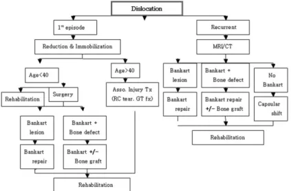
Fig. 1. Algorithm for treatment of shoulder dislocation
12. Kirkley A, Werstine R, Ratjek A, Griffin S: Prospective randomized clinical trial comparing the effectiveness of immediate arthroscopic stabilization versus immobilization and rehabilitation in first traumatic anterior dislocations of the shoulder: Long=term evaluation. Arthroscopy, 21:55-63, 2005 13. Kropf EJ, Tjoumakaris FP, Sekiya JK: Arthroscopic shoulder stabilization: Is there ever a need to open?
Arthroscopy, 23:779-784, 2007.
14. Lo IK, Parten PM, Burkhart SS: The inverted pear glenoid: An indicator of significant glenoid bone loss.
Arthroscopy, 20:169-174, 2004.
15. Meehan RE, Petersen SA: Results and factors affecting outcome of revision surgery or shoulder instability. J Shoulder Elbow Surg, 14:31-37, 2005.
16. Montgomery MH Jr, Wahl M, Hettrich C, Itoi E, Lippitt SB, Matsen FA III: Anteroinferior bone- grafting can restore stability in osseous glenoid defects. J Bone Joint Surg, 87-A:1972-1977, 2005.
17. Rowe CR, Patel D, Southmayd WW: The Bankart procedure: A long-term end-result study. J Bone Joint Surg, 60:1-16, 1978.
18. Simonet WT, Cofield RH: Prognosis in anterior shoulder dislocation. Am J Sports Med 12:19-24, 1984.
19. Stein DA, Jazrawi L, Bartolozzi AR: Arthroscopic stabilization of anterior shoulder instability: A review of the literature. Arthroscopy 18:912-924, 2002.
20. Uhurchak JM, Arciero RA, Huggard D, Taylor DC: Recurrent shoulder instability after open reconstruction in athletes involved in collision and contact sports. Am J Sports Med, 28:794-799, 2000.
21. Warner JJP, Gill TJ, O’Holleran JD, Pathare N, Millett PJ: Anatomical glenoid reconstruction for recurrent anterior glenohumeral instability with glenoid deficiency using an autogenous tricortical iliac crest bone graft. Am J Sports Med, 34:205-212, 2006.