ISSN 1225-6552, eISSN 2287-7630 http://dx.doi.org/10.7853/kjvs.2015.38.2.101
< Original Article >
Veterinary Service
Available online at http://kjves.org
*Corresponding author: Okjin Kim, Tel. +82-63-850-6668, Fax. +82-63-850-7308, E-mail. [email protected]
#
Both authors contributed equally to this work.
A 16S rDNA polymerase chain reaction assay to detect Mycoplasma pulmonis in rats model
Sunhwa Hong 1# , Hyun-A Lee 1# , Yeon-Shik Choi 2 , Yungho Chung 3 , Okjin Kim 1 *
1
Center for Animal Resource Development, Wonkwang University, Iksan 570-749, Korea
2
Department of Bio-Medical Analysis, Bio Campus of Korea Polytechnics, Nonsan 320-905, Korea
3
Department of Companion Animal and Animal Resources Science, Joongbu University, Geumsan-gun 312-702, Korea (Received 17 May 2015; revised 16 June 2015; accepted 23 June 2015)
Abstract
Murine mycoplasmosis, caused by Mycoplasma (M.) pulmonis, is a prominent disease in rodent animals.
The aim of this study was to develop a sensitive and specific PCR assay to detect M. pulmonis in ani- mals and to assess the suitability of this assay for the detection of mycoplasmal infection in rats ex- perimentally infected with M. pulmonis. A new PCR assay using the M. pulmonis-specific primer pairs MPul-F and MPul-R was developed. The primers and probe for the assay were designed from regions in the 16S rRNA gene that are unique to M. pulmonis. The novel PCR assay was very specific and sensitive for M. pulmonis, detecting the equivalent of 5 pg of target template DNA. It detected only M. pulmonis and no other Mycoplasma species or other bacterial species. The newly developed PCR assay also effectively detected M. pulmonis infection in rats. These results suggest that this PCR assay using M. pulmonis-specific primer pairs of MPul-F and MPul-R will be useful and effective for monitor- ing M. pulmonis infection in animals.
Key words : Mycoplasma, Mycoplasma pulmonis, 16S rRNA, Pneumonia, Rat
INTRODUCTION
Murine mycoplasmosis, caused by M. pulmonis, is a prominent disease in laboratory rats and mice. Because of its chronicity and slow cumulative mortality, M. pul- monis has profound impact on many parameters in ro- dent studies (Lindsey et al, 1971; Cassell et al, 1986).
One of the primary diseases caused by M. pulmonis is murine respiratory mycoplasmosis or chronic respiratory disease, which involves the nasal passages, middle ears, trachea, and lungs, and causes rhinitis, otitis media, tra- cheitis, and pneumonia (Lindsey et al, 1971; Cassell et al, 1981a). Apart from respiratory disease, this organism can also cause genital infections, resulting in reduced birth rates (Cassell et al, 1981a; Cassell et al, 1981b;
Cassell, 1982). Despite their pathogenic potential, myco- plasmal infections can remain imperceptible and be very dangerous because mycoplasmas possess immunomodu- latory activities that can influence the outcome of ex- periments (Cassell et al, 1981b; Cassell et al, 1986).
Identification of Mycoplasma species as the causative
agent of disease is often hindered by the lack of rapid
diagnostic tests, along with similarities in the clinical
features of the diseases caused by them. Conventional
methods of Mycoplasma species diagnosis are based on
microbiological and serological tests such as comple-
ment fixation test, enzyme-linked immunosorbent assay
(ELISA), and immunoblotting; these methods can be
time-consuming, insensitive, and nonspecific (Muthomi
and Rurangirwa, 1983; Ball and Finlay, 1998; Nicholas
et al, 1996). It is necessary to clarify the status of
Mycoplasma contamination in animal colonies because
of its prevalence in commercial and animal facilities
Fig. 1. Location of species-specific PCR primer pairs MPul-F and MPul-R designed from the Mycoplasma pulmonis 16S ribosomal RNA sequence.
(Lindsey et al, 1971). Polymerase chain reaction (PCR) is a powerful technique for identifying mycoplasmas and for studying homology between their nucleic acids (McAulifffe et al, 2003).
The aim of this study was to develop a sensitive and specific PCR assay to detect M. pulmonis in laboratory rodent animals and to assess the suitability of this assay for the detection of mycoplasmal infection in rats ex- perimentally infected with M. pulmonis.
MATERIALS AND METHODS
Microorganisms and growth conditions M. pulmonis (ATCC19612), M. hyopneumoniae (AT- CC25934), M. hyorhinis (ATCC27717), M. hominis (ATCC23114), and M. arthritiditis (ATCC19611) were obtained from the American Type Culture Collection (Rockville, Md.). Mycoplasmas were grown in modified Friis medium (Friis, 1973), containing 20% porcine se- rum (Gibco-BRL, USA), 5% fresh yeast extract (Gi- bco-BRL), 0.15 mgㆍmL
−1methicillin (Sigma-Aldrich, USA), 0.15 mgㆍmL
−1bacitracin (Sigma-Aldrich), and 0.08 mgㆍmL
−1thallium acetate (Sigma-Aldrich). Cells were harvested by centrifugation at 12,000 x g for 30 min at 4
oC, washed 3 times, and suspended in 0.1 M phosphate-buffered saline (PBS; pH 7.4).
Animals and infection
We acquired 5-week-old male specific pathogen-free Sprague Dawley rats from Samtako (Osan, Korea). Rats were acclimatized and kept in an isolated specific patho- gen-free (SPF) barrier room with regulated temperature (23
oC±1
oC), humidity (50%±5%), and light/dark cycle (12/12 h). The animals were fed sterilized pellet diet by 2 M rad radiation (Purina, Korea) and sterilized water ad libitum. After an adaptation period of 1 week, the animals were divided into 2 groups (infected and con- trol) and maintained in an opaque polypropylene cage in an isolated ventilated cage system. Five rats (rats 1 through 5) were experimentally infected with 10
6col- ony-forming units (CFU) of M. pulmonis (in 100 L of
culture medium) by intranasal inoculation. Five non-in- fected control rats (rats 6 through 10), housed separately from the infected rats, were used as controls. After 1 week, these rats were sacrificed by cervical dislocation.
Fresh pulmonary tissues were collected and subjected to the M. pulmonis-specific PCR assay developed in this study.
All studies were performed in accordance with the Guide for Animal Experimentation by Wonkwang Uni- versity and approved by the Institutional Animal Care and Use Committee of Wonkwang University (Approval No. WKU13-35). All efforts were made to minimize the pain or discomfort to the animals used.
Nucleic acid extraction and designation of the M. pulmonis-specific PCR primers
DNA was extracted from the M. pulmonis cultures. In addition, the pulmonary tissues were homogenized and resuspended in PBS and subjected to DNA extraction as described previously (Cho et al, 2011). Genomic DNA was isolated using an AccuPrep Genomic DNA ex- traction kit (Bioneer Corporation, Daejeon, Korea) ac- cording to the manufacturer's instructions. DNA was eluted in Tris-EDTA buffer (pH 8.0), and an aliquot was used for PCR amplification. All DNA samples were stored at −20
oC until the PCR assays were performed.
Species-specific PCR primer pairs were designed from
Mycoplasma 16S rRNA sequence for the detection of
M. pulmonis (Fig. 1). The forward primer MPul-F was
5'-CAG TAC TTG AGT TAG AAA ATG GA-3' (23-mer,
nucleotide 256498-256520). The reverse primer MPul-R
was 5'-ATC TGA AAG TTT TGA AGA GTT TTG-3'
(24-mer, nucleotide 257383-257405).
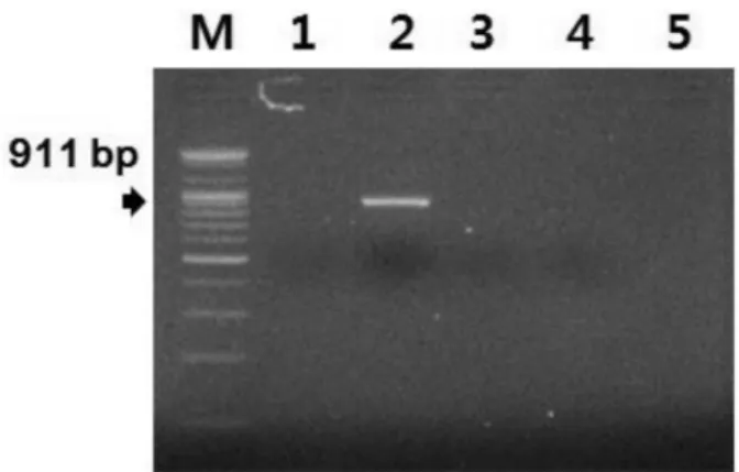
Fig. 2. Specificity of the developed species-specific PCR targeted with the Mycoplasma pulmonis 16S ribosomal RNA sequence. Lane M: 100-bp marker, N: negative control, 1: M. hyopneumoniae, 2: M.
pulmonis, 3: M. hyorhinis, 4: M. hominis, 5: M. arthritiditis.
Assay specificity
DNA was extracted from the cultures of M. hyopneu- moniae, M. hyorhinis, M. pulmonis, M. hominis, and M.
arthritiditis. Amplification of the M. pulmonis-specific gene was performed with the primer pairs of MPul-F and MPul-R designed from the Mycoplasma 16S rRNA sequence in this study. The template DNA (50 ng) and 20 pmol each primer were added to a PCR mixture tube (AccuPower PCR PreMix; Bioneer Corp., Korea) con- taining 2.5 U of Taq DNA polymerase, 250 M each deoxynucleoside triphosphate (dNTP), 10 mM Tris-HCl (pH 8.3), 40 mM KCl, 1.5 mM MgCl
2, and the gel loading dye. The volume was adjusted to 20 L with distilled water. The reaction mixture was subjected to denaturation at 94
oC for 5 min, followed by 30 cycles of 95
oC for 1 min, 60
oC for 1 min, and 72
oC for 1 min, and a final extension step of 72
oC for 3 min; the sam- ples were kept at 4
oC until analysis. Reactions were conducted using My Genie 32 Thermal Block PCR (Bioneer, Korea). After amplification, a 5-L aliquot of each PCR reaction mixture was separated by electro- phoresis on 2% agarose gels, followed by ethidium bro- mide (EtBr) staining and ultraviolet (UV) transillumination.
Assay sensitivity
DNA was extracted from M. pulmonis and M. homi- nis cultures. To determine the sensitivity of the newly developed PCR assay, serial dilutions of purified chro- mosomal DNA of M. pulmonis strains were tested.
Amplification of the M. pulmonis-specific gene was per- formed with the primer pairs of MPul-F and MPul-R designed from the Mycoplasma 16S rRNA sequence in this study. The reaction condition of PCR assay After amplification, a 5-L aliquot of each PCR reaction mix- ture was separated by electrophoresis on 1.5% agarose gels, followed by EtBr staining and UV transillumination.
Applicability to animal samples
To evaluate the PCR system under field conditions, pulmonary tissues were collected from the rats experi- mentally infected with M. pulmonis. The pulmonary tis-
sues were homogenized, resuspended in PBS, and sub- jected to DNA extraction, as described previously (Cho et al, 2011). The PCR assay was performed with the M.
pulmonis-specific primer pairs of MPul-F and MPul-R designed in this study. After amplification, a 5-L ali- quot of each PCR reaction mixture was separated by electrophoresis on 1.5% agarose gels, followed by ethi- dium bromide (EtBr) staining and UV transillumination.
RESULTS
Assay specificity
A PCR assay using the M. pulmonis-specific primer pairs MPul-F and MPul-R was developed and evaluated for its specificity and sensitivity. Because of PCR am- plification, 911-bp amplicons were detected from the ex- tracted DNA of M. pulmonis (Fig. 2). The targeted 911-bp of the 16S rRNA gene of M. pulmonis were specifically amplified by the optimized PCR system with the M. pulmonis-specific primer pairs MPul-F and MPul-R designed in this study. The specificity of the newly developed M. pulmonis-specific primer pairs was confirmed using other bacterial DNA with a high level of homology in their sequences (Fig. 2). No positive signals were observed in the template DNA samples of M. hyopneumoniae, M. hyorhinis, M. hominis, and M.
arthritiditis (Fig. 2). However, the DNA of M. pulmonis
Fig. 3. Sensitivity of the developed species-specific PCR targeted with the Mycoplasma pulmonis 16S ribosomal RNA sequence. Lane M: 100-bp marker, 1: 10
4pg of M. pulmonis DNA, 2: 5×10
3pg of M.
pulmonis DNA, 3: 10
3pg of M. pulmonis DNA, 4: 5×10
2pg of M. pul- monis DNA, 5: 10
2pg of M. pulmonis DNA, 6: 5×10 pg of M. pulmo- nis DNA, 7: 10 pg of M. pulmonis DNA, 8: 5 pg of M. pulmonis DNA, 9: 1 pg of M. pulmonis DNA.
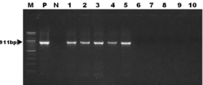
Fig. 4. Amplified products of sample DNAs obtained by the devel- oped species-specific PCR for Mycoplasma pulmonis were identified using 1.5% agarose gel electrophoresis. Lane M: 100-bp marker, P:
positive control, N: negative control, 1∼5: DNAs extracted from the pulmonary tissues of M. pulmonis-infected rats, 6∼10: DNAs ex- tracted from the pulmonary tissues of non-infected control rats.
yielded a strong positive signal (Fig. 2).
Assay sensitivity
To evaluate the sensitivity of the newly developed M.
pulmonis-specific PCR assay, serial dilutions of the DNA extracted from the cultured M. pulmonis were tested. Results revealed the presence of at the least 5 pg of genomic DNA in the 5-L aliquots of PCR product by EtBr staining (Fig. 3).
Applicability to animal samples
A newly developed PCR assay using primers de- signed from the Mycoplasma 16S rRNA sequence was employed to detect M. pulmonis infection in rats. The targeted 911-bp gene was specifically amplified by the PCR assay with the M. pulmonis-specific primer pairs MPul-F and MPul-R. The target nucleic acid fragments were specifically amplified in the pulmonary tissue sam- ples of M. pulmonis-infected rats by the newly devel- oped PCR analysis (Fig. 4). No positive signals were detected in the tissue samples of non-infected control rats (Fig. 4).
DISCUSSION
M. pulmonis can have a significant detrimental impact on research that utilizes infected mice and rats by caus- ing morbidity and mortality, and through interference
with respiratory function and altered respiratory carcino- genesis and immunity (Baker, 1998; Institute of Laboratory Animal Resources, 1991). Contamination of transplantable tumors, cell cultures, and other biological materials with M. pulmonis and other Mycoplasma spp. is quite com- mon and problematic for the maintenance and use of these materials (Collins and Parker, 1972; Nicklas et al, 1993).
Diagnosis of contamination by Mycoplasma species is usually accomplished by microbiological studies or im- munofluorescence tests performed on frozen, thin pul- monary tissue sections by using polyclonal antibodies (Kobisch and Friis, 1996; Maes et al, 1996). However, because of the fastidious nature of the Mycoplasma spp., its microbiological and serological identification may take up to 1 month. Serological detection is further hampered by cross-reactions, which have been reported among M. hyopneumoniae, M. hyorhinis, and M. floccu- lare (Freeman et al, 1984; Armstrong et al, 1987).
With the advances made in molecular biology during the last few years, more is known about Mycoplasma spp. genes, and other diagnostic tools have been devel- oped for this organism. Recently, PCR methods have been used to detect Mycoplasma spp. (Hong et al, 2011). PCR-based methods for the detection of certain regions in the Mycoplasma genome have proven to be both rapid and specific (Hu et al, 1995; Harasawa and Kanamoto, 1999; Kong et al, 2001; Loens et al, 2003;
Khanna et al, 2005).
In this study, a new PCR assay using the M. pulmo-
nis-specific primer pairs MPul-F and MPul-R was
developed. The primers and probe for the assay were designed from regions in the 16S rRNA gene that are unique to M. pulmonis. The novel PCR assay was very specific and sensitive for the detection of M. pulmonis.
The assay was able to detect the equivalent of 5 pg of target template DNA, indicating that the assay was very sensitive. The M. pulmonis PCR assay was shown to be highly specific as only M. pulmonis and no other Myco- plasma spp. or other bacterial species was detected. In addition, the newly developed PCR assay effectively de- tected M. pulmonis infection in rats.
Thus, results from this study suggest that this newly developed PCR assay using the M. pulmonis-specific primer pairs MPul-F and MPul-R will be useful and ef- fective for monitoring M. pulmonis infection in animals.
ACKNOWLEDGEMENTS
This study was supported by the research fund of Wonkwang University in 2015. We wish to appreciate Gi-Wook Oh, research assistants of Center for Animal Resources Development, Wonkwang University, for car- rying out the technical support.
REFERENCES
Armstrong CH, Freeman MJ, Sands-Freeman L. 1987. Crossreactions between Mycoplasma hyopneumoniae and Mycoplasma
flocculare: practical implications for the serodiagnosis of mycoplasmal pneumonia of swine. Isr J Med Sci 23:
654-656.
Baker DG. 1998. Natural pathogens of laboratory mice, rats, and rabbits and their effects on research. Clin Microbiol Rev 11: 231-266.
Ball HJ, Finlay D. 1998. Diagnostic application of monoclonal antibody (MAb)-based sandwich ELISAs. Methods Mol Biol 104: 127-132.
Cassell GH. 1982. The Derrick Edward Award Lecture: The pathogenic potential of mycoplasmas: Mycoplasma pul-
monis as a model. Rev Infect Dis 4(Suppl): 18-34.
Cassell GH, Davis JK, Simecka JW, Lindsey JR, Cox NR, Ross S, Fallon M. 1986. Mycoplasmal infections: disease pathogenesis, implications for biomedical research and control. pp. 87-130. In: Bhatt PN, Jacoby RO, Morse III HC, New AE (ed.). Viral and mycoplasmal infections of laboratory rodents effects on biomedical research. Aca-
demic Press, Orlando.
Cassell GH, Lindsey JR, Davis JK. 1981a. Respiratory and geni- tal mycoplasmosis of laboratory rodents: implications for biomedical research. Isr J Med Sci 17: 538-554.
Cassell GH, Wilborn WH, Silvers SH, Minion FC. 1981b.
Adherence and colonization of Mycoplasma pulmonis to genital epithelium and spermatozoa in rats. Isr J Med Sci 17: 593-598.
Cho SJ, Lee HA, Hong S, Kim O. 2011. Uterine adenocarcinoma with feline leukemia virus infection. Lab Anim Res 27:
347-351.
Collins MJ Jr, Parker JC. 1972. Murine virus contaminants of leukemia viruses and transplantable tumors. J Natl Cancer Inst 49: 1139-1143.
Freeman MJ, Armstrong CH, Freeman-Sands LL, Lopez-Osuna M. 1984. Serological cross-reactivity of porcine refer- ence antisera to Mycoplasma hyopneumoniae, M. floccu-
lare, M. hyorhinis and M. hyosynoviae indicated by the
enzyme-linked immunosorbent assay, complement fix- ation and indirect hemagglutination tests. Can J Comp Med 48: 202-207.Friis NF. 1973. The pathogenicity of Mycoplasma flocculare.
Acta Vet Scand 14: 344-346.
Harasawa R, Kanamoto Y. 1999. Differentiation of two biovars of Ureaplasma urealyticum based on the 16S-23S rRNA intergenic spacer region. J Clin Microbiol 37: 4135-4138.
Hong S, Lee HA, Park SH, Kim O. 2011. Sensitive and specific detection of Mycoplasma species by consensus polymer- ase chain reaction and dot blot hybridization. Lab Anim Res 27: 141-145.
Hu M, Buck C, Jacobs D, Paulino G, Khouri H. 1995.
Application of PCR for detection and identification of
mycoplasma contamination in virus stocks. In Vitro Cell
Dev Biol Anim 31: 710-715.Institute of Laboratory Animal Resources. 1991. Infectious dis- eases of mice and rats. National Academy Press, Wa- shington D.C.
Khanna M, Fan J, Pehler-Harrington K, Waters C, Douglass P, Stallock J, Kehl S, Henrickson KJ. 2005. The pneumo- plex assays, a multiplex PCR-enzyme hybridization as- say that allows simultaneous detection of five organisms,
Mycoplasma pneumoniae, Chlamydia (Chlamydophila) pneumoniae, Legionella pneumophila, Legionella micda- dei, and Bordetella pertussis, and its real-time counterpart.
J Clin Microbiol 43: 565-571.
Kobisch M, Friis NF. Swine mycoplasmoses. 1996. Rev Sci Tech 15: 1569-1606.
Kong F, James G, Gordon S, Zelynski A, Gilbert GL. 2001.
Species-specific PCR for identification of common con- taminant mollicutes in cell culture. Appl Environ Microbiol 67: 3195-3200.
Lindsey JR, Baker HJ, Overcash RG, Cassell GH, Hunt CE.
1971. Murine chronic respiratory disease. Significance as a research complication and experimental production with Mycoplasma pulmonis. Am J Pathol 64: 675-708.
Loens K, Ursi D, Goossens H, Ieven M. 2003. Molecular diag- nosis of Mycoplasma pneumoniae respiratory tract infections. J Clin Microbiol 41: 4915-4923.
Maes D, Verdonck M, Deluyker H, de Kruif A. 1996. Enzootic pneumonia in pigs. Vet Q. 18: 104-109.
McAulifffe L, Ellis RJ, Ayling RD, Nicholas RA. 2003.
Differentiation of Mycoplasma species by 16S ribosomal DNA PCR and denaturing gradient gel electrophoresis fingerprinting. J Clin Microbiol 41: 4844-4847.
Muthomi EK, Rurangirwa FR. 1983. Passive haemagglutination and complement fixation as diagnostic tests for conta-
gious caprine pleuropneumonia caused by the F-38 strain of mycoplasma. Res Vet Sci 35: 1-4.
Nicholas RA, Santini FG, Clark KM, Palmer NM, De Santis P, Bashiruddin JB. 1996. A comparison of serological tests and gross lung pathology for detecting contagious bo- vine pleuropneumonia in two groups of Italian cattle.
Vet Rec 139: 89-93.
Nicklas W, Kraft V, Meyer B. 1993. Contamination of trans- plantable tumors, cell lines, and monoclonal antibodies with rodent viruses. Lab Anim Sci 43: 296-300.