237 http://dx.doi.org/10.4196/kjpp.2013.17.3.237
ABBREVIATIONS: DMSO, dimethyl sulfoxide; D-e-Mapp, (1S,2R)-2- N-(tetradecanoylamino)-1-phenyl-1-propanol; B13, (1R,2R)-2-N-(tet- radecanoylamino)-1-(4'-nitrophenyl)-1,3-propanediol; PBS, phosphate- buffered saline; MTT, 3-[4,5-dimethylthiazol-2-yl]-2,5-diphenyltetra- zolium bromide; 3D-QSAR, three dimensional quantitative structure activity relationship; CoMSIA, comparative molecular similarity indices analysis.
Received November 22, 2012, Revised February 22, 2013, Accepted March 6, 2013
Corresponding to: Chaeuk Im, College of Pharmacy, Chung-Ang University, Heuksuk-dong, Dongjak-gu, Seoul 156-756, Korea. (Tel) 82-2-820-5603, (Fax) 82-2-816-7338, (E-mail) [email protected]
This is an Open Access article distributed under the terms of the Creative Commons Attribution Non-Commercial License (http://
creativecommons.org/licenses/by-nc/3.0) which permits unrestricted non-commercial use, distribution, and reproduction in any medium, provided the original work is properly cited.
Cytotoxic Activity and Quantitative Structure Activity Relationships of Arylpropyl Sulfonamides
Yu Jin Hwang, Sang Min Park, Chul Bu Yim, and Chaeuk Im College of Pharmacy, Chung-Ang University, Seoul 156-756, Korea
B13 is a ceramide analogue and apoptosis inducer with potent cytotoxic activity. A series of arylpropyl sulfonamide analogues of B13 were evaluated for their cytotoxicity using MTT assays in prostate cancer PC-3 and leukemia HL-60 cell lines. Some compounds (4, 9, 13, 14, 15, and 20) showed stronger activities than B13 in both tumor cell lines, and compound (15) gave the most potent activity with IC
50values of 29.2 and 20.7 μ M, for PC-3and HL-60 cells, respectively. Three-dimensional quantitative structure-activity relationship (3D-QSAR) analysis was performed to build highly reliable and predictive CoMSIA models with cross-validated q
2values of 0.816 and 0.702, respectively. Our results suggest that long alkyl chains and a 1R, 2R configuration of the propyl group are important for the cytotoxic activities of arylpropyl sulfonamides. Moreover, the introduction of small hydrophobic groups in the phenyl ring and sulfonamide group could increase biological activity.
Key Words: Arylpropanol, Ceramide, Cytotoxicity, QSAR
INTRODUCTION
Apoptosis is an endogenous program of cell death that is mediated through biochemical modulators, and it is in- duced by anticancer agents, tumor necrosis factor, and ion- izing radiation [1,2]. Because ceramide is known to be a key modulator of apoptosis and its accumulation in cells leads to apoptosis, many approaches have been explored to increase endogenous ceramide [3-5]. These approaches in- clude the application of short-chain ceramides (C
6-ceramide) [6-8] and induction of ceramide by modulation of ceram- ide-metabolizing enzymes (LCL 15 and LCL 16) [9-13].
In recent years, ceramide analogues have emerged as a new strategy for cancer therapy [14-17]. The ceramide ana- logues, such as sphingosine, sphinganine, and C
2-eramide, were known to induce apoptotic cell death in the malignant melanoma, colon cancer cells, and prostate cancer cells [18-21]. It was reported that the activity of ceramide ana- logues is influenced by the stereochemistry and chain length of the amide moiety. Analogues with a ceramide con- figuration (2S, 3R) are not active, whereas their stereo- isomers inhibit cell growth. The ceramides with medium size alkyl chains (C
11~C
15) are active compounds, but ce- ramides with shorter (C
1~C
9) and longer (C
17~C
23) alkyl
chains are less active [22]. The aromatic analogues of ce- ramide like D-e-Mapp with a 1S, 2R configuration increase endogenous ceramide to induce apoptosis in many cancer cell lines [23-26]. Another active analogue, B13 with a 1R, 2R configuration, exhibited cytotoxicity in malignant mela- noma, colon, and prostate cancer cells [27-29]. B13 also in- duced apoptosis in cancer cells, increased the cellular ce- ramide level, inhibited metastasis, and had no effect on nor- mal cells [29-31].
It has been reported that isosteric replacement of the amide group in a ceramide by urea or amine can increase the ceramidase inhibitory activity and cytotoxicity [32].
And the carboxyl moiety of ceramide can also be bioisosteri- cally replaced by a sulfone group. Sulfonamides are known to have various biological activities, including hypoglycemic, diuretic, antibacterial activity, and cytotoxicity [33-36].
Extending these findings to the ceramide, we suspected that introduction of a phenyl ring, as in D-e-Mapp and B13, altering alkyl chain lengths and stereochemistry, and bio- isosterical replacement of the carboxyl group with a sulfone group would improve and modify the biological properties of ceramide.
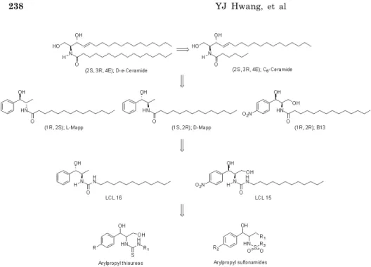
As a continuation of our previously reported work, in
which arylpropyl thiourea analogues (Fig. 1) were studied
[37], we determined the cytotoxic activity of 21 new ar-
ylpropyl sulfonamide analogues (Fig. 1) in prostate cancer
and leukemia cell lines to evaluate the effects of R and S
enantiomers in propyl moiety, a p-nitro group in the phenyl
Fig. 1. Ceramide, Mapp, B13, and arylpropyl compounds.
ring, and the alkyl chain length of the sulfonamide moiety.
We also perform QSAR analysis to investigate the relation- ship between the structural features and cytotoxicity of ar- ylpropyl sulfonamides and to develop more potent anti- cancer agents.
METHODS Materials
A series of arylpropyl compounds were previously syn- thesized in our lab. Phosphate-buffered saline (PBS) was purchased from Boehringer Mannheim. Dimethyl sulfoxide (DMSO), 3-[4,5-dimethylthiazol-2-yl]-2,5-diphenyltetrazolium bromide (MTT), and other reagents were obtained from Sigma.
In vitro cytotoxic assay
The cytotoxicity of arylpropyl compounds was evaluated in two human tumor cell lines: prostate cancer PC-3 cells and leukemic cancer HL-60 cells. The cytotoxicity was de- termined using a MTT-based colorimetric assay [38]. The cells were treated as described in the Table 1 legend and the results from the assay are shown in Table 1 [39].
Data sets
Twenty-one arylpropyl compounds with cytotoxic IC
50values ranging from 20.7 to 267.3μM were used to carry out 3D-QSAR analysis. Their molecular structures are il- lustrated in Table 1. The test set was selected based on their various configurations at C
1and C
2and used for ex- ternal validation of the 3D-QSAR models. The training set was consisted of seventeen compounds including B13 and the test set was made of the following four different config- uration structures, compound 2(1R,2S), 8(1S,2R), 12(1R,2R), and 17(1S,2S). All IC
50values were transformed into pIC
50(-log IC
50) values and used as the dependent variables in
the CoMSIA studies.
Molecular modeling and alignment
The modeling software Sybyl-X 1.3 was used for the structure building, molecules modeling, partial least squares, and conducting CoMSIA [40]. The structures of compounds were generated with a sketch tool and energy minimization was performed using a TRIPOS force field with the Powell method and conjugate gradient termination.
The atomic charges of molecules were calculated using the Gasteiger- Hückel charges. Simulated annealing was used to determine the low-energy conformations. One of the most important requirements for CoMSIA models is that the 3D structures of molecules are aligned to a suitable conforma- tional template. The molecular alignment was achieved by the fitting atoms method. In Table 1, the bold line repre- sents a common substructure and B13 was used as a tem- plate molecule in the alignment.
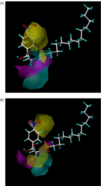
CoMSIA 3D-QSAR models
CoMSIA is based on the relationship between the bio- logical activity and structural properties of compounds when the receptor structure is not known. It evaluates the five physicochemical properties: steric, electrostatic, hydro- phobic, hydrogen bond acceptor, and hydrogen bond donor fields. The CoMSIA method involves a common probe atom and similarity indices determined in regularly spaced grid points for the aligned molecules. The common probe atom with a radius of 1.0 Å, charge of +1, hydrophobicity of +1, hydrogen bond donating of +1, and hydrogen bond accept- ing of +1 was used to calculate the five fields. A default value of 0.3 was used for the attenuation factor.
Partial least squares (PLS) analysis
In the PLS analysis, three CoMSIA descriptors (electro-
static, hydrophobic, and hydrogen bond acceptor fields)
were used as independent variables, and the pIC
50values
Table 1. Structures and cytotoxic activities of arylpropyl sulfonamides
R1 OH
HNSR3 O R2 O
1 23
Compounds
R
1R
2R
3Configuration
Cytotoxicity IC
50(μM) Prostate cancer
(PC-3)
Leukemia (HL-60) 1
2 3 4 5 6 7 8 9 10 11 12 13 14 15 16 17 18 19 20 B13
H H H H H H H H H H OH OH OH OH OH OH OH OH OH OH
H H H H H H H H H H NO
2NO
2NO
2NO
2NO
2NO
2NO
2NO
2NO
2NO
2C
7H
15C
8H
17C
9H
19C
11H
23C
13H
27C
7H
15C
8H
17C
9H
19C
11H
23C
13H
27C
7H
15C
8H
17C
9H
19C
11H
23C
13H
27C
7H
15C
8H
17C
9H
19C
11H
23C
13H
271R, 2S 1R, 2S 1R, 2S 1R, 2S 1R, 2S 1S, 2R 1S, 2R 1S, 2R 1S, 2R 1S, 2R 1R, 2R 1R, 2R 1R, 2R 1R, 2R 1R, 2R 1S, 2S 1S, 2S 1S, 2S 1S, 2S 1S, 2S 1R, 2R
189.3 95.3 81.7 44.9 45.1 188.5 80.8 64.3 40.5 67.8 98.4 52.8 31.8 39.1 29.2
> 267.3 75.1 51.0 56.0 35.9 79.3
129.8 77.6 53.7 28.5 34.3 130.4 62.8 44.9 33.1 44.4 116.6 54.9 27.6 24.7 20.7 160.6 67.6 41.4 37.7 23.0 33.6 The cells were plated at a density of approximately 1×10
4cells/well in 96-well plates. Each well contained 180μl of medium and 20μl of 10×concentration of prepared compounds or PBS were added. After 96 h of culture, 0.1 mg of MTT was added to each well and incubated at 37
oC for 4 h. The plates were centrifuged at 450×g to precipitate the formazan crystals. The medium was rem9oved and 150μL of DMSO was added to each well to dissolve the formazan. In this assay, MTT was converted to blue formazan by mitochondrial dehydrogenase. The intensity of the blue color was measured with a microplate reader at a wavelength of 540 nm. The measured mean values were expressed as the IC
50, the concentration that reduced the optical density of the treated cells by 50% with respect to the untreated controls.
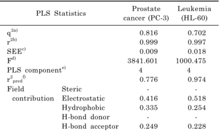
Table 2. PLS analysis of CoMSIA 3D-QSAR models PLS Statistics Prostate
cancer (PC-3)
Leukemia (HL-60) q
2a)r
2b)SEE
c)F
d)PLS component
e)r
2predf)0.816 0.999 0.009 3841.601
4 0.776
0.702 0.997 0.018 1000.475
4 0.974 Field
contribution Steric Electrostatic Hydrophobic H-bond donor H-bond acceptor
- 0.416 0.335 -
0.249
- 0.518 0.254 -
0.228
a)
q
2, cross-validated correlation coefficient from leave-one- out (LOO);
b)r
2, non-cross-validated correlation coefficient;
c)SEE, standard error of estimate;
d)F, F-test value;
e)PLS component, opti- mum number of components;
f)r
2pred, predicted correlation coefficient.
were used as dependent variables. The predictive pIC
50val- ues of the models were evaluated by leave-one-out (LOO) cross-validation. In the LOO method, one compound is re-
moved from the data set and its biological activity is pre- dicted with the model derived from the rest of data set.
The LOO method determines the optimum number of com- ponents, which are then used for the non-cross-validated analysis. To test the utility of the model as a predictive tool, the test set compounds that were not used in the model generation were predicted.
RESULTS In vitro cytotoxic activity
The cytotoxicities of 21 compounds were evaluated in vitro and presented in Table 1. B13 gave moderate cytotox- icity with IC
50values of 79.3 and 33.6μM for prostate can- cer PC-3 and leukemia HL-60 cells, respectively. The IC
50values of the other structures ranged from 29.2 to 267.3μM for PC-3 cells and 20.7 to 160.6μM for HL-60 cells.
For prostate cancer PC-3 cells, the long alkyl chain (C
13H
27and C
11H
23) compounds (4, 5, 9, 10, 14, 15, 19, and 20) exhibited more potent activities than B13 to give IC
50values of 44.9, 45.1, 40.5, 67.8, 39.1, 29.2, 56, and 35.9 μM,
respectively. However the short alkyl chain (C
7H
15and
C
8H
17) compounds (1, 2, 6, 7, 11, and 16) showed less potent
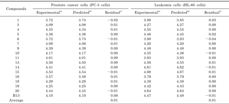
Table 3. Residuals of the predicted cytotoxicities (pIC50) of training set
Compounds Prostate cancer cells (PC-3 cells) Leukemia cells (HL-60 cells)
Experimental
a)Predicted
b)Residual
c)Experimental
a)Predicted
b)Residual
c)1
3 4 5 6 7 9 10 11 13 14 15 16 18 19 20 B13 Average
3.72 4.09 4.35 4.36 3.72 4.09 4.39 4.17 4.01 4.50 4.41 4.53 3.57 4.29 4.25 4.44 4.10
3.74 4.08 4.34 4.36 3.73 4.08 4.39 4.17 4.01 4.50 4.41 4.54 3.56 4.28 4.25 4.45 4.10
- 0.02 0.01 0.01 0.00
- 0.01 0.01 0.00 0.00 0.00 0.00 0.00
- 0.01 0.01 0.01 0.00
- 0.01 0.00 0.01
3.89 4.27 4.55 4.46 3.88 4.20 4.48 4.35 3.93 4.56 4.61 4.68 3.79 4.38 4.42 4.64 4.47
3.85 4.27 4.55 4.45 3.93 4.20 4.48 4.36 3.93 4.55 4.62 4.67 3.79 4.38 4.43 4.64 4.48
0.03 0.00 0.00 0.02
- 0.04 0.00 0.00
- 0.01 0.00 0.01
- 0.01 0.01 0.00 0.00 0.00 0.00
- 0.01 0.01
a)
Experimental cytotoxic activity;
b)predicted activity by the CoMSIA model with electrostatic, hydrophobic, and hydrogen bond accept- or fields;
c)difference between the experimental and predicted activities; The pIC
50(-log IC
50) values were converted from IC
50values.
Table 4. Residuals of the predicted cytotoxicities (pIC
50) of test set
Compounds Prostate cancer cells (PC-3 cells) Leukemia cells (HL-60 cells)
Experimental
a)Predicted
b)Residual
c)Experimental
a)Predicted
b)Residual
c)2
8 12 17 Average
4.02 4.19 4.28 4.12
4.02 4.17 4.27 4.21
0.00 0.02 0.01
-0.08 0.03
4.11 4.35 4.26 4.17
4.12 4.34 4.23 4.14
-0.01 0.01 0.03 0.03 0.02
a)