
Treatment of skeletal Class II adult patient with vertical and transverse problems caused by nasal airway obstruction using microimplant anchorage
16
0
0
전체 글
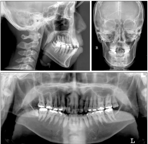
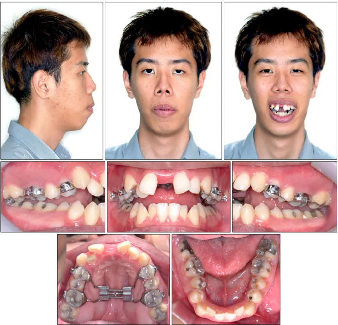
(2) Chae JM, Chang NY, Cho JH, Kang KH, Kim SC. anterior facial height, incompetent lip posture, excessive appearance of the maxillary anterior teeth, narrow external nares, steep mandibular plane angle, and a “V” shaped maxillary arch. Correction of transverse maxillary width discrepancies by opening of the midpalatal suture has been an integral part of orthodontic therapy for many years. Rapid maxillary expansion (RME) has been recommended for the correction of maxillary transverse deficiency with the additional benefit of increasing nasal airflow.6-9 Evidence to support this comes both from. Fig 1. Pretreatment facial and intraoral photographs.. 258. 대치교정지 39권 4호, 2009년. patients’ self-reported perceptions and from radiographic images. In children and adolescents, conventional orthodontic RME has been successful when used prior to sutural closure. On the other hand, in skeletally mature patients, the possibility of successful maxillary expansion decreases as the sutures close and the resistance to mechanical force increases. After sutural closure or the completion of transverse growth, orthopedic transverse maxillary expansion is largely unsuccessful because the expansion is composed primarily of alveolar or dental.
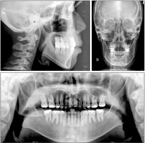
(3) Vol. 39, No. 4, 2009. Korean J Orthod. Treatment of skeletal Class II adult patient using microimplant anchorage. 10. tipping with little or no basal skeletal movement. In mature patients, RME also causes severe pain, periodontal complications, and gingival recession of the maxillary posterior teeth. Surgically-assisted RME (SARME) has been proposed to produce better treatment results in adults as well as to prevent complications by surgically releasing the closed sutures resisting the expansion forces.10 The SARME procedure decreases the risk of aseptic necrosis and relapse, particularly in patients with a major transverse discrepancy. SARME is a useful and predictable technique for achieving transverse maxillary expansion.11 As above-mentioned, RME increases the transverse dimension of the maxillary arch by separating the suture. However, mandibular expansion effects are localized to the alveolar bone and primarily induce tooth inclination. These effects are believed to be related to relapse.12-14 The addition of a lower Schwarz appliance to the treatment protocol allows significant extrusion of the lower molars and incisors. These changes correlate with the increase in vertical skeletal dimension.15. Surgical superior impaction of the maxilla has become an accepted treatment for the correction of vertical maxillary excess. Orthognathic surgery, which vertically repositions the maxilla, does predictably reduce nasal resistance, which intuitively should increase the percentage of nasal flow.16 On the other hand, directional force technology reinforced with MIA (microimplant anchorage)17 is a useful treatment approach for correcting a Class I or Class II dentoalveolar protrusion malocclusion. In particular, it is effective in treating patients with vertical problems, such as clockwise rotation of the dentoalveolar complex. In addition, it can create a favorable counterclockwise skeletal change as well as a balanced face minimizing surgical treatment.17,18. DIAGNOSIS AND ETIOLOGY A 22-year-old male presented with the chief complaint of lip protrusion and difficulty in nasal breathing. Facially, he exhibited a convex profile due to a. Fig 2. Pretreatment radiographs.. 259.
(4) Chae JM, Chang NY, Cho JH, Kang KH, Kim SC. retrognathic mandible. The facial photographs showed a convex facial profile with marked protrusion of the lips, mentalis strain, an excessive lower anterior facial height and a typical “adenoid face”. Intraorally, he had a Class I canine and molar relationship with moderate crowding, severe constricted “V” shaped arch forms of both jaws, an anterior openbite and poor oral hygiene (Fig 1). The panoramic radiograph revealed the presence of upper left and lower right third molars as well as restorations on several teeth. The lateral cephalogram and its tracing confirmed the severe skeletal problem (Fig 2). The skeletal pattern was deemed hyperdivergent as evidenced by a FMA (Frankfort-mandibular plane angle) of 33.5o and a FHI (facial height ino dex) of 0.61%. The ANB angle of 4.5 reflected the class II skeletal problem. The Z-angle of 51.5o quantified the facial imbalance (Table 1). The posteroanterior cephalometric radiograph showed a 2-mm deviation of the mandibular dental midline to the left relative to the maxillary dental midline with slight canting of the maxilla and transverse contriction of upper and lower arches (Fig 2). There were no significant signs or symptoms of any temporomandibular disorders.. TREATMENT OBJECTIVES. 대치교정지 39권 4호, 2009년. stricted upper and lower arches with SARME and a Schwarz appliance, respectively, followed by orthodontic treatment with extraction of the 4 first premolars and 2 third molars, and directional force technology with MIA. As described previously,17 MIA can provide absolute anchorage not only in the. Table 1. Cephalometric measurements Pretreatment. Post-. 13-month. treatment. retention. o. FMIA ( ). 53.5. 73.0. 71.5. FMA (o). 33.5. 30.5. 32.0. o. IMPA ( ). 93.0. 76.5. 76.5. SNA (o). 84.0. 83.0. 83.0. SNB ( ). 79.5. 80.0. 79.5. ANB (o). 4.5. 3.0. 3.5. AO-BO (mm). −2.0. −1.0. −1.5. Occlusal plane. 10.0. 8.0. 10.0. 112.0. 103.0. 98.0. o. o. angle ( ) FH to U1 (o) o. Z angle ( ). 51.5. 61.5. 60.5. PFH (mm). 54.5. 57.5. 57.0. AFH (mm). 91.0. 88.0. 89.0. FHI (PFH/AFH) (%). 0.61. 0.65. 0.64. (54.5/91.0) (57.5/88.0) (57.0/89.0). FMIA indicates angle between Frankfort plane and. The treatment objectives were to (1) align and level the teeth in both arches and establish a functional occlusion, (2) normalize the overjet and overbite relationships, (3) obtain a balanced facial profile, and (4) control the mouth breathing habit.. between SN and NA; SNB, angle between SN and NB;. TREATMENT ALTERNATIVES. AOBO, distance between perpendiculars drawn from. mandibular incisor axis; FMA, angle between Frankfort plane and mandibular plane; IMPA, angle between lower incisor axis and mandibular plane; SNA, angle ANB, difference between the SNA and SNB angles; point A and point B onto the occlusal plane; FH,. The first alternative was orthognathic surgery. Twojaw surgery including counterclockwise differential impaction of the maxilla and concurrent mandibular advancement surgery would be followed after expanding both constricted arches. Genioplasty would be necessary to reduce the long lower facial height and advance the chin along the facial midline. However, the patient requested minimal surgery so another treatment plan was chosen. The second alternative was expansion of the con-. 260. Frankfort horizontal plane; UI, maxillary incisor axis; FH to UI, angle between Frankfort plane and maxillary incisor axis; Z angle, angle between FH and Z line; Z line, profile line tangent to the chin and the more anterior vermilion border of both lips; FHI, ratio of PFH to AFH; PFH, linear measurement from articulare, along a line tangent to the posterior border of the mandible, to the intersection with the mandibular plane; and AFH, linear measurement from palatal plane to mention, measured perpendicular to palatal plane..
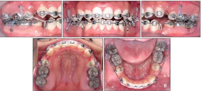
(5) Vol. 39, No. 4, 2009. Korean J Orthod. Treatment of skeletal Class II adult patient using microimplant anchorage. retraction of the maxillary and mandibular anterior teeth, but also in the intrusion of the maxillary anterior and posterior teeth, and mandibular posterior teeth. This would induce a horizontal mandibular response followed by a balanced facial profile.. TREATMENT PROGRESS The protocol began with a phase I treatment in which the Schwarz appliance was activated once per week for approximately 5 months. SARME was followed concurrently with mandibular expansion. A Hyrax palatal expansion appliance was cemented to the maxillary first molars and first premolars. The patient was then referred to the oral surgery department for the surgically assisted expansion procedure. At the conclusion of surgery, the surgeon turned the expansion appliance until the papillary gingival tissue blanched between the central incisors (8 turns or approximately 2 mm). The patient returned to the orthodontic department 7 days later (Fig 3). The patient was instructed to turn the appliance two turns per day (0.5 mm) for 20 days and then 1 turn per week for a further 28 days until the desired level of expansion was achieved (Fig 4). After the expansion, a hyrax palatal expander remained in place for an additional 3.5. months while the mandibular expansion was completed (Fig 5). The phase II treatment plan involved TweedMerrifield directional force technology with MIA, after extracting the maxillary and mandibular first premolars, as well as the third molars. After the extractions, 0.022 × 0.028-in nontipped, nontorqued edgewise appliances were placed in both arches, and leveling began with a 0.014-in nickel-titanium archwire and 0.018 stainless steel arch wires. Maxillary posterior MIs (microimplants, 1.3 1.2 mm in diameter, 8 mm in length; Absoanchor SH1312-08, Dentos, Taegu, South Korea) were implanted into the buccal alveolar bone between the maxillary second premolars and first molars. The mandibular posterior MIs (1.3 - 1.2 mm in diameter, 7 mm in length; Absoanchor SH1312-08, Dentos) were implanted into the buccal alveolar bone between the mandibular first and second molars. An elastic chain force was loaded immediately after placing the MIs, from the maxillary and mandibular posterior MIs to the canine brackets, in order to retract the maxillary and mandibular canines to level the six anterior teeth (Fig 6A). Two months into the treatment, 0.019 × 0.025-in stainless steel archwire with closing loops was placed. Fig 3. Surgical assisted rapid maxillary expansion on the maxilla and Schwarz appliance on the mandible.. 261.
(6) Chae JM, Chang NY, Cho JH, Kang KH, Kim SC. 대치교정지 39권 4호, 2009년. Fig 4. Photographs after 28 day expansion of the maxilla and mandible.. in the lower arch to retract the six anterior teeth. An intruding force was applied from the mandibular posterior MIs to the omega stop loop areas of the mandibular archwire in order to prevent mesial tipping and extrusion of the mandibular posterior teeth. Three months into the treatment, a 0.017 × 0.022-in stainless steel archwire was placed in the upper arch to level the six anterior teeth and an intruding force was applied from the maxillary posterior MIs to the maxillary archwire to intrude the maxillary posterior teeth. An. 262. additional unilateral Class III elastic force was applied to the loop of the mandibular archwire from the maxillary posterior MIs in order to reduce the dental asymmetry (Fig 6B). Four months into the treatment, 0.020 × 0.025-in stainless steel archwire with closing loops was placed in the upper arch to retract the six anterior teeth. This was supported by the maxillary anterior MIs (microimplants, 1.3 - 1.2 mm in diameter, 8 mm in length; Absoanchor SH1312-08, Dentos) that were implanted.
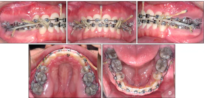
(7) Vol. 39, No. 4, 2009. Korean J Orthod. Treatment of skeletal Class II adult patient using microimplant anchorage. Fig 5. Photographs after 3.5 months of retention of the maxilla and 5 months of expansion of the mandible.. into the labial alveolar bone between the maxillary central incisors for torque control, bodily movement and intrusion of the maxillary anterior teeth (Fig 6C). After en masse movement, directional force control was performed to promote a mandibular response. The treatment was completed with ideal archwires (0.020 × 0.025-in stainless steel archwire in the upper arch and 0.019 × 0.025-in stainless steel archwire in the lower arch) and cusp-seating elastics (Fig 6D). Fixed lingual retainers were bonded to the lingual sides of the six anterior teeth and circumferential clear retainers were. placed on both arches, immediately before and after removing the appliances for retention, respectively. The total treatment time of phase II was 24 months.. RESULTS At phase I of the treatment, considerable maxillary and mandibular expansion was achieved in the anterior and posterior segments, and the expansion provided the space to align the maxillary and mandibular anterior teeth. The posteroanterior cephalometric radiograph. 263.
(8) Chae JM, Chang NY, Cho JH, Kang KH, Kim SC. showed an increase in transverse dimension of intermolar width (measured from the buccal surface of the maxillary left first molar to the buccal surface of the maxillary right first molar) by 10.0 mm after 1 month expansion with SARME and an increase in transverse dimension of intermolar width in the mandibular arch by 5.5 mm after 5 months of expansion with the plane. 대치교정지 39권 4호, 2009년. o. angle) was increased by 1 due to molar extrusion as a result of the expansion of both arches but was recovered during the retention period of phase I. The facial appearance was worsened slightly by the clockwise rotation of the mandible. However, the patient was pleased with the slightly peaceful looking facial appearance along with easier nasal breathing.. Fig 6. A, Denture preparation: retraction of the maxillary and mandibular canines to level six anterior teeth.. Fig 6. B, Denture correction: en masse retraction with a closing loop archwire supported by mandibular posterior MIs, intruding forces with elastomeric chain between the main archwires and maxillary and mandibular posterior MIs, and unilateral Class III elastics to correct asymmetry.. 264.
(9) Vol. 39, No. 4, 2009. Korean J Orthod. Treatment of skeletal Class II adult patient using microimplant anchorage. Fig 6. C, Denture correction: en masse retraction with closing loop archwire supported by maxillary posterior and anterior microimplants.. Fig 6. D, Denture completion: directional force and finishing with Class II and cusp seating elastics, and elastomeric chain and microimplants.. After phase II treatment, the post-treatment facial photographs (Fig 7) and lateral cephalometric radiograph (Fig 8) showed a well balanced and harmonious face through retraction of the upper and lower lips, reduction of the mentalis muscle strain, and decrease in the lower anterior facial height. The intrusive forces used on the maxillary anterior segment successfully re-. duced the excessive gingival display during smiling. The intraoral photograph showed good interdigitation of the teeth, an acceptable arch form and a normal overjet-overbite relationship (Fig 7). The post-treatment panoramic radiograph showed good overall root parallelism with no significant resorption of root and alveolar bone (Fig 8). The post-treatment posteroanterior. 265.
(10) Chae JM, Chang NY, Cho JH, Kang KH, Kim SC. 대치교정지 39권 4호, 2009년. Fig 7. Post-treatment facial and intraoral photographs.. cephalometric radiograph showed a coincident dental midline but a slight canting remained, and relapse of transverse dimension of the maxillary intermolar width by 5.5 mm and an additional increase of the mandibular width by 3.5 mm (Fig 8). The thirteen-month retention records showed good retention without any obvious relapse (Fig 9). As shown on the pretreatment and post-treatment cephalometric superimposition (Fig 10A), the maxillary anterior teeth were bodily retracted with intrusion, the maxillary posterior teeth were intruded, the mandibular. 266. anterior teeth were retracted with uprighting, and the mandibular posterior teeth were uprighted. A good mandibular response was achieved through the counterclockwise directional forces. Chin advancement was obtained by autorotation of the mandible, which was facilitated by vertical control of the dentition, resulting in a 3.0o decrease in the Frankfort-mandibular plane ano gle, and a 1.5 reduction of the ANB angle. The mandibular incisors were uprighted from 93.0o to 76.5o. The Z-angle was improved from 51.5o to 61.5o, the Frankfort-mandibular incisor angle was increased by.
(11) Vol. 39, No. 4, 2009. Korean J Orthod. Treatment of skeletal Class II adult patient using microimplant anchorage. Fig 8. Post-treatment radiographs.. 19.5o, and the anterior facial height was reduced by 3 mm. All these changes contributed to improving the facial profile. The post-treatment and 13-month retention cephalometric superimposition (Fig 10B) showed slight relapse, such as extrusion of the posterior teeth, a 1.5o increase in the Frankfort-mandibular angle, a 0.5o increase in ANB angle, and 1 mm lengthening of the anterior facial height (Table 1).. DISCUSSION The typical dental and morphological characteristics associated with nasal impairment have been reviewed in detail elsewhere and can be summarized as follows: an increase in the total anterior face height that is primarily due to a more vertical development of the lower anterior face. Concomitantly, an increase in the mandibular plane and gonial angles as well as tipping of the palate can be observed.2,19 Therefore, mouth breathing caused by a nasal airway obstruction must be considered a major etiological factor for the induction. of excessive vertical growth.20 3 The causes of nasal obstruction followed by oral respiration can be adenoid or tonsil hypertrophy, nasal injury, congenital nasal deformities, foreign bodies, polyps, and neoplasms. Procedures, such as intranasal surgery, maxillary expansion, and medication for the control of mucosal swelling, are used to improve the airflow through the nose. The patient in this case report had undergone intranasal surgery several years ago earlier but the symptom had not improved. The influence of an airway obstruction on the development pattern of the face is controversial. As shown in this patient, a nasopharyngeal obstruction could be associated with the downward and backward rotation of the mandible resulting in an anterior openbite and a long anterior facial height. On the other hand, Vig 16 et al. reported that patients with a long vertical face height had higher nasal resistance than normal patients, however, these individuals did not necessarily have the lowest nasal airflow.. 267.
(12) Chae JM, Chang NY, Cho JH, Kang KH, Kim SC. 대치교정지 39권 4호, 2009년. Fig 9. 13-month retention photographs and radiographs.. Rapid palatal expansion (RPE) has become an established, proven method for treating children and adolescents with severe transverse maxillary deficiencies combined with a crossbite. However, in adults, this technique is frequently associated with complications, such as buccal tipping, extrusions, root resorption and fenestrations of the alveolar process of the supporting teeth 21 absorbing the force. Therefore, surgically assisted rapid palatal expansion (SARPE) is more likely to be considered for adults. SARPE is a simple, effective and stable procedure. 268. for correcting severe maxillary transverse deficien22,23 cies. It not only increases the intermolar distance and palatal width but, in certain cases, can improve nasal respiration.6-9 Therefore, we decided to expand the maxilla with SARPE to increase the nasal airflow and recommended that he undergo nasal breathing exercises. However, this technique is occasionally associated with complications, such as devitalization of teeth and altered pulpal blood flow,24 and periodontal breakdown.25 The patient also showed a loss of vitality in the maxillary right central incisor and a slight alveo-.
(13) Vol. 39, No. 4, 2009. Korean J Orthod. Treatment of skeletal Class II adult patient using microimplant anchorage. lar bone resorption. The Schwarz appliance is a horseshoe-shaped acrylic appliance that fits along the lingual border of the man-. dibular dentition, extending to the distal aspect of the permanent second molars. The Schwarz appliance is used typically in patients with mandibular incisor. Fig 10. A, Pretreatment and post-treatment cephalometric superimposition; B, post-treatment and 13-month retention cephalometric superimposition.. Fig 11. Schematic illustration of the contemporary directional technology using MIA in the case of Class II div.1 bialveolar protrusion. A, Before treatment; B, placement of posterior MIs, canine retraction for leveling in both arches; C, placement of maxillary anterior MI, en masse retraction of six anterior teeth, using loop mechanics in both arches; D, directional forces; E, Tweed occlusion; F, denture recovery.. 269.
(14) Chae JM, Chang NY, Cho JH, Kang KH, Kim SC. crowding or lingually inclined mandibular posterior teeth.15,26,27 The patient had mandibular incisor crowding and lingually inclined mandibular posterior teeth. Therefore, a decision was made to expand the lower arch with the Schwarz appliance to upright posterior teeth. Contemporary directional force technology with MIA17 is quite useful, particularly for dentoalveolar protrusion and Class II malocclusion corrections. The key to successful orthodontic treatment is control of the vertical dimensions through anchorage preparation. Control of the horizontal movement of the dentition depends on how the vertical dimensions of the maxillomandibular complex are managed. Vertical control can allow horizontal correction (Fig 11). A hallmark of Tweed-Merrifield treatment strategy is a directional force system that controls the mandibular anterior and posterior teeth and the maxillary anterior teeth. The resultant vector of all orthodontic forces should be counterclockwise so that the opportunity for a favorable skeletal change is enhanced. Such a force system requires that the mandibular incisors be upright over basal bone so the maxillary incisors can be moved distally and superiorly. For the counterclockwise force system to be a reality, vertical control is critical. To control the vertical dimension, one must control the mandibular plane, the occlusal plane, and the palatal plane. If Point B drops down and back, the face becomes lengthened, the mandibular incisor is tipped forward off basal bone, and the maxillary incisor drops down and back instead of being moved up and back.17,28 Maxillary and mandibular posterior MIs were effective in maintaining or closing the occlusal plane and mandibular plane angle, as well as in inducing forward and upward movement of the chin by intruding the maxillary and mandibular posterior teeth during treatment. In the cephalometric superimposition, intrusion of the maxillary molars was more than the mandibular molars. It can be explained by the different mesio-distal angulation of upper and lower molars. Mandibular molars should be uprighted at the same time as intrusion, and extrusion is inevitable during uprighting. Therefore, the total amount of intrusion of mandibular molars was less than maxillary molars.. 270. 대치교정지 39권 4호, 2009년. 29,30. showed that the center of resistSeveral studies ance (CR) of the 6 maxillary anterior teeth was close sagittally and vertically to the cervical one third area of cuspid roots. In en masse retraction, the point of force application is usually above the CR of the upper anterior segment. Therefore, during en masse retraction, uprighting and extrusion of the upper six anterior teeth are inevitable causing a deep bite and a gummy smile. To prevent these detrimental results, intrusive forces can be supplemented in front of the estimated CR of the upper six anterior teeth inducing lingual root torque and intrusion. From this point, maxillary anterior MI is absolutely essential for supporting the maxillary anterior teeth and the anterior segment of the maxilla. Control of the anterior vertical dimensions during treatment inhibits the increase in anterior facial height and allows a large increase in the horizontal mandibular response for a Class II correction, chin en17 hancement and Z-angle improvement. 31 Lee and Park reported the relapse rate was 10.36% after intrusion of maxillary posterior teeth and Suga32 wara et al. reported that one third of mandibular molar intrusions relapse during the first year of retention. The patient also showed one third of maxillary molar intrusion relapse during the first year of retention. This is due to the following reasons. One involves the relapse potential that occurs whenever a tooth is intruded. The principal fibers of the periodontal ligament change their orientation and become oblique causing the teeth to extrude.33,34 Another reason is due to the change in facial vertical dimension. Maxillary impaction surgery for correction of skeletal open bite is not stable long-term. A possible reason for instability could be the maxillary osteotomy procedure. But it is due to dentoalveolar changes, not skeletal changes. It seems reasonable that this could be the pattern of physiologic adaptation to the surgery. When the maxilla is moved superiorly, the postural length of the mandibular elevator muscle changes, the mandible rotates upward and forward to a new “rest” (really postural) position, and the freeway space between the posterior teeth remains about the same. Consequently, these would induce clockwise rotation of the mandible and molar extrusion followed by an increase in facial vertical dimension. The tongue posture and hypotonic buccal musculature.
(15) Vol. 39, No. 4, 2009. Korean J Orthod. Treatment of skeletal Class II adult patient using microimplant anchorage. 35,36. also can aggravate such relapse. In this patient, the treatment was successful by reduction of the facial vertical dimension by intrusion of the maxillary and mandibular posterior teeth. The posttreatment changes in the periodontal ligament and mandibular elevator muscle length, and hypotonic musculature are considered as important factors of relapse. In order to prevent relapse of the intruded posterior teeth, a retainer with a posterior bite block was provided, and chewing and nasal breathing exercises were prescribed to strengthen the masticatory muscles and minimize oral breathing.. CONCLUSION A nasal airway obstruction may be an important factor of arch constriction and long face syndrome. In phase I treatment, correction of the transverse deficiencies was successful by using SARME and Schwarz appliance to expand the constricted arches of the maxilla and mandible caused by the nasal airway obstruction. However, it aggravated the vertical problem. Consequently, in phase II treatment, a contemporary directional force system with MIA was used to control the vertical and horizontal dimensions and an acceptable facial balance was obtained. - 국문초록 -. 비강기도 협소에 의한 수직 및 횡적 문제를 가진 골격성 II급 성인환자에서의 마이크로임플란트를 이용한 증례보고 채종문a†ㆍ장나영bㆍ조진형aㆍ강경화cㆍ김상철d. 본 증례보고는 I급 견치 및 구치 관계를 보이나, 비강기도 협소에 따른 하안면 고경의 과도한 증가와 양 악궁의 폭경 감소 뿐만 아니라, 하악골의 후방위치 및 현저한 입술의 돌 출로 인해 돌출된 안모를 지닌 성인 환자의 치료를 소개하고 자 한다. 협착된 양 악궁은 수술을 동반한 급속 구개확장과 슈발츠 장치로써 확장되었으며, 마이크로임플란트를 이용한 directional force technology를 적용하여 상하악 구치부에서 의 고정원 보강과 상악 전치부의 토크 조절 및 시계 반대 방 향으로의 양호한 mandibular response를 얻음으로써 양호 한 안모의 균형을 얻을 수가 있었다. 총 치료기간은 29개월. 소요되었고, 장치 제거 후 13개월 유지 후에도 양호한 결과 가 유지되었다. 주요 단어: 기도, 마이크로임플란트, II급 부정교합, 확장. REFERENCES 1. Quinn GW. Airway interference syndrome. Clinical identification and evaluation of nose breathing capabilities. Angle Orthod 1983;53:311-9. 2. Bresolin D, Shapiro PA, Shapiro GG, Chapko MK, Dassel S. Mouth breathing in allergic children: its relationship to dentofacial development. Am J Orthod 1983;83:334-40. 3. Schlenker WL, Jennings BD, Jeiroudi MT, Caruso JM. The effects of chronic absence of active nasal respiration on the growth of the skull: a pilot study. Am J Orthod Dentofacial Orthop 2000;117:706-13. 4. McNamara JA. Influence of respiratory pattern on craniofacial growth. Angle Orthod 1981;51:269-300. 5. Ricketts RM. Respiratory obstruction syndrome. Am J Orthod 1968;54:495-507. 6. Northway WM, Meade JB Jr. Surgically assisted rapid maxillary expansion: a comparison of technique, response, and stability. Angle Orthod 1997;67:309-20. 7. Hartgerink DV, Vig PS, Abbott DW. The effect of rapid maxillary expansion on nasal airway resistance. Am J Orthod Dentofacial Orthop 1987;92:381-9. 8. Basciftci FA, Mutlu N, Karaman AI, Malkoc S, Küçükkolbasi H. Does the timing and method of rapid maxillary expansion have an effect on the changes in nasal dimensions? Angle Orthod 2002;72:118-23. 9. Bicakci AA, Agar U, Sökücü O, Babacan H, Doruk C. Nasal airway changes due to rapid maxillary expansion timing. Angle Orthod 2005;75:1-6. 10. Betts NJ, Vanarsdall RL, Barber HD, Higgins-Barber K, Fonseca RJ. Diagnosis and treatment of transverse maxillary deficiency. Int J Adult Orthodon Orthognath Surg 1995;10: 75-96. 11. Lanigan DT, Mintz SM. Complications of surgically assisted rapid palatal expansion: review of the literature and report of a case. J Oral Maxillofac Surg 2002;60:104-10. 12. Haas AJ. Rapid expansion of the maxillary dental arch and nasal cavity by opening the mid-palatal suture. Angle Orthod 1961;31:73-90. 13. Wertz RA. Skeletal and dental changes accompanying rapid midpalatal suture opening. Am J Orthod 1970;58:41-66. 14. Bishara SE, Staley RN. Maxillary expansion: clinical implications. Am J Orthod Dentofacial Orthop 1987;91:3-14. 15. Wendling LK, McNamara JA Jr, Franchi L, Baccttti T. A prospective study of the short term treatment effects of the acrylic-splint rapid maxillary expander combined with the lower Schwarz appliance. Angle Orthod 2004;75:7-14. 16. Vig Ps, Sarver DM, Hall DJ, Warren DW. Quantitative evaluation of nasal airway in relation to facial morphology. Am J Orthod 1981;79:263-72. 17. Chae JM. A new protocol of Tweed-Merrifield directional. 271.
(16) Chae JM, Chang NY, Cho JH, Kang KH, Kim SC. 18.. 19.. 20. 21.. 22.. 23.. 24.. 25.. 26.. 27.. force technology with microimplant anchorage. Am J Orthod Dentofacial Orthop 2006;130:100-9. Park HS, Kwon TG, Kwon OW. Treatment of open bite with microscrew implant anchorage. Am J Orthod Dentofacial Orthop 2004;126:627-36. Trask GM, Shapiro GG, Shapiro PA. The effects of perennial allergic rhinitis on dental and skeletal development: a comparison of sibling pairs. Am J Orthod Dentofacial Orthop 1987;92:286-93. Tourne LP. The long face syndrome and impairment of the nasopharyngeal airway. Angle Orthod 1990;60:167-76. Langford SR, Sims MR. Root surface resorption, repair, and periodontal attachment following rapid maxillary expansion in man. Am J Orthod 1982;81:108-15. Bays RA, Greco JM. Surgically assisted rapid palatal expansion: an outpatient technique with long-term stability. J Oral Maxillofac Surg 1992;50:110-3. Berger JL, Pangrazio-Kulbersh V, Borgula T, Kaczynski R. Stability of orthopedic and surgically assisted rapid palatal expansion over time. Am J Orthod Dentofacial Orthop 1998;114: 638-45. Ozturk M, Doruk C, Ozec I, Polat S, Babacan H, Bicakci AA. Pulpal blood flow: effects of corticotomy and midline osteotomy in surgically assisted rapid palatal expansion. J Craniomaxillofac Surg 2003;31:97-100. Cureton SL, Cuenin M. Surgically assisted rapid palatal expansion: orthodontic preparation for clinical success. Am J Orthod Dentofacial Orthop 1999;116:46-59. Motoyoshi M, Shirai S, Yano S, Nakanishi K, Shimizu N. Permissible limit for mandibular expansion. Eur J Orthod 2005;27:115-20. O'Grady PW, McNamara JA Jr, Baccetti T, Franchi L. A. 272. 대치교정지 39권 4호, 2009년. 28. 29.. 30.. 31.. 32.. 33. 34.. 35.. 36.. long-term evaluation of the mandibular Schwarz appliance and the acrylic splint expander in early mixed dentition patients. Am J Orthod Dentofacial Orthop 2006;130:202-13. Merrifield LL, Cross JJ. Directional forces. Am J Orthod 1970;57:435-64. Vanden Bulcke MM, Burstone CJ, Sachdeva RC, Dermaut LR. Location of the center of resistance for anterior teeth during retraction using the laser reflection technique. Am J Orthod Dentofac Orthop 1987;91:375-84. Jeong GM, Sung SJ, Lee KJ, Chun YS, Mo SS. Finite-element investigation of the center of resistance of the maxillary dentition. Korean J Orthod 2009;39:83-94. Lee HA, Park YC. Treatment and posttreatment changes following intrusion of maxillary posterior teeth with miniscrew implants for open bite correction. Korean J Orthod 2008; 38:31-40. Sugawara J, Baik UB, Umemori M, Takahashi I, Nagasaka H, Kawamura H, et al. Treatment and posttreatment dentoalveolar changes following intrusion of mandibular molars with application of a skeletal anchorage system (SAS) for open bite correction. Int J Adult Orthod Orthognath Surg 2002;17:243-53. Reitan K. Principles of retention and avoidance of posttreatment relapse. Am J Orthod 1969;55:776-90. Reitan K, Kvam E. Comparative behavior of human and animal tissue during experimental tooth movement. Angle Orthod 1971;41:1-14. Denison TF, Kokich VG, Shapiro PA. Stability of maxillary surgery in open bite versus nonopen bite malocclusions. Angle Orthod 1989;59:5-10. Proffit WR, Bailey LJ, Phillips C, Turvey TA. Long-term stability of surgical open-bite correction by Le Fort I osteotomy. Angle Orthod 2000;70:112-7..
(17)
수치



+7


관련 문서
Telavancin versus standard therapy for treatment of complicated skin and skin structure infections caused by gram-positive bacteria: FAST 2 study..
After first field tests, we expect electric passenger drones or eVTOL aircraft (short for electric vertical take-off and landing) to start providing commercial mobility
In addition to the problem of this bias, the problem caused by using the time variable with a weighted wage rate may be a multicollinearity between time value (=time x
Percutaneous polymethylmethacrylate vertebroplasty in the treatment of osteoporotic vertebral body compression fractures: technical aspects. Pulmonary embolism caused
Time series of vertical cross section of potential vorticity and wind vector calculated by Case 1 along the A-A' line indicated at Fig.. Same
First, the rhythmic gymnastics exercise program improves the skeletal muscle mass of adult women in office workers and reduces body fat percentage,
Cephalometric comparisons of craniofacial and upper airway structure by skeletal subtype and gender in patients with obstructive sleep apnea.. Nasal
82 year-old female patient with traffic accident, (A,B,C) Initial x-rays show open fracture of left distal femur(AO 33-C3, Gustilo-Anderson II) with patella
