Korean Circulation Journal
Introduction
Despite remarkable advances in cardiac surgery and intervention
that have allowed repair of congenital heart defects, pulmonary arterial hypertension (PAH) associated with congenital heart disease remains a major problem. In principle, closure of a large shunt lesions in patients with severe pulmonary hypertension should be performed during early childhood because the patients will progress to an irreversible state with time.
In the past decade, several types of pulmonary vasodilators that can decrease pulmonary arterial pressure have been introduced and have shown promising results.
1-5)These pulmonary vasodilators appear to be effective in reducing the pulmonary vascular resistance and improving symptoms. Interestingly, this might reverse pulmonary vascular remodelling and enable closure of congenital septal defects in patients previously thought to be in an irreversible or an inoperable state.
We retrospectively analyzed our experience of the stepwise management using targeted medical therapy and surgical/
Print ISSN 1738-5520 • On-line ISSN 1738-5555
Stepwise Approach Using Combined Management in Patients with Congenital Heart Disease and Borderline Pulmonary Vascular Disease
Sang-Yun Lee, MD 1 , Soo-Jin Kim, MD 2 , Jae Sung Son, MD 2 , Seong-Ho Kim, MD 1 , and Chang-Ha Lee, MD 1
1
Department of Pediatrics, Department of Thoracic and Cardiovascular Surgery, Sejong Cardiovascular Institute, Bucheon,
2Department of Pediatrics, Konkuk University School of Medicine, Seoul, Korea
Background and Objectives: Despite remarkable advances in pediatric cardiology, pulmonary arterial hypertension associated with congenital heart disease remains a major problem. In the past decade new vasodilators have been introduced and appear to be effective in reducing pulmonary vascular resistance (PVR).
Subjects and Methods: From 2000 to 2011, we retrospectively reviewed the records of 22 patients who had congenital septal defects and borderline pulmonary vascular disease (PVD). The PVR in these patients was from 6 to 16 wood units ∙ m
2, and/or the systolic pulmonary arterial pressure was more than 2/3 of the systemic arterial pressure.
Results: The median age was 16 years (range, 9 months-46 years). The median duration of follow-up was 7.4 years (range, 1.4-11.7 years). According to hemodynamic data and clinical symptoms, the initial management comprised targeted medical therapy in four (18%), complete closure in four (18%), and partial closure in 14 patients (63.6%). In the four patients who had a high PVR and negative vasoreactivity, the PVR decreased and vasoreactivity increased after targeted medical therapy; three of these patients underwent cardiac surgery later. Finally, 11 (50%) received targeted medical therapy and 21 patients (95.4%) underwent cardiac surgery. Complete closure resulted in six patients and partial closure in 17 patients. Mortality was observed in two patients. The other 19 patients (91%) had New York Heart Association functional class I.
Conclusion: Targeted medical therapy may be effective in reducing PVR in patients with congenital heart disease and borderline PVD.
A stepwise approach may help to achieve improved outcomes in these patients. (Korean Circ J 2015;45(5):408-415) KEY WORDS: Pulmonary hypertension; Congenital heart defect; Vasodilators; Pulmonary vascular resistance.
Received: September 26, 2014 Revision Received: January 6, 2015 Accepted: April 28, 2015
Correspondence: Soo-Jin Kim, MD, Division of Pediatric Cardiology, Department of Pediatrics, Konkuk University Hospital, 120-1, Neungdong-ro, Gwangjin-gu, Seoul 05030, Korea
Tel: 82-2-2030-7678, Fax: 82-2-2030-7748 E-mail: [email protected]
• The authors have no financial conflicts of interest.
This is an Open Access article distributed under the terms of the Creative Commons Attribution Non-Commercial License (http://creativecommons.
org/licenses/ by-nc/3.0) which permits unrestricted non-commercial use,
distribution, and reproduction in any medium, provided the original work
is properly cited.
interventional closure in patients with congenital heart disease and severe PAH.
Subjects and Methods
From February 2000 to March 2011, 22 patients with congenital septal defects and severe PAH were managed in Sejong General Hospital. They had borderline pulmonary vascular disease and the pulmonary vascular resistance (PVR) was from 6 to 16 wood units (WU∙m
2) and/or the pulmonary arterial pressure was more than two-thirds that of the systemic pressure. In cardiac catheterization, pulmonary flow (Qp) was calculated by oxygen consumption
6)divided by oxygen content difference between pulmonary vein and pulmonary atery. PVR was calculated by transpulmonar pressure gradient divided by pulmonary flow (Qp). The diagnosis was atrial septal defect (ASD) in 7 patients, ventricular septal defect (VSD) in 6 patients, patent ductus arteriosus (PDA) in 4 patients, atrioventricular septal defect in 4 patients, and VSD type of double outlet right ventricle in 1 patient.
The median age of patients was 16 years (range, 9 months–46 years). Eleven patients (50%) of patients had Down’s syndrome. The median follow-up duration from the time of dianosis was 7.4 years (range, 1.4–11.7 years).
Cardiac catheterization was performed in all patients to evaluate the pulmonary hypertension and to determine operability or acceptability of undergoing defect closure.
Based on the biventricular physiology, a patient with a PVR
<about 6 WU was considered operable or acceptable for undergoing closure of the defects at our center. In patients with a PVR >6 WU, we usually performed the pulmonary vasoreactivity test with
oxygen and patients who showed a decrease in the final PVR to
<about 6 WU were considered operable or acceptable for undergoing closure of the defects. The vasoreactivity test was done using oxygen (10 L/min) supplied by mask for 10 min and PVR was calculated in consideration of dissolved oxygen in plasma.
Sometimes the balloon occlusion test was performed immediately before complete closure of the defects to confirm the hemodynamic stability after closure and the possible post-procedural outcome.
Every decision was taken after an in-depth disscusion with pediatric cardiologists and cardiac surgeons at our center. If it was difficult to arrive at a decision of closing the defect, we avoided prompt closure of the defect and repeated cardiac catheterization .
Initial management was selected on an individual basis according to the clinical symptoms and hemodynamic data, such as pulmonary arterial pressure (PAP), PVR, and ratio of pulmnary flow/systemic flow (Qp/Qs). The stepwise approach was performed very slowly and cautiously using targeted medical therapy and surgical/
interventional closure (Fig. 1). The targeted medical therapy included phosphodiesterase type-5 inhibitor (sildenafil), endothelin receptor antagonist (bosentan), and/or oral prostanoid (beraprost) as monotherapy or combined therapy.
In this study, we analysed the outcome after dividing the patients into 3 groups according to the initial management. Group 1 included 4 patients (18%) who underwent complete closure of the septal defect. Group 2 included 14 patients (63.6%) who underwent partial closure of the septal defect or patch closure of the septal defect leaving a fenestration. Group 3 included 4 patients (18%) who received medical therapy with pulmonary vasodilators.
This study was approved by the Institutional Review Board of Sejong General Hospital and the requirement for informed consent was waived. Data were expressed as mean±standard deviation or median with ranges for continuous variables.
Results
The diagnoses and the age of patients in each group are presented in Table 1. The patients with a right-to-left shunt or a bidirectional shunt suggesting a more advanced pulmonary vascular disease were more frequently observed in Group 3 compared to the other groups. The patients who had Down’s syndrome were more frequently observed in Group 1 and Group 2 than in Group 3.
PVR in patients of Group 1 was relatively lower when measured in room air than that in other groups, and decreased markedly to less than 6 WU on the vasoreactivity test (Fig. 2A and B). However, Group 2 and Group 3 showed higher PVR and a partial or no response to pulmonary vasodilator. The amount of a left-to-right shunt (Qp/Qs) was higher in Group 1 compared to Group 2 and Fig. 1. Management summary according to the treatment strategy for
PAH in the borderline group. PAH: pulmonary arterial hypertension, PA:
pulmonary artery, FU: follow up, ED: early death, LD: late death, VSD:
ventricular septal defect.
Total (n=22)
n=3
n=3
Group 2 (n=14)
n=4 n=8
FU (n=1)
ED (n=1)
LD (n=1)
Group 1 (n=4)
n=4
Long-term medication (n=4)
*Spontaneous closure of VSD(n=1) Partial repair or fenestration (n=17)
Complete repair (n=4) PA banding (n=1)
Targeted medical therapy (n=4)
Targeted medical therapy (n=8)
No medication (n=16) Group 3
(n=4)
Device closure
(n=2)
Group 3 (Fig. 2C).
Among the patients in Group 1 who underwent complete closure of septal defects, 3 patients underwent surgical closure of ventricular septal defect (VSD), atrioventricular septal defect (AVSD),
and PDA; and 1 patient underwent percutaneous closure of PDA.
There was no mortality and the New York Heart Association (NYHA) Functional Class was improved in all of the patients. After defect closure, the mean ratio of peak right ventricular pressure/peak aortic
Rp in oxygen (WU · m
2)
Qp/Qs in room air Rp in room air (WU · m
2)
Treatment group
Treatment group Total correction
Total correction 30.00
25.00
20.00
15.00
10.00
5.00
0.00
3.00
2.50
2.00
1.50
1.00
0.50
0.00
30.00
25.00
20.00
15.00
10.00
5.00
0.00
Total correction Partial closure
Partial closure
Partial closure Targeted medical therapy
Targeted medical therapy
Targeted medical therapy Treatment group
A
C
B
Fig. 2. Comparison of pulmonary vascular resistance between the three groups. (A) Pulmonary vascular resistance measured in room air. (B) Pulmonary vascular resistance after 100 % oxygen inhalation. (C) Amount of left-to-right shunt (Qp/Qs). Qp/Qs: systemic flow amount/pulmonary flow amount, Rp: pulmonary resistance, WU: wood unit.
Table 1. Group characteristics
Total (n=22) Group 1 (n=4)
Complete closure Group 2 (n=14)
Partial closure Group 3 (n=4)
Targeted medical therapy
Median age (years) 21.3 18.8 19.3
Diagnosis (n) PDA (2), AVSD (1),
VSD (1) ASD (6), VSD (5),
AVSD (2), DORV (1) PDA (2), VSD (1), AVSD (1) Shunt direction on echo Bidirectional (25%)
L→R (75%) Bidirectional (29%)
L→R (71%) Bidirectional (50%)
L→R (50%)
Down syndrome, n (%) 3 (75) 7 (50) 1 (25)
PDA: patent ductus arteriosus, AVSD: atrioventricular septal defect, VSD: ventricular septal defect, ASD: atrial septal defect; DORV: double outlet right ven-
tricle, L→R, left to right
pressure {p(RV/Ao)} or ratio of peak pulmonary arterial pressure/
aortic pressure {p(PA/Ao)} was <0.3 (range, 0.32–0.35). The echocardiographic findings did not show any evidence of PAH or right ventricular enlargement during the mean follow-up period of 7.4 years (range, 1.4–11.7 years). In two Group 1 patients, follow-up cardiac catheterization showed a normal PVR (<3 WU) and PAP at 4.2 years later after complete closure.
In Group 2, 14 patients underwent partial closure of septal defects or patch closure of orginal defects leaving a fenestration that functioned as a pop-off valve. Fenestration creation of atrial septum was performed in 5 patients with VSD or PDA. Partial ASD closure was performed in 9 patients, partial VSD closure was performed in 2 patients, and partial closure of PDA by banding was performed in 1 patient. There was one case of early mortality that developed bacterial pneumonia and severe pulmonary hypertension after surgery and the patient eventually died of septic shock. The other patients showed a postoperative p(PA/Ao) or p(RV/Ao) of
<0.5. Still persistent PAH after surgical management necessitated subsequent medical therapy in 5 patients who underwent follow-up
cardiac catheterization at 4.45 years later after partial closure in Group 2 (Fig. 3).
Group 3 included one case of complete AVSD, 2 cases of PDA, and one case of VSD. Because these patients had unfavorable hemodynamic data and poor vasoreactivity, and seemed to be in an inoperable clinical state, they initially received targeted medical therapy without cardiac surgery (Table 2). In this group, the patients showed a low Qp/Qs and a high PAH that did not change with administration of pulmonary vasodilation. They also showed mild resting desaturation, consistent with right-to-left shunting on echocardiography before the initiation of targeted medical therapy.
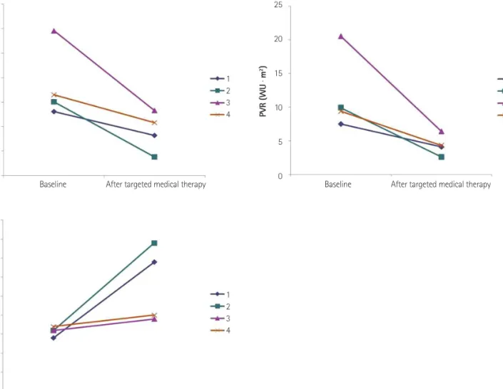
Generally, we performed follow-up cardiac catheterization annually or biennially. The mean duration from targeted medical therapy to closure of defects was 3.6 years (range, 15–96 months). Several years later, the direction of the shunt changed to a purely left-to- right shunt on echocardiography and the PVR decreased and Qp/Qs increased on serial cardiac catheterizations. These patients underwent successful closure of defects and subsequent long-term targeted medical therapy with the expectation of a further decrease in the PVR. All patients showed a decrease in PVR after targeted medical therapy (Fig. 4A) compared to baseline data. As well, PVR after administration of pulmonary vasodilators for vasoreactivity test was decreased markedly up to the acceptable level to perform the septal defect closure (Fig. 4B). In addition, the mean Qp/Qs increased from 1.6 to 3.6 (Fig. 4C), and chest X ray showed increase in the cardiomegaly. Of these patients, 3 underwent surgical partial closure of defects (PDA and VSD) or ASD fenestration, and 1 underwent pulmonary atery banding. The p(PA /Ao) or p(RV/Ao) was 0.3 in the PDA patient, 0.5 in the VSD patient, and 0.7 in the PA banding patient. Patients who underwent cardiac surgery showed improvement in the NYHA functional class.
Two years later, 2 patients underwent percutaneous closure using the Amplatzer device (Mendelssohn Avenue Golden Valley, MN, USA) after confirmation of safety with balloon test occlusion of the Table 2. Baseline demographic and clinical characteristics of Group 3 (n=4)
Case Age
(years) Diagnosis Shunt direction on echo
Cardiac catheterization (vasodilator)
Drug (medication duration until first surgey) SpO2 (%) p(RV/Ao) Qp/Qs PVR
(WU∙m
2)
1 21 AVSD, FC II, Down Bidirectional shunt 88 0.95 1.4 (2.5) 13.1 (7.5) Beraprost (3 years, 4 months) 2 38 PDA, FC III Bidirectional shunt, RV
dysfunction 94 1 1.6 (3) 29 (20) Sildenafil+beraprost
(1 year, 3 months)
3 12 PDA Bidirectional shunt 90 0.9 1.6 (-) 15.1 (-) Beraprost (8 years)
4 7 VSD Bidirectional shunt 96 1 1.7 (3.2) 16.5 (9.4) Sildenafil, bosentan, beraprost
PA banding (3 years 10 months) SpO
2: oxygen saturation, p(RV/Ao): ratio of peak pulmonary arterial pressure/ peak aortic pressure, Qp/Qs: systemic flow amount/pulmonary flow amount, PVR: pulmonary vascular resistance, WU: wood unit, AVSD: atrioventricular septal defect, FC: functional class, PDA: patent ductus arterriosus, RV: right ventricle, VSD: ventricular septal defect, PA: pulmonary artery
0 2 4 6 8 10 12 14
Baseline After partial closure and targeted medical therapy
ASD ASD ASD ASD VSD