Original Articles 순환기:순환기:제순환기:순환기:제제제 2 8 권권권 제권 제제 3 호제 호호 1998호
Manually Crimped NIR Stent의 초기결과
포천 중문대학교 의과대학 분당차병원 심장내과학교실*
연세대학교 의과대학 심장내과학교실
김태용
*·이동일·홍범기·최동훈·임세중·김명곤 김동수·장양수·정남식·심원흠·조승연
=
=
=
= Abstract = = = =
Immediate Results of Manually Crimped NIR Stent
Tae Yong Kim, M.D.,* Dong Il Lee, M.D., Bum Kee Hong, M.D., Dong Hoon Choi, M.D., Se Joong Rim, M.D., Myeong Kon Kim, M.D.,
Dong Soo Kim, M.D., Yangsoo Jang, M.D., Namsik Chung, M.D., Won Heum Shim, M.D., Seung Yun Cho, M.D.
Cardiology Division,* Pundong CHA Hospital, College of Medicine, Pochon CHA University, Sungnam, Korea
Cardiology Division, College of Medicine, Yonsei University, Seoul, Korea
Background:The several kinds of coronary stents havve proven successful in theirrole to treat acute or subacute closures after balloon angioplasty as well as to reduce the restenosis rate in de novo lesions. However, investigations continue in order to develop an ideal stent with a strong, highly flexible, radial force, especially useful in cases of tortuous vessels, lesions at bends, and lesions distal to previously deployed stents. The NIR stent is a recently developed balloon-expandable, stainless- steel, slotted tube stent; it is designed for improved flexibility with a higher radial force when compared with the traditional Palmaz-Schatz stent. We report the immediate results of our experience with the NIR stent. The purpose of the present study was to assess the feasibility, safety and efficacy of the deployment of manually crimped NIR stents in patients with complex coronary anatomy as well as the clinical outcomes within the first month.
Methods:Between January and July 1997, 143 NIR stents were implanted in the coronary arteries of 124 patients(male 76%, mean age 56±10 years). Sixty-one patients had UAP, 43 had SA, and 20 patients had AMI.
Results:
1) Indications of stenting were de novo lesions in 123(95%) and restenosis lesion in 6(5%).
2) Frequency of used stent length was 16mm in 65 cases(46%), 32mm in 60 cases(42%), 25 mm in 12 cases(8%), and 9mm in 6 cases(4%).
3) Single stents were implanted in 115(89%) lesions, and overlapping stenting with 2nd NIR stents in 14(11%) lesions.
교신저자:조승연, 120-752 서울 서대문구 신촌동 전화) (02) 361-7071, 전송) (02) 393-2041
4) Procedural success rate(defined as the angiographically residual stenosis of <30% immediately after the procedure with no major clinical events within 4 weeks after the procedure) was 95.2%
(118 / 124 pts). Angiographic success rate(defined as a residual stenosis of <30% without major dissection) was 96.1%(124 / 129 lesion). The procedural success rate and the angiographic success rate in calcified lesions and/or thrombi containing lesions were 100%. The procedural success rate and the angiographic success rate in cases of tortuous proximal vessels to the lesion were 91% and 91%, respectively. The procedural success rate and the angiographic success rate in more than 45 degrees angulated lesions were 98% and 94%, respectively.
5) The mean lumen diameter of target lesions was increased from 0.6±0.4mm to 3.1±
0.5mm(p<0.001) after stent implantation. The percent of diameter stenosis was decreased from 82±
12% to -1±13%(p<0.001) after stent implantation. The mean diameter of the reference artery was 3.1±0.6mm.
6) Incidence of peristent dissection after stenting was 6.2%(8 / 129 lesion).
7) The rate of stenting failure was 4.8%(6 pts). There were 2 cases of stent migration, 2 cases of failure to cross the lesion and 2 cases of procedure-related emergency CABG.
Conclusion:There is a higher tendency for stent migration with manually crinped stents compared with that of premounted stents. However, coronary stenting with manually-crimped NIR stents can be safely performed and may be particularly useful in patients with unfavorable clinical and angiographic characteristics for percutaneous coronary intervention. Follow-up data is needed to assess long term patency of this stent.
KEY WORDS:Coronary artery disease·NIR stent·Immediate results.
서 론
이상적인 stent가 가져야할 중요한 특성중 하나로 유연성이 좋으면서 radial support가 강해야한다는 점 을 들 수 있다1). Coil 형태와 slotted tube 형태의 대 표적인 stent인 Gianturco-Roubin stent와 Palmaz- Schatz stent는 유연성과 radial support에 있어서 하 나는 좋고 하나는 나쁘다는 각각 서로 상반되는 특성을 지니고있다. Palmaz-Schatz stent는 radial support 가 강하다는 장점이 있으나 유연성이 나쁘고 단일길 이이며 stent중앙에 1mm의 연결부가 있기 때문에 시 술후 1mm의 bare area가 생긴다는 단점이 있다.
NIR stent는 Palmaz-Schatz stent의 단점을 개선한 최근에 개발된 여러 balloon expandable stainless steel slotted tube stent중의 하나이다. 이 stent는 tracking하는 동안 구불구불한 혈관을 용이하게 통과 하여 병변까지 stent를 무사히 도달시킬수 있도록 유연 성이 개선되었으며, stent를 편 후에는 radial support 가 더욱 강하게 되도록 도안되어졌다. 따라서, Palmaz-
Schatz stent의 단점인 1mm의 연결부가 필요없게 되었으며 25mm, 32mm등 긴 stent의 사용이 가능하 게 되었다. 이에 저자등은 124명의 관동맥질환 환자 에서 다양한 길이의 manually crimped NIR stent를 시술하고 이 stent의 안정성 및 시술결과를 분석하여 보고하는 바이다.
대상 및 방법
1. 대 상
1997년 1월 30일부터 7월 15일까지 연세대학교 의과대학 세브란스병원에서 NIR stent가 시술된 124 명의 환자, 129병변을 대상으로 하였다.
2. 정 의
혈전:여러 각도의 관동맥조영술상 관동맥내에 둥 근 filling defect가 관찰되거나, 조영제가 수 차례의 심주기가 지날때까지 stain 되어 남아있는 경우, 또는 급성 심근경색환자에서 병변이 완전폐쇄소견을 보일 때 혈전이 있다고 하였다2-4).
석회화:여러 각도에서 보아 fluoroscope상 radio- pacity가 관찰될 경우 석회화 병변으로 정의 하였다.
Peristent dissection:NHLBI 분류에 의한 type A-F의 내막박리가 stent 삽입후 stent의 원위부 또 는 근위부에 생긴 경우로 정의 하였다5).
3. NIR stent의 구조 및 특성
NIR stent는 경도의 radiopacity를 가진 balloon expandable stainless steel tube stent이다. 이 stent 는 펴졌을때 다이아몬드형태의 cell이 stent구성의 기 본단위가 되며, stent횡축으로 7개 또는 9개의 cell이 겹쳐지면서 한바퀴 도는 형태가 되도록 도안되어졌다.
이 다이아몬드형태의 cell은 4개의 변과 2개의 bridge 로 구성되는데 stent가 풍선도자에 mount되어졌을때 4변은 horizontal loop, bridge는 vertical loop를 이 루게 된다. Mount된 stent를 각이진 혈관에 tracking 할 때 각진부위의 안쪽면과 바깥쪽면에 있는 bridge 는 각각 서로 다른 정도로 늘어나도록 도안되어져 Palmaz-Schatz stent에 비하여 유연성을 향상시킴으 로 25mm, 32mm등 긴 stent사용이 가능하게 되었다.
또, stent가 펴진 후에는 cell의 횡으로 연결되는 부위가 서로 직각을 이루게 도안되어져 삽입후 radial force 가 강하다는 장점이 있다. 삽입후 stent가 recoil 되는 정도는 2%이며 금속/혈관의 면적은 혈관의 크기에 따 라 14~19%정도로 알려져 있다. 삽입후 stent 길이 의 축소는 stent가 펴질 경우 4변에 의하여 발생되는 길이축소를 각이 진 bridge가 펴지면서 상쇄하도록 하는 self compensatory cell design으로 되어있기
때문에 3% 미만으로 거의 없다고 한다(Fig. 1).
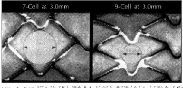
이 stent의 길이는 9mm, 16mm, 25mm, 32mm로 다양하며, cell이 7개인 7 cell type과 9개인 9 cell type이 공급되고 있다. 일반적으로 7 cell type은 혈 관직경이 2.5~3.5mm, 9 cell type은 혈관직경이 3.5
~5mm인 경우에 시술하도록 권장하고 있다. 직경이 3mm인 혈관에 7 cell type을 삽입할 경우 cell의 직 경은 9 cell type을 삽입할 경우에 비하여 두배로 (1.2mm vs. 0.6mm) 증가되고 cell의 크기는 83%증 가되기 때문에 stenting범위 내에 분지가 존재할 경우 는 분지의 보호를 위하여 7 cell type의 사용이 권장 되고있다(Fig. 2). Non-expanded profile과 가능최 대기압은 stent가 mount되는 풍선도자에 따라 다양 하다.
4. Stent시술 및 약물요법
저자들의 경우 전확장 후 stent를 삽입할때 풍선도 자는 stent길이보다 4mm정도 긴 길이의 양쪽 끝에 금속 표시가 부착된 것을 주로 사용하였다. Stent밖으 로 양끝이 각각 2mm정도 나오도록 풍선도자를 위치 시킨 후 stent strut이 흐트러지지 않도록 유의하면서 stent 양끝에서부터 중앙으로 서로 직각이 되는 방향으 로 여러번 눌러 고정하는 manually crimped method 를 이용하여 시술하였다. 유도도자는 주로 8F를 사용 하였으며 stent 삽입후 필요한 경우 다른 풍선도자를 이용하여 재확장을 하였다. Stent시술전 약물투여는 응급인 경우를 제외하고는 aspirin(100mg qd)과 ti-
Fig. 1. A:Vertical loops of each cell allow high flexibi- lilty by differential elongation during insertion(note the difference in the opening of the vertical loop inside(1) and outside(2) of the curve). B:
Self-compensatory cell design provides minimal foreshortening.
Fig. 2. NIR stents, containing seven attached cells in cir- cumference, are appropriate for implantation in smaller diameter vessels(2.5-3.5mm), whereas the nine-cell NIR stents are recommended for optimal structural support and larger(3.5-5.0 mm) arteries. When the NIR stent is expanded to 3mm, the cell diameter of the seven cell-NIR stent is double(1.2 vs. 0.6mm) and 83% increase in cell size then that of the nine-cell NIR stents, permitting better access to side branches co- vered by seven-cell NIR stents.
clopidine(250mg bid)을 시술 2~3일 전부터 투약하 고 ticlopidine은 2주 이상, aspirin은 계속 투약하였 다. 시술중 heparin은 10,000단위를 정주하였으며 시 술동안 ACT는 250 이상을 유지하였다. 병변내에 혈 전이 있거나 시술후 내막박리 등의 합병증으로 혈전 이 생길 가능성이 높은 경우에는 시술후 24시간 동안 heparin을 시간당 1,000단위로 연속점적하였다.
5. 자료분석 및 통계
병변의 형태는 AHA/ACC분류에 따라 정리하였고, 시술 전후의 관동맥조영술소견은 empty catheter를 기준으로 electric caliper를 사용하여 협착의 정도를 분석하였다. 통계처리는 연속변수를 평균 ± 표준편차 로 표현하였고, 각 결과간의 비교는 Student t-test 를 이용하여 p값이 0.05이하인 경우 통계학적으로 의 미있다고 하였다.
결 과
1. 대상환자
대상환자의 임상적 특징은 다음과 같다(Table 1).
대상환자는 남자가 76%였으며, 평균연령은 56세였다.
동맥경화에 대한 위험인자는 흡연이 64%로 가장 많 았으며 고혈압, 당뇨병, 고콜레스테롤혈증 등의 빈도 순으로 나타났다. 임상적 진단은 불안정성 협심증이
49%로 가장 많았으며, 안정성 협심증 35%, 심근경색 16%였다. 관동맥조영술 진단은 2 혈관질환이 40%로 가장 많았으며 좌주간부 병변은 세 환자에서 있었다.
2. 병변의 특징
병변의 분포는 좌전하지동맥에 48%로 가장 많았으 며, 우관동맥에 33%, 좌회선지동맥에 17%였다. ACC/
AHA분류에 의한 병변의 형태는 C형이 43%로 가장 많았으며 B2, B1, 그리고 A형의 빈도순으로 나타났다.
병변이 45° 이상 각진 곳에 위치한 경우는 39%였으 며 병변 근위부가 굴곡이 졌던 경우는 9%였다. 병변
Table 1. Clinical characteristics of the patients Number(%) Sex(male/female) 94(76%)/30(24%)
Age(years, mean±SD) 56 ± 10
Risk factors
Smoking 79 (64%)
Hypertension 54 (44%)
Diabetes mellitus 25 (20%) Hypercholesterolemia(>240mg%) 15 (12%) Clinical diagnosis
Stable angina pectoris 43 (35%) Unstable angina pectoris 61 (49%) Acute myocardial infarction 20 (16%) Angiographic diagnosis
One vessel disease 46 (37%) Two vessel disease 50 (40%) Three vessel disease 25 (20%) Left main disease 3 ( 3%)
Table 2. Angiographic characteristics of the lesions Number(%) Target lesion vessel
LAD 62 (48%)
LCX 21 (17%)
RCA 43 (33%)
Left main 3 ( 2%)
Lesion type(ACC/AHA classification)
A 6 ( 5%)
B1 15 (12%)
B2 52 (40%)
C 56 (43%)
Angulation >45° 50 (39%)
Tortuosity* 11 ( 9%)
Thrombi 16 (12%)
Calcification 12 ( 9%)
Indications De novo lesion 123 (95%)
Restenotic lesion 6 ( 5%)
*Moderate tortuosity:lesion is distal to two bands >75°
Severe tortuosity:lesion is distal to three bands >75°
or two bands >90°
Table 3. Stent size and number
Number(%) Length(mm)
9mm 6 (4%)
16mm 65 (46%)
25mm 12 (8%)
32mm 60 (42%)
No. of stents by lesion vessel
Single stent 115 (89%)
Overlapping with NIR stent 14 (11%) (with other stents 13(10%))
에 혈전이 있었던 경우는 12%, 석회화가 있었던 경우는 9%였다. Stent삽입의 적응증은 de novo 병변에 삽입 한 경우가 95%였고 재협착병변은 5%였다(Table 2).
3. 시술결과
모든 stent는 manually crimped형태로 삽입되었 다. 사용된 stent는 143개로 16mm 65개(46%), 32mm 60개(42%), 25mm 12개(8%), 9mm 6개 (4%)였다. 한 병변에 한 개의 NIR stent가 삽입된 경우는 89%였으며 두 개의 NIR stent가 겹쳐져 삽 입된 경우는 11%였다(Table 3). 병변의 평균MLD는 시술전 0.6±0.4 mm에서 시술후 3.1±0.5mm로 증 가하였고 병변의 협착정도는 시술전 82±12%에서 시술후 -1±13%로 감소(p<0.001)하였다. 표준혈관 크기는 평균 3.1±0.6mm였다. Stent 삽입후 stent 양끝에서 발생한 내막박리는 8예(6.2%)에서 관찰되
었는데 이 중 stent의 원위부에 발생한 경우는 일곱 으로 대부분을 차지하였다. Stenting 범위 내에 존재 하였던 직경 1.5 mm이상의 분지 52개 중 stenting 후 폐쇄된 경우는 5개(9.6%)였다(Table 4).
Procedural success rate는 95.2%, angiographic success rate는 96.1%였다(Table 5). 시술실패는 6 예에서 있었다(Table 6). 시술과 관련되어 응급 관동 맥우회로술을 시행한 경우가 2예 있었는데, 관동맥 천 공과 관동맥내에서 stent가 migration되어 응급 관동 맥우회로술을 시행한 경우가 각각 1예씩 있었다. 2예 모두 stent길이는 32mm였다. 관동맥 천공이 있었던 1예는 좌전하지 중간부에 만성 폐쇄가 있었던 환자로 유도철선에 의하여 subintima에 손상을 받은 상태에 서 ballooning을 시행하여 천공된 것으로 추정된다.
유도철선이 통과된 후 6기압으로 전확장을 하였고 8 기압으로 stent를 삽입한 직후 시행한 관동맥조영술 상 조영제가 심낭내로 새는 것이 관찰되었다. 즉시 Table 4. Angiographic result
Reference diameter(mm) 3.1 ± 0.6
MLD(mm)
Pretent 0.6 ± 0.4
Poststent 3.1 ± 0.5
Percent diameter stenosis
Prestent 82 ± 12
Poststent -1 ± 13
Peri-stent dissection 8 (6.2%) Proximal to stent 1
Distal to stent 7 Proximal and distal to stent 0
Compromised side branch 5/52 (9.6%) Mean ± S.D.
Pre vs poststent:p<0.001
Table 5. Success rate
Number(%) Procedural success* 118/124 (95.2%)
Stenting failure 6 ( 4.8%) Clinical event at 4 week** 0 ( 0 %) Angiographic success*** 124/129 (96.1%)
CABG or residual stenosis of >30% 3 ( 2.3%) Major edge dissection 2 ( 1.6%)
* Procedural success defined as residual stenosis of
<30% without occurrence of major clinical events within 4 weeks after procedure
** Death, Myocardial infarction, Revascularization
*** Angiographic success defined as residual stenosis of <30% without major dissection
Table 6. Procedure related complications
Number(%) Stenting failure 6 ( 4.8%)
Emergency CABG 2
Coronary artery perforation 1 Stent migration in coronary artery* 1 Stent migration(extra-coronary) 2 Failure to cross the lesion 2 Stenting success 118 (95.2%)
Q-myocardial infarction 0 ( 0%) Bleeding requiring transfusion 0 ( 0%) Acute and subacute closure 0 ( 0%)
* Post operative acute myocardial infarction
Fig. 3. A case of overlapping stent with two 32mm NIR stents. A:Right coronary angiogram with LAO view showed total obstruction of proximal RCA.
B:Angiogram after successful PTCA with over- lapping two 32mm NIR stents.
A A
A A B B B B
perfusion balloon을 삽입하여 심낭내로 피가 새는 것 을 막는 한편 antegrade flow가 유지되도록 하면서 응급 관동맥우회로술을 성공적으로 시행하였다. Stent 가 migration되어 stent가 좌주간부에서 좌회선지에 걸쳐져있어 응급 관동맥우회로술을 시행하였던 경우 는 수술후 Q파 급성 심근경색이 발생하였다. 관동맥 밖에서 stent가 migration된 경우는 2예 있었다. 한 예는 9mm 길이의 stent가 좌측신동맥내로 색전되었 던 예이며, 다른 한 예는 32mm 길이의 stent가 굴곡 진 우관동맥근위부를 통과하지 못한 채 stent의 원위 1/3정도는 관동맥내에 위치하고 근위 2/3정도는 대동 맥내에 위치된 상태로 migration되었던 예이다.
전자의 경우 시술후 여러번 시행한 요검사상 적혈 구수는 정상범위에 있었으며 혈청 creatinine의 상승 은 관찰되지 않았다. 후자의 경우는 반대측 대퇴동맥 을 통하여 우측 Judgkin도자를 우관동맥개구부에 삽 입시켜 stent를 고정시킨 후 snare wire를 이용하여 stent를 제거하였으며 Micro-Ⅱ stent를 성공적으로 시술하였다(Fig. 4). Stent가 병변을 통과하지 못하였 던 경우는 두 예 있었다. Stenting에 성공하였던 예에 서는 시술후 4주 이내에 사망, 급성 심근경색, 재관류 요법시행등은 없었다.
고 안
Stent삽입은 허혈성 심질환환자의 중재적 치료후 약 4-12%에서 발생할 수 있는 급성폐쇄의 예방 및 치료에 효과적일 뿐만 아니라 Benestent6), Bene- stent Ⅱ7) 및 STRESS8) 연구에 의하면 de novo 병 변에 시술한 경우 풍선도자를 이용한 경피적 경혈관 관동맥확장술에 비하여 재협착율을 감소시킨다고 알
려져, 현재 stent는 여러 가지 new device에 의한 중 재적 치료법 중 가장 선호되는 방법으로 쓰여지고있 다. 그러나, stent시술에도 몇 가지 문제점은 남아있는 데 구조 및 기능적으로 이상적인 stent의 개발, 시술 후 일부에서 발생하는 stent내 혈전형성, 그리고 재협 착등을 들 수 있으며 현재 이 문제점들을 해결하기 위 하여 각 방면에서 많은 연구가 진행되고 있다.
일반적으로 이상적인 stent가 가져야할 특성은 tr- acking하는 동안은 유연성이 좋아야하며, stent가 펴 진 후에는 radial force가 강해야한다고 알려져있다.
또한 fluoroscope상에서 stent의 위치식별이 용이하 여야하며 펴진 후에는 stent 길이의 축소가 적고, 적 당한 metal coverage를 가져 stent cell사이로 plaque 나 dissection flap이 삐져나오지 않도록 도안되어져야 한다1). NIR stent는 Palmaz-Schatz stent의 단점을 개선한 최근에 개발된 balloon expandable stainless steel slotted tube stent중 하나로서 기존 Palmaz- Schatz stent의 장점인 강한 radial force는 그대로 유지하면서 유연성이 개선되도록 도안되어졌다.
시술에 성공하고 잔여협착이 30%미만인 경우를 stent삽입술의 성공으로 정의할때 본 보고의 성공률은 95.2%였으며, 시술에 성공하고 4주 이내에 사망, 급성 심근경색, 재관류요법시행등이 없는 경우를 procedural success rate로 정의할 경우 본원의 manually crimped NIR stent의 procedural success rate는 95.2%였다.
이는 255환자, 341병변을 대상으로 9 cell type의 manually crimped NIR stent를 시술하고 95%의 procedural success rate(시술후 잔여협착은 50%
미만)를 보고한 FINESS study9)와 유사한 결과이며, 201환자를 대상으로 혈관직경이 평균 2.4±0.24mm 인 병변에 225개의 7 cell type NIR stent를 시술하 Fig. 4. A case of stent migration. A:Right coronary angiogram with LAO view showed
tortuous proximal RCA lesion(Pre-PTCA). B:Successful stent removal was per- formed with snare wire(black arrow). C:Tortuous proximal lesion was successfully dilated with Micro-Ⅱ stent after removal of migrated NIR stent.
고 96%의 clinical success rate와 2%의 아급성 혈 전형성을 보고한 Chevalier등10)의 결과와도 유사한 성적이다. 저자들의 경우 표준혈관이 3.5mm미만인 경우는 7 cell type을 사용하였으며 3.5mm이상인 경 우는 9 cell type을 사용하였다. 표준혈관의 크기는 평 균 3.1±0.6 mm였으며 시술후 병변의 평균 최소혈관 직경(MLD, mm)은 시술전 0.6±0.4에서 시술후 3.1
±0.5로 의미있게 증가하여 이미 발표된 FINESS study 성적과 유사하였다. 사용된 stent길이는 16mm 46%
(65개), 32mm 42%(60개), 25mm 8%(12개), 9mm 4%(6개)로 16mm 59.5%, 32mm 26%, 9mm 14.4%
가 사용된 FINESS study에 비하여 긴 stent의 사용 이 많았다.
금속성 stent삽입의 가장 큰 제한점은 stent내 혈전 형성이다. 환자가 심도자실을 떠나기 전에 발생하는 급성 혈전의 발생은 1%미만으로 드물다고 알려져있 으며, 시술후 3~5일 사이에 가장 많이 발생하며11) 시 술후 1개월까지 발생의 위험이 있는12) 아급성 혈전형 성의 빈도는 stent종류와 보고자에 따라 3~21%8,13-17) 로 알려져있다. 현재 아급성 혈전형성의 발생원인은 금속성 이물질인 stent에 대한 반응에 의하여 생기는 것인지, stent와 혈관벽 사이에 존재할 수 있는 조그 만 사강(dead space)에 의하여 생기는 것인지 명확 히 밝혀져 있지 않다. 하지만, 최근 Colombo등18)은 Palmaz-Schatz stent시술후 강력한 항응고요법 대신 항혈소판요법과 고압의 병행확장요법을 시행한 결과 아급성 혈전형성 발생율과 항응고요법에 의한 혈관 및 출혈의 합병증 발생율이 모두 감소하였다고 보고하면 서 시술전후 혈관내초음파(intravascular ultrasound) 를 이용하여 표준혈관의 크기를 정확히 결정하고 stent 가 혈관벽에 잘 apposition 되도록 시술함으로서 아급 성 혈전형성 발생율을 감소시킬 수 있다고 하였다. 그 러나, 고압 풍선확장요법은 stent를 혈관벽에 잘 ap- position시켜 아급성 혈전형성 발생율을 감소시킬 수 는 있지만, 과연 재협착율을 감소시키는가하는 점에 대해서는 현재 논란이 많은 상태로19-23) 계속 연구되 어져야할 문제라고 생각되며 현재로서는 병변의 특성 에 적합한 stent를 선택하여 적절한 압력으로 시술하 고, 필요한 경우 시술전후 혈관내초음파를 이용하여 stent가 혈관벽에 잘 apposition 되도록 시술하는 것
이 아급성 혈전형성 발생율과 재협착율을 감소시킬 수 있는 방법이라 사료된다. 본 보고에서는 stent 시 술후 고압 풍선확장요법은 모든 예에서 시행되지는 않 았으며 14기압 이상 고압의 병용풍선확장요법은 60%
에서만 시행되었다.
Stent삽입후 내막박리는 10%미만24,25)으로 PTCA 의 20~40%26-29)보다 낮다고 보고되어 있다. 저자들 의 경우 stent 양끝에서 발생한 내막박리는 6.2%(8 예)에서 관찰되었는데 이 중 stent의 원위부에서 발생 한 경우가 88%(7예)로 대부분을 차지하였다. Type C 이상의 major 내막박리로 인하여 stent를 겹쳐서 시술한 경우는 2병변이었으며 Type A, B의 minor 내막박리가 있으면서 TIMI 3의 혈류가 유지되어 그 대로 두었던 경우는 5병변이었다. 시술된 stent의 길 이에 따른 내막박리의 발생율은 16mm 3%(2/65), 32mm 8%(5/60)로 stent길이가 긴 경우에 내막박리 가 더 많이 발생하였으며 이는 류 등30)의 보고와 같 았다. 혈관별 내막박리의 빈도는 RCA 9.3%(4/43), LAD 4.8%(3/62)로 RCA에서 높았다. 이는 RCA의 경우 LAD보다 혈관이 더 구불구불하기 때문으로 생 각되는데, 내막박리가 있었던 경우를 분석해 보면 stent 원위부가 위치하는 혈관에 10~20%의 협착이 있었 던 경우가 2예 였으며 stent원위부와 혈관이 45° 이 상 각이진 경우가 5예 있었다. 따라서, stent 원위부에 서의 내막박리 발생은 stent 원위부에서 풍선 / 혈관 비율이 중요한 인자이지만, 이 점외 본 보고에서는 NIR stent삽입시 stent원위부가 위치할 혈관이 각이져있는 경우와 이 부위에 약간의 협착이 있었던 경우에 stent 원위부 내막박리가 더 조장된 것으로 여겨진다. 또한, 저자들의 경험으로는 stent원위부가 위치할 혈관이 각 이져있는 경우에는 stent와 혈관이 연결되는 부분이 심박동에 따라 kingking되는 경우도 있었기 때문에 이점에 유의하여 시술해야 할 것으로 생각된다. 본원 에서 이미 보고한 Micro-Ⅱ의 초기결과30)의 peris- tent dissection 발생율은 14%로서 본 보고보다 오히 려 높았는데, stent의 유연성과 peristent dissection 발생율과의 상관관계에 대해서는 보다 많은 연구가 필 요하리라 사료된다.
Stent삽입후 분지의 fate에 대한 보고로서 Mazur 등31)은 G/R stent삽입후 분지의 폐쇄율은 풍선을 이
용한 관동맥성형술(6%, 7/108개)에서 단지 1%(8/
108개)가 추가될 뿐이라 하였으며 폐쇄된 경우의 88%
(7/8개)는 시술전 이미 개구부에 50%이상 협착이 있 었던 것이라 하였고 시술전 50%이상 협착이 있었던 경우에서 시술후 폐쇄된 것은 21%(7/33개)라 보고 하였다. 또한, Fischman등32)은 P/S stent 삽입후 분 지의 폐쇄율은 풍선을 이용한 관동맥성형술(9%, 6/66 개)에서 5%(9/66개)가 추가되었으며 추가된 3개 모 두 시술전 개구부에 50%이상의 협착이 있었다고 하 였다. 본 보고에서 stenting범위 내에 존재하였던 직 경 1.5mm이상의 분지 52개 중 stenting후 폐쇄된 경우는 9.6%(5개)로 Mazur등과 Fischman등의 보 고와 유사하였다. 폐쇄된 분지중 시술전 분지의 개구 부에 협착이 관찰되지 않았던 경우는 1예(20%)이며 4예(80%)에서는 시술전 개구부에 50~80%의 협착 이 관찰되었다. 또, 시술전 분지의 개구부에 50%이상 의 협착이 있었던 경우 중 stenting후 분지가 폐쇄된 경우는 27%(4/15개)였으며, 시술전 분지의 개구부가 정상이거나 50%이하의 협착이 관찰되었던 경우에서 stenting후 분지가 폐쇄된 경우는 2.7%(1/37개)였다.
따라서, 시술전 분지의 개구부에 50%이상의 협착이 있었던 경우에서 50%이하의 협착이 있었던 경우에 비하여 stenting후 분지 폐쇄율이 10배 높았으며 이 역시 Mazur등과 Fischman등의 보고와 유사하였다.
병변근위부가 구불구불했던 경우는 9%(11/129병 변)였다. 32mm 길이의 stent가 구불구불한 우관동맥 근위부를 통과하지 못한채 migration된 예가 1예 있 어 이 경우 시술성공율은 91%(10/11)였다. 따라서, NIR stent는 P/S stent에 비하여 유연성이 개선되었 지만 manually crimped형태의 긴 stent를 굴곡이 심 한 부위를 통과시킬 경우 여전히 stent migration의 위험이 남아있다고 생각된다. 본 보고에서는 32mm stent 2개, 9mm stent 1개가 migration되었다.
병변이 45° 이상 각진 곳에 위치한 경우는 39%(50/
129병변)였다. 90° 각도로 분지 되는 첫번째 obtuse marginal branch를 16mm 길이 stent가 통과하지 못 하였던 경우가 1예 있었고, 시술후 stent원위부에 major dissection이 관찰되어 stent를 겹쳐 시술한 경우가 2예 있어 이 경우 시술성공율은 98%(49/50), angio- graphic success rate는 94%(47/50)였다. 병변 내에
석회화가 있었던 경우는 9%(12병변)였으며 이 경우 시술성공율은 100%였다.
급성 심근경색을 포함하여 병변 내에 혈전이 존재 하는 경우에도 stent를 이용한 PTCA는 풍선도자를 이용한 PTCA에 비하여 안전하며 유용하다고 보고되 어 있는데33-36), Suryapranata등33)은 204명의 급성 심근경색환자를 102명씩 나누어 stent와 풍선도자를 이용하여 PTCA한 결과 시술성공율은 stent군이 98%
로 풍선도자군의 96%에 비하여 높았던 반면 아급성 폐쇄는 stent군이 1%로서 풍선도자군의 5%에 비하 여 낮았다고 보고하였다. 또, 시술 후 30일의 cardiac event free survival rate 역시 stent군이 97%로서 풍선도자군의 87%에 비하여 유의하게(p=0.02) 높 았다고 하였다. 저자들의 경우 병변 내에 혈전이 관찰 되었던 경우는 direct PTCA 7예를 포함하여 12%
(16병변)였으며 이 경우 시술성공율은 100%였고 시 술후 4주 이내에 major clinical event는 없었다.
결론적으로 stent migration율이 1% 이내로 보고되 는 premounted stent에 비하여 본 보고에서는 stent migration율이 2%로 manually crimped NIR stent 에서 migration이 다소 많다고 생각되나 관동맥질환의 치료에 있어 비교적 안전하고 유용하였다. 이 stent의 장기 개존율은 향후 추적관찰이 필요하겠다.
요 약
연구배경:
NIR stent는 Palmaz-Schatz stent의 단점을 개선한 최근에 개발된 balloon expandable stainless steel slotted tube stent중의 하나이다. 이 stent는 병변까 지 stent를 tracking하는 동안은 유연성이 좋고 stent 를 풍선도자로 편후에는 radial support가 강하게 되 도록 도안되어져 있다. 본 연구에서는 다양한 길이의 manually crimped NIR stent를 이용하여 혈관확장술 을 시행할 경우 이 stent의 안전성과 feasibility를 보 고자 하였다.
방 법:
1997년 1월 30일부터 7월 15일까지 연세대학교 의과대학 부속 세브란스병원에서 124명의 환자(남자 76%, 평균연령 56±10세), 129병변에 143개의 ma-
nually crimped NIR stent를 시술하고 그 결과를 분 석하였다.
결 과:
1) Stent삽입의 적응증은 de novo 병변이 123병변 (95%)이었고 재협착병변은 6병변(5%)이었다.
2) 사용된 stent의 빈도는 16mm 65개(46%), 32 mm 60개(42%), 25mm 12개(8%), 9mm 6개(4%) 순이었다.
3) 한 병변에 한 개의 NIR stent를 삽입한 경우는 89%였으며 두 개의 NIR stent를 겹쳐서 삽입한 경우 는 11%였다.
4) Procedural success rate는 95.2%, angiogra- phic success rate는 96.1%였다. 석회화된 병변과 병 변내에 혈전이 관찰된 경우의 procedural success rate 와 angiographic success rate는 각각 모두 100%였 으며, 병변근위부가 구불구불하였던 경우의 procedural success rate와 angiographic success rate는 각각 91%였다. 병변이 45° 이상 각진 곳에 위치한 경우의 procedural success rate와 angiographic success rate는 98%와 94%였다.
5) 병변의 평균MLD는 시술전 0.6±0.4mm에서 시술후 3.1±0.5mm로 증가(p<0.001)하였고 병변의 협착정도는 시술전 82±12%에서 시술후 -1±13%
로 감소(p<0.001)하였다. 표준혈관 크기는 평균 3.1
±0.6mm였다.
6) Stent 삽입후 stent 양끝에서 발생한 내막박리 는 6.2%(8예)에서 관찰되었는데 이 중 stent의 원위 부에서 발생한 경우가 7예로 대부분을 차지하였다.
7) 시술실패는 6예(4.8%)에서 있었다. Stent mi- gration 2예, stent 병변통과실패가 2예 있었으며 시 술과 관련된 응급 관동맥우회로술시행이 2예 있었다.
성공적인 시술후 1개월 내에 사망, 급성 심근경색, 재 관류요법시행등은 없었다.
결 론:
결론적으로 stent migration율이 1% 이내로 보고 되는 premounted stent에 비하여 본 보고에서는 stent migration율이 2%로 manually crimped NIR stent 에서 migration이 다소 많다고 생각되나 관동맥질환의 치료에 있어 비교적 안전하고 유용하였다. 이 stent의 장기 개존율은 향후 추적관찰이 필요하겠다.
References
1) 장양수 : 이상적인 스텐트 선택과 디자인. 순환기 27:1055-1060, 1997
2) Ambrose JA, Winters SL, Stern A, et al:Angiographic morphology and the pathogenesis of unstable angina pectoros. J Am Coll Cardiol 5:609-616, 1985
3) Fuster V, Badimon L, Badimon J, et al:The pathoge- nesis of coronary artery disease and the acute coronary syndromes. N Engl J Med 326:242-250, 1992
4) Cowley MJ, DiSciascio G, Vetrovec GW:Coronary thrombus in unstable angina;Angiographic observations and clinical relevance. In Hugenholtz PG and Goldman BD(eds):Unstable angina;Current concept and ma- nagement. Schattauer Press, Stuttgart, pp95-102, 1985 5) Huber M, Mooney J, Madison J, Mooney M:Use of a
morphologic classification to predict clinical outcome after dissection from coronary angioplasty. Am J Cardiol 68:467-471, 1991
6) Serruy PW, de Jaegere D, Kiemeneij F, et al:A comparision of balloon expandable stent implantation with balloon angioplasty in patients with coronary artery disease. N Engl J Med 331:489-495, 1994
7) BENESTENT Ⅱ study group:Heparin-coated Palmaz- Schatz stents in human coronary arteries. The European Society of Cardiology Meeting(Stockholm, Sweden), August, 1997
8) Fischman DL, Leon MB, Baim DS, et al:A randomized comparision of coronary stent placement and balloon angioplasty in the treatment of coronary artery disease.
N Engl J Med 331:496-501, 1994
9) Yaron A, Steven F, Ferdinand K, et al:First Interna- tional New Intravascular Rigid-Flex Endovascular Stent Study(FINESS) :Clinical and Angiographic Rresults After Elective and Urgent Stent Implantation. J Am Coll Cardiol 30:847-854, 1997
10) Chevalier B, Lefevre T, Meyer P, Corcos T, Bille J, Bendaas D, Louvard Y, Guyon P, Sainsous J, Royer T, Barragan P, Guerin Y:French registry of seven cells NIR stent implantation in <2.5mm coronary arteries(Abstr). Circulation 96(Suppl):I-274, 1997
11) Holmes D, Garratt K, Schwartz R:Timing of stent occlusion/thrombosis after stent placement. J Am Coll Cardiol 23:70A, 1994
12) Ueda Y, Nanto S, Komamura K, Kodama K:Neointimal coverage of stents in human coronary arteries observed by angioscopy. J Am Coll Cardiol 23:341-346, 1994 13) Barragan P, Sainsous J, Silvestri M, Bouvier JL, Comet
B, Simeoni JB, Charmasson C, Bremondy M:Ticlopidine and subcutaneous heparin as an alternative regimen
following coronary stenting. Cathet Cardiovasc Diagn 32:133-138, 1994
14) Muller DWM, Shamir KJ, Ellis SG, Topol EJ:Peripheral vascular complications after cinventional and complex percutaneous coronary intervention procedures. Am J Cardiol 69:63-68, 1992
15) Herrmann HC, Buchbinder M, Clemen MW, Fischman D, Goldberg S, Leon M, Schatz RA, Tierstein P, Walker CM, Hirschfield JW:Emergency use of balloon expandable coronary artery stenting for failed percutaneous transl- uminal coronary angioplasty. Circulation 86:812-819, 1992
16) Sutton JM, Ellis SG, Roubin GS, et al:Major clinical events after coronary stenting:The multicenter registry of acute and elective Gianturco-Roubin stent placement.
Circulation 89:1126-1137, 1994
17) Haude M, Erbel R, Issa H, Straub U, Rupprecht HJ, Treese N, Heyer J:Subacute thrombotic complications after intracoronary implantation of Palmaz-Schatz stents.
Am Heart J 126:15-22, 1993
18) Colombo A, Hall P, Nakamura S, Aalmagor Y, Maiello L, Martini G, Saglione A, Goldberg SL, Tobias JM:Intra- coronary stenting without anticoagulation accomplished with intravascular ultrasound guidance. Circulation 91: 1676-1688, 1995
19) 이남호·장양수·홍범기·최동훈·하종원·임세 중·김태용·심원흠·조승연:Long Lesion의 관동 맥 협착에서 Coronary Less Shortening Wallstent의 6 개월 Follow-up Results. 순환기 27:1443-1451, 1997 20) Hall P, Nakamura L, Blengino S, Martini G, Finci L,
Colombo A:Factors associated with late angiographic outcome after intravascular ultrasound guided Palmaz- Schatz coronary stent implantation. J Am Coll Cardiol 1995;(in presss)
21) 최동훈·장양수·홍범기·이남호·김태용·하종 원·임세중·정남식·심원흠:AVE Micro Ⅱ stent: 6 month follow up. 순환기 27:1280-1288, 1997 22) Yang P, Hassen A, Heyer G, Klein W, Luha O, Mue-
hlberger V, Pachinger O, Sochor H, Sykora J, Weidinger F, Glogar D : High pressure post dilatation of the Medtronic Wictor stent:intravascular ultrasound and quantitative angiographic data from the Austrian Multi- center trial(Abstr). Circulation 94(Suppl):I-207, 1996 23) Ozaki Y, Violaris AG, Hamburger JAAP, Melkert R,
Foley D, Keane D, et al:Short and long clinical and quantitative angiographic results with the new, less shortening Wallstent for vessel reconstruction in chronic total occlusion:A quantitative angiographic study. J Am Coll Cardiol 28:345-360, 1996
24) Mehta S, Popma J, Margolis J, et al:Angiographic complications after new device angioplasty in native coronary arteries:A NACI Angiographic Core Laboratory
Report. The proceedings of TCT Meeting(Washington DC), February, 1995
25) Bailey S, Ricci D, Kiesz S, et al:Incidence and clinical impact of dissections after PTCA and stent placement: Results from the Randomized Stent REStenosis Study.
The proceedings of TCT Meeting(Washington DC), Fe- bruary, 1995
26) Tan K, Sulke N, Taub N, Sowton E:Clinical and lesion morphologic determinants of coronary angioplasty success and complications:Current experience. J Am Coll Cardiol 25:855-865, 1995
27) Hermans WR, Foley DP, Rensing BJ, Rutsch W : Usefulness of quantitative and qualitative angiographic lesion morphology, and clinical characteristics in predi- cting major adverse cardiac events during and after native coronary balloon angioplasty. Am J Cardiol 72:14-20, 1993
28) Hermans WR, Rensing BJ, Foley DP, Deckers JW:
Therapeutic dissection after successful coronary balloon angioplasty:No influence on restenosis or on clinical outcome in 693 patients. J Am Coll Cardiol 20:767- 780, 1992
29) Sharma SK, Israel DH, Kamean JL, Bodian CA:Clinical, angiographic and procedural determinants of major and minor coronary dissection during angioplasty. Am Heart J 126:39-47, 1993
30) 류종철·장양수·김건영·이승환·김종현·전동 운·심원흠·조승연 :Micro-Ⅱ의 초기결과. 순환기 27:532-540, 1997
31) Mazur W, Grinstead WC, Hakim AH, Dabaghi SF, Abukhalil JM, Ali NM, Joseph J, French BA, Raizner AE:Fate of side branches after intracoronary implan- tation of the Gianturco-Roubin flex-stents for acute or threatened closure after percutaneous trransluminal coronary angioplasty. Am J Cardiol 74:1207-1210, 1994 32) Fischman DL, Savage MP, Leon MB, Schatz RA, Ellis S,
Cleman MW, Hirshfeld JW, Tierstein P, Baley S, Walker CM, Goldberg S:Fate of lesion related side branches after coronary artery stenting. J Am Coll Cardiol 22: 1641-1646, 1993
33) Suryapranata H, Hoorntje JCA, de Boer MJ, Zijlstra F:
Randomized comparision of primary stenting with primary balloon angioplasty in acute myocardial infarction(Abstr). Circulation 96(Suppl):I-327, 1997
34) Antoniucci D, Santoro GM, Bolognese L Valenti R, Trapani M, Moschi G, Taddeucci E, Dovellini EV, Fazzini PF:A prospective randomized trial of elective senting in acute myocardial infarction-priliminary reaults of the FRESCO study(Abstr). Circulation 96(Suppl):
I-327, 1997
35) Stone GW, Brodie BR, Griffin JJ, Morice MC, Costa- ntini C, Overlie PA, St Goar FG, Popma JJ, McDonell J,
Jones D, Grine CL:Improved short term ooutcomes of primary stenting compared to primary angioplasty in acute myocardial infarctiion:The PAMI Stent pilot trial (Abstr). Circulation 96(Suppl):I-594, 1997
36) Saito S, Hosokawa G:Primary Palmaz-Schatz stent implantation for acute myocardial infarction:The final results of Japanese PASTA trial(Abstr). Circulation 96 (Suppl):I-595, 1997