Received on November 29, 2013. Revised on February 3, 2014. Accepted on February 4, 2014.
CC This is an open access article distributed under the terms of the Creative Commons Attribution Non-Commercial License (http://creativecommons.org/licenses/by-nc/3.0) which permits unrestricted non-commercial use, distribu- tion, and reproduction in any medium, provided the original work is properly cited.
*Corresponding Author. Sung-Gyoo Park, School of Life Sciences, Gwangju Institute of Science and Technology (GIST), 123 Cheomdan Gwagiro, Oryong-dong, Buk-gu, Gwangju, Korea. Tel: 82-62-715-2511; Fax: 82-62-715-2484; E-mail:
Keywords: NF-κB, Th17, T cell receptor, Autoimmune
Abbreviations: RHD, rel homology domain; TAD, transactivation domain; TCR, T cell receptor; PI3K, phosphoinositide 3-kiase; PDK1, phosphoinositide-dependent kinase 1; CBM, Carma1-Bcl10-Malt1; GLK, GCK-like kinase; Th, T helper; RA, rheumatoid arthritis; IBD, inflammatory bowel disease; SLE, systemic lupus erythematous; PGE2, prostaglandin E2
Figure 1. NF-κB family. The mammalian NF-κB protein family consists of five members: p65 (RelA), RelB, c-Rel, NF-κB2 (precursor, p100; mature form, p52), and NF-κB1 (precursor, p105; mature form, p50).
NF-κB Activation in T Helper 17 Cell Differentiation
Sang-Heon Park, Gabi Cho and Sung-Gyoo Park*
School of Life Sciences, Gwangju Institute of Science and Technology (GIST), Gwangju 500-712, Korea
CD28/T cell receptor ligation activates the NF-κB signaling cascade during CD4 T cell activation. NF-κB activation is re- quired for cytokine gene expression and activated T cell sur- vival and proliferation. Recently, many reports showed that NF-κB activation is also involved in T helper (Th) cell differ- entiation including Th17 cell differentiation. In this review, we discuss the current literature on NF-κB activation pathway and its effect on Th17 cell differentiation.
[Immune Network 2014;14(1):14-20]
INTRODUCTION
NF-κB is activated during immune responses and is important for the expression of immune response related genes includ- ing cytokine, chemokine, and adhesion molecule genes (1-3).
The NF-κB family is composed of RelA, RelB, c-Rel, p50 (NF- κB1), and p52 (NF-κB2) subunits. The NF-κB transcription factor binds to κB sites as dimers, either homodimers or heterodimers. The NF-κB protein contains N-terminal Rel ho- mology domain (RHD), which makes contact with DNA and supports subunit dimerization. Of the NF-κB subunits, only RelA, RelB, and c-Rel have transactivation domain (TAD) at C-terminus and this TAD domain is important for initiation of target gene transcription (1-3). However, p50 and p52 lack TAD domain. Thus, p50 and p52 can positively regulate gene expression through heterodimerization with TAD containing NF-κB subunits or other regulators (Fig. 1).
NF-κB complexes are inactive in most cells, and these complexes are located in the cytoplasm in a complex with inhibitory IκB proteins (IκBα, IκBβ, IκBε, IκBζ p100, p105, Bcl3, and IκBns). NF-κB pathway activating signals including cytokine receptor signals and antigen receptor signals activate the IκB kinase (IKK) complex, which phosphorylates IκB.
This phosphorylation induces IκB degradation, which leads to NF-κB complex translocation to the nucleus. Once in the nucleus, the NF-κB complex activates target gene tran- scription (1-3).
NF-κB PATHWAY IN T CELL ACTIVATION
Antigen recognition by T cell receptor (TCR) induces activa- tion of many transcription factors including NF-κB, NF-AT,
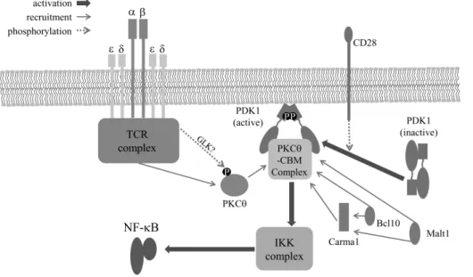
Figure 2. T cell receptor-mediated NF-κB activation. T cell receptor complex and CD28-mediated PDK1 activation are important for signal- ing complex formation composed of PKCθ, Carma1, Bcl10, and Malt1. This signaling complex leads to IKK complex activation and sub- sequently activates NF-κB during T cell activation by antigen.
and AP-1 (2,4), which are important for the induction of pro- liferation of activated T cells and their differentiation into Th1, Th2, Th17 and other Th cells (2,5). Ligation of CD28 co-re- ceptor, along with the TCR, is essential for full activation of T cells (6,7). Especially, CD28 co-receptor ligation is required for efficient activation of NF-κB. It is well known that CD28 greatly enhances phosphoinositide 3-kinase (PI3K) activity and this activity is required for many cellular responses in- cluding cell survival, and cell proliferation (8). The CD28 cy- toplasmic tail contains a PI3K binding motif such as YMNM motif (9). PI3K has been reported to bind to the YMNM phos- photyrosine, which leads to PI3K activation. CD28-mediated PI3K activation is involved in phosphoinositide-dependent kinase 1 (PDK1) and AKT recruitment into the immunological synapse (10-12), which activates PDK1 and AKT (13). These processes are important for PKCθ and Carma1-Bcl10-Malt1 (CBM) complex-mediated NF-κB activation during T cell acti- vation (13-15). However, the concept of NF-κB activation by YNMN-mediated PI3K activation has been challenged because YNMN motif mutation had no significant effect on T cell pro- liferation and IL-2 production (16,17). The report suggested that another region of the CD28 cytoplasmic tail is responsible for PDK1 activation and subsequently this induces PKCθ- mediated NF-κB activation (17). A recent in vivo infection experiment suggested that another CD28 cytoplasmic tail re- gion is responsible for NF-κB activation (10). Even though the exact CD28 cytoplasmic tail region for NF-κB activation is controversial, CD28-mediated PDK1 activation and sub-
sequent PKCθ-mediated NF-κB activation pathway is well supported by previous studies (13,17,18).
During T cell activation, CD28 recruits PDK1 into the im- munological synapse where the recruited PDK1 is converted into competent states for binding to down-stream molecules such as PKCθ and Carma1 (13,19). During this process, phos- phorylation of threonine 513th on PDK1 is important for the conversion of PDK1 heterotypic dimer to homotypic dimer, which enables the formation of the PDK1-PKCθ-CBM com- plex. In addition, recently, GCK-like kinase (GLK) was sug- gested as the kinase for PKCθ phosphorylation at threonine 538th (20). Thus, it is possible that PDK1 works as a scaffold for the complex formation. Subsequently, IκB kinase (IKK) is activated by the signaling complex (2), and the IKK com- plex activates NF-κB during T cell activation (Fig. 2).
Th17 CELLS
CD4 T cells play an essential role in the adaptive immune response. During the adaptive immune response, activated CD4 T cells differentiate into T helper (Th) 1, Th2 or Th17 effector cells. Th1 cells are important in host defense against intracellular pathogens and Th2 cells are involved in allergic immune responses and defense against parasite infections.
Th17 cells play an important role in host defense against ex- tracellular pathogens and fungal infections (21,22). Further- more, Th1 and Th17 cells are important in intestinal immune responses. During CD4 T cell differentiation, IL-6 and TGF-β
are important for Th17 cell differentiation. IL-23 is also in- volved in Th17 cell differentiation (23). In addition to the di- rect effect of IL-23 on Th17 cell differentiation, IL-23 stim- ulates intestinal TCRγδ T cells, invariant natural killer T cells (iNKT), and intestinal innate-like T cells to secrete cytokines related to Th17 differentiation (24). In addition to TGF-β and IL-23, IL-6 is also important for Th17 differentiation. However, while TGF-β negatively regulates human Th17 cell differ- entiation, this cytokine is important for Th17 cell differ- entiation in murine Th17 differentiation (25-27). During Th17 cell differentiation, the transcription factors RORγt, RORγ, RORα, IRF4, and STAT3 are important for effector T cell differentiation. However, IFN-γ, IL-2, and IL-4 negatively reg- ulate Th17 cell differentiation (28,29).
Th17 CELLS IN AUTOIMMUNE DISEASE
Differentiated Th17 cells produce proinflammatory cytokines such as IL-17A, IL-17F, IL-21, TNF, and GM-CSF (5,30,31).
These cytokines are important in host defense against ex- tracellular bacteria through acute immune responses (32). In addition, Th17 cells are involved in the development of auto- immune diseases including rheumatoid arthritis (RA), inflam- matory bowel disease (IBD), and multiple sclerosis (26,33).
It has been suggested that unbalanced immune responses can induce inflammatory diseases such as IBD. The detailed mechanism of IBD induction, including Crohn’s disease and ulcerative colitis, has not been clarified (34-36); however, un- controlled T cell activation and biased effector T cell (Th1, Th2, and Th17 cells) differentiation have been suggested as causative factors. Unbalanced production of Th17-related cy- tokines is also involved in the induction of IBD and other autoimmune disease. Th17 cells produce cytokines including IL-17A, IL-17F, IL-21, and IL-22 (37,38). IL-17 is the repre- sentative cytokine produced by Th17 cells and is involved in RA, asthma, and systemic lupus erythematous (SLE) develop- ment. A number of studies have investigated the role of IL-17A in intestinal inflammation, and showed that IL-17A is overproduced in patients with Crohn’s disease and ulcerative colitis (39-42). In addition, IL-17 family cytokines are also in- creased in patients with autoinflammatory diseases including RA, asthma, and SLE (43-45). IL-17 and IL-23R genomic DNA sequence analysis found polymorphic regions related to IBD induction (46,47). In addition, IL-21 produced by Th17 cells was found to be involved in exacerbation of IBD (48,49).
Furthermore, IL-21 gene deleted mice are resistant to Th1/Th17
cell-mediated colitis induction (50).
EXTRINSIC EFFECT OF NF-κB ACTIVATION ON Th17 CELL DIFFERENTIATION
The NF-κB pathway regulates antigen presenting cell func- tions and affects CD4 T cell differentiation into Th effector cells (51,52). Dendritic cells are the most important antigen presenting cells for Th cell differentiation. RelA deficiency re- duced IL-1α, IL-1β, and IL-6 production from dendritic cells in response to LPS stimulation (53). In fact, these cytokines are involved in Th17 cell differentiation (25). In inflammatory responses, prostaglandin E2 (PGE2), an endogenous lipid me- diator, enhances the production of IL-1β and TNF-α from bone marrow derived dendritic cells. In addition, PGE2 re- duces the level of IL-12, but increases the level of IL-23 production. In addition to changes in cytokine production, PGE2 affects the expression of TLR-4, 2, and 9, IL-1R-asso- ciated kinase 1 (IRAK1), IKK, and p38 activator (MKK3), which are important components of the NF-κB activation pathway in dendritic cells. In addition, PGE2 reduces the level of IL-12, but increases the level of IL-23 production. In addi- tion to changes in cytokine production, PGE2 affects the ex- pression of TLR-4, 2, and 9, IL-1R-associated kinase 1 (IRAK1), IKK, and p38 activator (MKK3), which are important components of the NF-κB activation pathway in dendritic cells. Moreover, PGE2 treatment of bone marrow derived den- dritic cells increases Th17 cell differentiation in vitro (54).
Thus, it is suggested that NF-κB modulation in antigen pre- senting cells can also affect Th17 cell differentiation.
INTRINSIC EFFECT OF NF-κB ACTIVATION ON Th17 CELL DIFFERENTIATION
Binding of RORγt and RORγ to IL-17A and IL-17F promoters regulates their expression, which is important for Th17 differentiation. It has been shown that NF-κB subunits such as p65 and c-Rel are localized in the RORγt and RORγ pro- moter regions and affect RORγt and RORγ gene expression.
Thus, it has been suggested that NF-κB activation is im- portant for Th17 differentiation (55). Also, IKKβ can stim- ulate PKCθ-mediated STAT3 promoter activation. This pro- moter activation is essential for Th17 cells differentiation (56).
During T cell activation, Malt1 is an important component of the NF-κB activation pathway through regulation of IKK activation. In in vitro conditions, naïve T cell differentiation
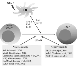
Figure 3. Effect of NF-κB activation on Th17 cell differentiation. NF- κB activation in antigen presenting cells is important for production of Th17 cell differentiation cytokines. Some research groups report that NF-κB activation in CD4 T cells positively regulates Th17 cell differentiation while others showed that NF-κB activation does not affect or negatively regulates Th17 cell differentiation.
into Th17 cells is decreased by Malt1 deficiency (57). In addi- tion, Carma1, which is adaptor protein for TCR-mediated NF- κB activation, is needed for expressions of IL-17A, IL-17F, IL-21, IL-22, IL-23R, and CCR6. Carma1 deficiency also blocks Th17 cell development because chromatin loci of Th17 effec- tor molecules cannot form open conformation, but tran- scription factors, which are needed for Th17 cells develop- ment, were normally expressed (58). In addition, over- expression of calpastatin minimal domain, which is a natural inhibitor of calpain, also decreased Th17 cell differentiation through stabilization of IκBα and subsequent inhibition of NF-κB activation (59). In addition, one recent report de- scribed an important role for IκBζ in Th17 differentiation, and showed that IκBζ acts with the nuclear orphan re- ceptors RORγ and RORα to promote IL-17A gene expression (60). Thus, many data support the importance of NF-κB acti- vation in Th17 cell differentiation.
However, recently, negative results were also reported.
IL-2 is secreted from activated T cells, and acts as a negative regulator of Th17 cell differentiation (61). In the above report, c-Rel was suggested as a positive regulator of Th17 cell differentiation. c-Rel deficiency in T cells decreases IL-2 pro- duction from activated T cells. However, this condition did not affect Th17 cell differentiation. In the report, even though IL-2 was added in Th17 cell differentiation conditions, it did not affect Th17 cell differentiation. IRF-4 is a positive regu- lator of Th17 cell differentiation; however, it was not affected by c-Rel deficiency. Thus, the report argued that c-Rel is not involved in Th17 cell differentiation (62). It has been reported that USP18 regulates the TAK1-TAB1 complex, which is known as NF-κB pathway. USP18 deficient T cells showed NF-κB hyperactivation, and subsequently increased the level of IL-2 secretion. This dysregulation of NF-κB reduced Th17 cell differentiation (63).
CONCLUSION
NF-κB activation is important during T cell activation and for cytokine gene expression in antigen presenting cells. NF-κB activation-mediated Th17 cell cytokine gene expression is im- portant for Th17 cell differentiation; however, different ex- perimental systems showed different roles of antigen re- ceptor-mediated NF-κB activation in Th17 cell differentiation (Fig. 3). Thus, deciphering the role of NF-κB in each of the Th17 cell differentiation conditions, such as different disease states, is an area of great interest.
ACKNOWLEDGEMENTS
This work was supported by a grant of the Korean Health Technology R&D Project, Ministry of Health & Welfare, Republic of Korea (A111838) and by the National Research Foundation of Korea funded by the Ministry of Education, Science and Technology (NRF-2013R1A1A2010995).
CONFLICTS OF INTEREST
The authors have no financial conflict of interest.
REFERENCES
1. Hayden, M. S. and S. Ghosh. 2004. Signaling to NF-kappaB.
Genes Dev. 18: 2195-2224.
2. Hayden, M. S. and S. Ghosh. 2011. NF-kappaB in immuno- biology. Cell Res. 21: 223-244.
3. Hayden, M. S., A. P. West, and S. Ghosh. 2006. NF-kappaB and the immune response. Oncogene 25: 6758-6780.
4. Schulze-Luehrmann, J. and S. Ghosh. 2006. Antigen-receptor signaling to nuclear factor kappa B. Immunity 25: 701-715.
5. Oh, H. and S. Ghosh. 2013. NF-kappaB: roles and regulation in different CD4(+) T-cell subsets. Immunol. Rev. 252:
41-51.
6. Alegre, M. L., K. A. Frauwirth, and C. B. Thompson. 2001.
T-cell regulation by CD28 and CTLA-4. Nat. Rev. Immunol.
1: 220-228.
7. Kane, L. P., J. Lin, and A. Weiss. 2002. It's all Rel-ative:
NF-kappaB and CD28 costimulation of T-cell activation.
Trends Immunol. 23: 413-420.
8. Frauwirth, K. A., J. L. Riley, M. H. Harris, R. V. Parry, J.
C. Rathmell, D. R. Plas, R. L. Elstrom, C. H. June, and C.
B. Thompson. 2002. The CD28 signaling pathway regulates glucose metabolism. Immunity 16: 769-777.
9. Pages, F., M. Ragueneau, R. Rottapel, A. Truneh, J. Nunes, J. Imbert, and D. Olive. 1994. Binding of phosphatidylinosi- tol-3-OH kinase to CD28 is required for T-cell signalling.
Nature 369: 327-329.
10. Pagan, A. J., M. Pepper, H. H. Chu, J. M. Green, and M.
K. Jenkins. 2012. CD28 promotes CD4+ T cell clonal ex- pansion during infection independently of its YMNM and PYAP motifs. J. Immunol. 189: 2909-2917.
11. Sanchez-Lockhart, M., E. Marin, B. Graf, R. Abe, Y. Harada, C. E. Sedwick, and J. Miller. 2004. Cutting edge: CD28-medi- ated transcriptional and posttranscriptional regulation of IL-2 expression are controlled through different signaling path- ways. J. Immunol. 173: 7120-7124.
12. Yokosuka, T., W. Kobayashi, K. Sakata-Sogawa, M.
Takamatsu, A. Hashimoto-Tane, M. L. Dustin, M. Tokunaga, and T. Saito. 2008. Spatiotemporal regulation of T cell cos- timulation by TCR-CD28 microclusters and protein kinase C theta translocation. Immunity 29: 589-601.
13. Park, S. G., J. Schulze-Luehrman, M. S. Hayden, N.
Hashimoto, W. Ogawa, M. Kasuga, and S. Ghosh. 2009. The kinase PDK1 integrates T cell antigen receptor and CD28 cor- eceptor signaling to induce NF-kappaB and activate T cells.
Nat. Immunol. 10: 158-166.
14. Narayan, P., B. Holt, R. Tosti, and L. P. Kane. 2006. CARMA1 is required for Akt-mediated NF-kappaB activation in T cells.
Mol. Cell. Biol. 26: 2327-2336.
15. Matsumoto, R., D. Wang, M. Blonska, H. Li, M. Kobayashi, B. Pappu, Y. Chen, D. Wang, and X. Lin. 2005. Phosphory- lation of CARMA1 plays a critical role in T Cell receptor-medi- ated NF-kappaB activation. Immunity 23: 575-585.
16. Garcon, F., D. T. Patton, J. L. Emery, E. Hirsch, R. Rottapel, T. Sasaki, and K. Okkenhaug. 2008. CD28 provides T-cell costimulation and enhances PI3K activity at the immune syn- apse independently of its capacity to interact with the p85/p110 heterodimer. Blood 111: 1464-1471.
17. Dodson, L. F., J. S. Boomer, C. M. Deppong, D. D. Shah, J. Sim, T. L. Bricker, J. H. Russell, and J. M. Green. 2009.
Targeted knock-in mice expressing mutations of CD28 reveal an essential pathway for costimulation. Mol Cell Biol. 29:
3710-3721.
18. Villalba, M., K. Bi, J. Hu, Y. Altman, P. Bushway, E. Reits, J. Neefjes, G. Baier, R. T. Abraham, and A. Altman. 2002.
Translocation of PKC[theta] in T cells is mediated by a non- conventional, PI3-K- and Vav-dependent pathway, but does not absolutely require phospholipase C. J. Cell Biol. 157:
253-263.
19. Kang, J. A., S. P. Jeong, D. Park, M. S. Hayden, S. Ghosh, and S. G. Park. 2013. Transition from heterotypic to homo- typic PDK1 homodimerization is essential for TCR-mediated NF-kappaB activation. J. Immunol. 190: 4508-4515.
20. Chuang, H. C., J. L. Lan, D. Y. Chen, C. Y. Yang, Y. M.
Chen, J. P. Li, C. Y. Huang, P. E. Liu, X. Wang, and T. H.
Tan. 2011. The kinase GLK controls autoimmunity and NF-kappaB signaling by activating the kinase PKC-theta in T cells. Nat. Immunol. 12: 1113-1118.
21. Romagnani, S. 1994. Lymphokine production by human T cells in disease states. Annu. Rev. Immunol. 12: 227-257.
22. Korn, T., E. Bettelli, M. Oukka, and V. K. Kuchroo. 2009.
IL-17 and Th17 Cells. Annu. Rev. Immunol. 27: 485-517.
23. Ahern, P. P., A. Izcue, K. J. Maloy, and F. Powrie. 2008.
The interleukin-23 axis in intestinal inflammation. Immunol.
Rev. 226: 147-159.
24. Cua, D. J. and C. M. Tato. 2010. Innate IL-17-producing cells:
the sentinels of the immune system. Nat. Rev. Immunol. 10:
479-489.
25. Laurence, A. and J. J. O'Shea. 2007. T(H)-17 differentiation:
of mice and men. Nat. Immunol. 8: 903-905.
26. Acosta-Rodriguez, E. V., G. Napolitani, A. Lanzavecchia, and F. Sallusto. 2007. Interleukins 1beta and 6 but not trans- forming growth factor-beta are essential for the differentiation of interleukin 17-producing human T helper cells. Nat.
Immunol. 8: 942-949.
27. Chen, Z., C. M. Tato, L. Muul, A. Laurence, and J. J. O'Shea.
2007. Distinct regulation of interleukin-17 in human T helper lymphocytes. Arthritis Rheum. 56: 2936-2946.
28. Harrington, L. E., R. D. Hatton, P. R. Mangan, H. Turner, T. L. Murphy, K. M. Murphy, and C. T. Weaver. 2005.
Interleukin 17-producing CD4+ effector T cells develop via a lineage distinct from the T helper type 1 and 2 lineages.
Nat. Immunol. 6: 1123-1132.
29. Park, H., Z. Li, X. O. Yang, S. H. Chang, R. Nurieva, Y.
H. Wang, Y. Wang, L. Hood, Z. Zhu, Q. Tian, and C. Dong.
2005. A distinct lineage of CD4 T cells regulates tissue in- flammation by producing interleukin 17. Nat. Immunol. 6:
1133-1141.
30. Littman, D. R. and A. Y. Rudensky. 2010. Th17 and regu- latory T cells in mediating and restraining inflammation. Cell 140: 845-858.
31. El-Behi, M., B. Ciric, H. Dai, Y. Yan, M. Cullimore, F. Safavi, G. X. Zhang, B. N. Dittel, and A. Rostami. 2011. The ence- phalitogenicity of T(H)17 cells is dependent on IL-1- and IL-23-induced production of the cytokine GM-CSF. Nat.
Immunol. 12: 568-575.
32. Cooke, A. 2006. Th17 cells in inflammatory conditions. Rev.
Diabet. Stud. 3: 72-75.
33. Kramer, J. M. and S. L. Gaffen. 2007. Interleukin-17: a new paradigm in inflammation, autoimmunity, and therapy. J.
Periodontol. 78: 1083-1093.
34. Kaser, A., S. Zeissig, and R. S. Blumberg. 2010. Inflammatory bowel disease. Annu. Rev. Immunol. 28: 573-621.
35. Chebotar, I. V., M. I. Zaslavskaia, T. M. Konyshkina, and A.
N. Maianskii. 1991. IgG- and C3-dependent adhesion of neu- trophils in systems with allogeneic and xenogeneic ligands.
Biull. Eksp. Biol. Med. 112: 403-404.
36. Xavier, R. J. and D. K. Podolsky. 2007. Unravelling the pathogenesis of inflammatory bowel disease. Nature 448:
427-434.
37. Zhou, L., I. I. Ivanov, R. Spolski, R. Min, K. Shenderov, T.
Egawa, D. E. Levy, W. J. Leonard, and D. R. Littman. 2007.
IL-6 programs T(H)-17 cell differentiation by promoting se- quential engagement of the IL-21 and IL-23 pathways. Nat.
Immunol. 8: 967-974.
38. Dong, C. 2008. TH17 cells in development: an updated view of their molecular identity and genetic programming. Nat.
Rev. Immunol. 8: 337-348.
39. Kobayashi, T., S. Okamoto, T. Hisamatsu, N. Kamada, H.
Chinen, R. Saito, M. T. Kitazume, A. Nakazawa, A. Sugita, K. Koganei, K. Isobe, and T. Hibi. 2008. IL23 differentially regulates the Th1/Th17 balance in ulcerative colitis and Crohn's disease. Gut 57: 1682-1689.
40. Fujino, S., A. Andoh, S. Bamba, A. Ogawa, K. Hata, Y. Araki, T. Bamba, and Y. Fujiyama. 2003. Increased expression of interleukin 17 in inflammatory bowel disease. Gut 52: 65-70.
41. Zhang, Z., M. Zheng, J. Bindas, P. Schwarzenberger, and J.
K. Kolls. 2006. Critical role of IL-17 receptor signaling in acute TNBS-induced colitis. Inflamm. Bowel Dis. 12: 382-388.
42. Park, S. G., R. Mathur, M. Long, N. Hosh, L. Hao, M. S.
Hayden, and S. Ghosh. 2010. T regulatory cells maintain in- testinal homeostasis by suppressing gammadelta T cells.
Immunity 33: 791-803.
43. Kotake, S., N. Udagawa, N. Takahashi, K. Matsuzaki, K. Itoh, S. Ishiyama, S. Saito, K. Inoue, N. Kamatani, M. T. Gillespie, T. J. Martin, and T. Suda. 1999. IL-17 in synovial fluids from patients with rheumatoid arthritis is a potent stimulator of osteoclastogenesis. J. Clin. Invest. 103: 1345-1352.
44. Wong, C. K., C. Y. Ho, F. W. Ko, C. H. Chan, A. S. Ho, D. S. Hui, and C. W. Lam. 2001. Proinflammatory cytokines (IL-17, IL-6, IL-18 and IL-12) and Th cytokines (IFN-gamma, IL-4, IL-10 and IL-13) in patients with allergic asthma. Clin.
Exp. Immunol. 125: 177-183.
45. Wong, C. K., L. C. Lit, L. S. Tam, E. K. Li, P. T. Wong, and C. W. Lam. 2008. Hyperproduction of IL-23 and IL-17 in patients with systemic lupus erythematosus: implications for Th17-mediated inflammation in auto-immunity. Clin.
Immunol. 127: 385-393.
46. Kim, S. W., E. S. Kim, C. M. Moon, J. J. Park, T. I. Kim, W. H. Kim, and J. H. Cheon. 2011. Genetic polymorphisms of IL-23R and IL-17A and novel insights into their associations with inflammatory bowel disease. Gut 60: 1527-1536.
47. Glas, J., J. Stallhofer, S. Ripke, M. Wetzke, S. Pfennig, W.
Klein, J. T. Epplen, T. Griga, U. Schiemann, M. Lacher, S.
Koletzko, M. Folwaczny, P. Lohse, B. Goke, T. Ochsenkuhn, B. Muller-Myhsok, and S. Brand. 2009. Novel genetic risk markers for ulcerative colitis in the IL2/IL21 region are in epistasis with IL23R and suggest a common genetic back- ground for ulcerative colitis and celiac disease. Am. J.
Gastroenterol. 104: 1737-1744.
48. Monteleone, G., I. Monteleone, D. Fina, P. Vavassori, G. Del Vecchio Blanco, R. Caruso, R. Tersigni, L. Alessandroni, L.
Biancone, G. C. Naccari, T. T. MacDonald, and F. Pallone.
2005. Interleukin-21 enhances T-helper cell type I signaling and interferon-gamma production in Crohn's disease.
Gastroenterology 128: 687-694.
49. Sarra, M., I. Monteleone, C. Stolfi, M. C. Fantini, P. Sileri, G. Sica, R. Tersigni, T. T. Macdonald, F. Pallone, and G.
Monteleone. 2010. Interferon-gamma-expressing cells are a major source of interleukin-21 in inflammatory bowel dis-
eases. Inflamm. Bowel Dis. 16: 1332-1339.
50. Stolfi, C., A. Rizzo, E. Franze, A. Rotondi, M. C. Fantini, M.
Sarra, R. Caruso, I. Monteleone, P. Sileri, L. Franceschilli, F.
Caprioli, S. Ferrero, T. T. MacDonald, F. Pallone, and G.
Monteleone. 2011. Involvement of interleukin-21 in the regu- lation of colitis-associated colon cancer. J. Exp. Med. 208:
2279-2290.
51. Ouaaz, F., J. Arron, Y. Zheng, Y. Choi, and A. A. Beg. 2002.
Dendritic cell development and survival require distinct NF-kappaB subunits. Immunity 16: 257-270.
52. O'Keeffe, M., R. J. Grumont, H. Hochrein, M. Fuchsberger, R. Gugasyan, D. Vremec, K. Shortman, and S. Gerondakis.
2005. Distinct roles for the NF-kappaB1 and c-Rel tran- scription factors in the differentiation and survival of plasma- cytoid and conventional dendritic cells activated by TLR-9 signals. Blood 106: 3457-3464.
53. Gerondakis, S. and U. Siebenlist. 2010. Roles of the NF-kappaB pathway in lymphocyte development and function. Cold Spring Harb. Perspect. Biol. 2: a000182.
54. Khayrullina, T., J. H. Yen, H. Jing, and D. Ganea. 2008. In vitro differentiation of dendritic cells in the presence of pros- taglandin E2 alters the IL-12/IL-23 balance and promotes dif- ferentiation of Th17 cells. J. Immunol. 181: 721-735.
55. Ruan, Q., V. Kameswaran, Y. Zhang, S. Zheng, J. Sun, J.
Wang, J. DeVirgiliis, H. C. Liou, A. A. Beg, and Y. H. Chen.
2011. The Th17 immune response is controlled by the Rel-RORgamma-RORgamma T transcriptional axis. J. Exp.
Med. 208: 2321-2333.
56. Kwon, M. J., J. Ma, Y. Ding, R. Wang, and Z. Sun. 2012.
Protein kinase C-theta promotes Th17 differentiation via upre- gulation of Stat3. J. Immunol. 188: 5887-5897.
57. Brustle, A., D. Brenner, C. B. Knobbe, P. A. Lang, C.
Virtanen, B. M. Hershenfield, C. Reardon, S. M. Lacher, J.
Ruland, P. S. Ohashi, and T. W. Mak. 2012. The NF-kappaB regulator MALT1 determines the encephalitogenic potential of Th17 cells. J. Clin. Invest. 122: 4698-4709.
58. Molinero, L. L., A. Cubre, C. Mora-Solano, Y. Wang, and M.
L. Alegre. 2012. T cell receptor/CARMA1/NF-kappaB signal- ing controls T-helper (Th) 17 differentiation. Pro. Natl. Acad.
Sci. USA 109: 18529-18534.
59. Iguchi-Hashimoto, M., T. Usui, H. Yoshifuji, M. Shimizu, S.
Kobayashi, Y. Ito, K. Murakami, A. Shiomi, N. Yukawa, D.
Kawabata, T. Nojima, K. Ohmura, T. Fujii, and T. Mimori.
2011. Overexpression of a minimal domain of calpastatin sup- presses IL-6 production and Th17 development via reduced NF-kappaB and increased STAT5 signals. PloS one 6: e27020.
60. Okamoto, K., Y. Iwai, M. Oh-Hora, M. Yamamoto, T. Morio, K. Aoki, K. Ohya, A. M. Jetten, S. Akira, T. Muta, and H.
Takayanagi. 2010. IkappaBzeta regulates T(H)17 develop- ment by cooperating with ROR nuclear receptors. Nature 464:
1381-1385.
61. Stockinger, B. 2007. Good for Goose, but not for Gander:
IL-2 interferes with Th17 differentiation. Immunity 26: 278- 279.
62. Visekruna, A., M. Huber, A. Hellhund, E. Bothur, K. Reinhard, N. Bollig, N. Schmidt, T. Joeris, M. Lohoff, and U. Steinhoff.
2010. c-Rel is crucial for the induction of Foxp3(+) regu- latory CD4(+) T cells but not T(H)17 cells. Eur. J. Immunol.
40: 671-676.
63. Liu, X., H. Li, B. Zhong, M. Blonska, S. Gorjestani, M. Yan, Q. Tian, D. E. Zhang, X. Lin, and C. Dong. 2013. USP18
inhibits NF-kappaB and NFAT activation during Th17 differ- entiation by deubiquitinating the TAK1-TAB1 complex. J.
Exp. Med. 210: 1575-1590.