High tibial osteotomy (HTO) is a procedure that realigns the weight-bearing line from the affected medial compart- ment to the relatively unaffected lateral compartment of the knee.1) Interest in HTO as an adjunctive procedure has increased recently due to technical advances in carti- lage healing procedures and meniscal transplantation for which malalignment would otherwise represent a contra- indication.2) Achieving an accurate correction angle is a key factor for long-term survival of HTO because a small alteration of the limb alignment may change the load dis- tribution of the knee and cause early degenerative changes and dysfunction.3) Although there is no general consensus concerning the precision of the correction of the align- ment for HTO, the postoperative mechanical axis of ± 3°
that has been generally accepted for total knee arthroplasty may be too wide to achieve good long-term results fol- lowing HTO.4) It is recommended that the weight-bearing line pass through a point at 60%–70% of the tibial plateau width when measured from the medial cortex,5,6) which
appears to correspond to a tolerance level of ± 1° from the desired weight-bearing mechanical axis.4)
Conventional HTO has demonstrated quite a high variability regarding postoperative alignment due to im- precise preoperative planning, inaccurate wedge closing or opening, or poor control of intraoperative realignment.7,8) Despite various conventional methods using a cable method or a grid with radio-opaque reference lines, it is difficult to obtain long leg views intraoperatively, and the accuracy can be affected by limb rotation, a bent cable, the alignment guide position, and the quality of the intensi- fied image. In addition, an inadvertent change in the tibial posterior slope angle is a common problem. Noyes et al.9) reported that a gap error of 1 mm could result in a change of the posterior slope of approximately 2°. Other problems with conventional HTO are that a poorly located hinge axis and incorrect orientation of the saw blade or chisel can lead to intraoperative tibial plateau fractures and in- jury to neurovascular structures.1)
Computer-assisted orthopedic surgery (CAOS) aims to improve both the accuracy and precision of orthopedic surgery. Accuracy refers to the degree of closeness to the target. Precision refers to the reproducibility or repeat- ability of obtaining this position, and increased precision means a reduction in outliers.10) However, it remains debatable whether improvements in the accuracy and
Computer-Assisted Navigation in High Tibial Osteotomy
Sang Jun Song, MD, Dae Kyung Bae, MD
Department of Orthopaedic Surgery, Kyung Hee University College of Medicine, Seoul, Korea
Computer-assisted navigation is used to improve the accuracy and precision of correction angles during high tibial osteotomy. Most studies have reported that this technique reduces the outliers of coronal alignment and unintended changes in the tibial posterior slope angle. However, more sophisticated studies are necessary to determine whether the technique will improve the clinical re- sults and long-term survival rates. Knowledge of the navigation technology, surgical techniques and potential pitfalls, the clinical results of previous studies, and understanding of the advantages and limitations of the computer-assisted navigation are crucial to successful application of this new technique in high tibial osteotomy. Herein, we review the evidence concerning this technique from previous studies.
Keywords: Knee, Tibia, Osteotomy, Computer-assisted surgery
Copyright © 2016 by The Korean Orthopaedic Association
This is an Open Access article distributed under the terms of the Creative Commons Attribution Non-Commercial License (http://creativecommons.org/licenses/by-nc/4.0) which permits unrestricted non-commercial use, distribution, and reproduction in any medium, provided the original work is properly cited.
Clinics in Orthopedic Surgery • pISSN 2005-291X eISSN 2005-4408 Received February 18, 2016; Accepted April 18, 2016
Correspondence to: Dae Kyung Bae, MD
Department of Orthopaedic Surgery, Kyung Hee University College of Medicine, 26 Kyungheedae-ro, Dongdaemun-gu, Seoul 02447, Korea Tel: +82-2-958-9489, Fax: +82-2-964-3865
E-mail: [email protected]
precision will lead to enhanced clinical results and long- term survival rates in various orthopedic procedures.10,11) Knowledge of technology concerning computer-assisted navigation, surgical techniques and potential pitfalls, the clinical results of previous studies, and understanding of the advantages and limitations of computer-assisted navi- gation are crucial to the successful application of this new technique in HTO. Herein, we review the evidence con- cerning this technique from previous studies.
CLASSIFICATION OF NAVIGATION SYSTEMS
Computer-assisted navigations can be broadly divided into two types according to the registration methods: image- based and imageless systems.11) SurgiGATE (Medivision, Oberdorf, Switzerland) for HTO is a fluoroscopy-based system. OrthoPilot (Aesculap AG, Tuttlingen, Germany) and VectorVision (BrainLab, Heimstetten, Germany) provide the software for HTO using the imageless system, which have become popular due to the benefits of the im- ageless system.
SURGICAL METHOD
There are two basic techniques available: medial open- wedge and lateral closed-wedge osteotomy. We will focus
on our methods of the open- and closed-wedge HTOs us- ing the VectorVision system.12)
Closed-Wedge HTO
A transverse skin incision and subfascial dissection is made in a similar manner to the conventional technique.
Partial resection of the fibular head or fibular osteotomy is performed to avoid the tethering effect of the fibula dur- ing wedge closing. Two separate dynamic reference bases (DRBs) are fixed to the distal femur and middle tibia with a pair of half-pins. Two pins, 3 mm in diameter, are pre- ferred to single 5-mm pins with bicortical fixation to avoid an incidental fracture. The hip center is determined using kinematic referencing. Other anatomic landmarks are reg- istered by point referencing, including medial and lateral malleoli, medial and lateral tibial plateau points, medial and lateral femoral epicondyles, and anteroposterior tibial rotation. A surgeon determines the start and end points of an osteotomy to be 10–15 mm distal to the tibial plateau.
The software calculates the length of the osteotomy leaving a 3- to 5-mm medial cortical hinge. The far cortical hinge can be best tailored to help the surgeon close the wedge easily. The software sets the cut plane of the osteotomy perpendicular to the sagittal mechanical axis. An addi- tional slope can be fine-tuned in the planned osteotomy to be consistent with the previously measured posterior slope
A B
C D
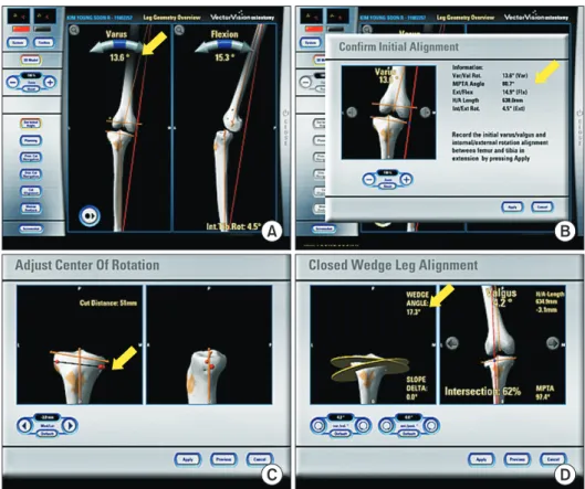
Fig. 1. Computer-assisted closed-wedge high tibial osteotomy. The navigation provides information on the deformity (A), medial proximal tibial angle (B), level of osteotomy (C), correction angle (D), and wedge size (arrows).
angle shown on the preoperative radiograph. For the opti-
mum degree of correction, the postoperative mechanical axis percentage, which will be obtained via the navigation, should be 62% from the medial cortex.
The navigation provides information about the de- formity, level of osteotomy, correction angle, and wedge size (Fig. 1). A precalibrated navigated drill guide is used to place two K-wires in the proximal plane of the osteot- omy. Another two K-wires are inserted in the distal plane of the osteotomy in the same manner (Fig. 2). Proximal and distal osteotomies are carried out using a sharp elec- tric saw over two K-wires. The wedge is removed, and the medial far cortex is then carefully decorticated using the sharp electric saw or osteotome. A valgus force is applied slowly to the extremity until the proximal and distal oste- otomy surfaces are firmly attached. Next, the osteotomy site can be rigidly fixed using various fixatives; we prefer to use a Miniplate staple (U&I Co., Uijeongbu, Korea) (Fig. 3).
The final alignment is confirmed on the computer screen (Fig. 4).
Open-Wedge HTO
For computer-assisted open-wedge HTO, there are several factors that need to be considered including the preven- tion of unintentional changes in the tibial posterior slope angle and autogenous tricortical iliac bone graft or allog- Fig. 2. Four-pin guide technique for wedge resection. Two proximal and
two distal K-wires are inserted using a precalibrated navigated drill guide. The anteroposterior image shows the accurate placement of the K-wires, and the two pairs of K-wires are completely parallel.
A B
Fig. 3. Wedge resection and stabilization.
(A) The wedge is removed, and the medial far cortex is then carefully decor- ticated using a sharp electric saw or an osteotome. A valgus force is applied slowly to the extremity until the proximal and distal osteotomy surfaces are firmly attached. (B) Next, the osteotomy site is rigidly fixed using a Miniplate staple (U&I Co.) of an appropriate size.
A B
Fig. 4. Confirmation of the correction angle assessed under navigation guidance and in postoperative weight-bearing radiography.
The postoperative alignment assessed under navigation (A) is well correlated with the radiographic measurement obtained in the standing position (B).
enous chip bone graft for filling of the opening gap and enhancing bone union.
After a skin incision and subfascial dissection, the superficial medial collateral ligament and underlying peri- osteum are reflected with a periosteal elevator, considering the amount of medial opening and size of the fixatives.
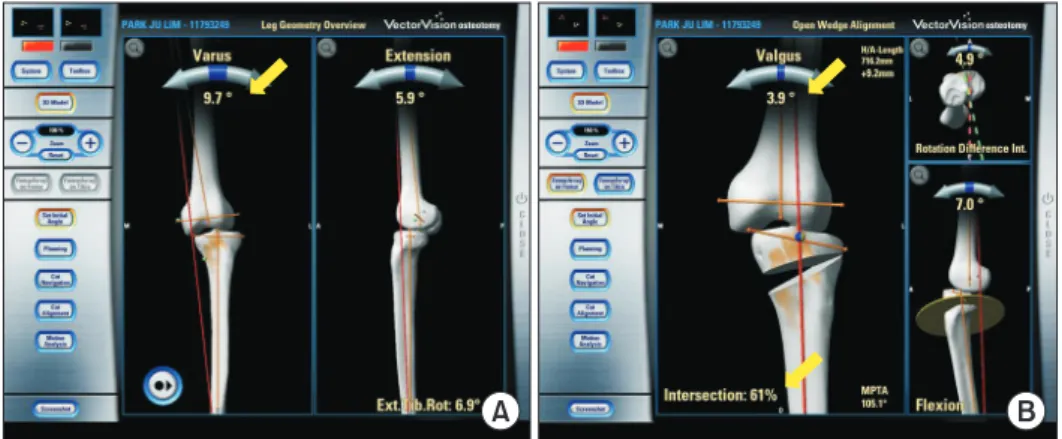
Two separate DRBs are fixed, and the registration is per- formed in the same manner as the above described closed- wedge HTO using the navigation. The starting point of the K-wire on the medial side of the tibia is placed at the level of the tibial tuberosity approximately 3- to 4-cm distal to the medial joint line. The end point on the lateral side of the tibia is placed at the upper portion of the fibular head about 1.5 cm below the joint line. Under navigation guid- ance, two parallel K-wires are inserted obliquely with a precalibrated navigated drill guide. The osteotomy is per- formed using an electric saw while taking care to protect the patellar tendon anteriorly and neurovascular structure posteriorly using right-angle or Hohmann retractors. A tapered wedge osteotome is inserted into the osteotomy site, and the medial opening is slowly and carefully pro- duced, leaving the lateral 5 mm of the posterolateral cortical hinge. Careful valgization through stepwise inser- tion of three coupled chisels is performed to avoid tibial plateau fractures. When the desired mechanical axis is obtained with real-time monitoring of the postoperative mechanical axis on the navigation (Fig. 5), a temporary metal block, bioactive material, or harvested tricortical iliac bone is impacted according to the surgeon’s prefer- ence. An unintended increase in the tibial posterior slope angle after open-wedge HTO is thought to be caused by the triangular configuration of the proximal tibia. The fol- lowing procedures should be performed to avoid changes in the tibial posterior slope angle: (1) osteotomy should be performed parallel to the joint line in the sagittal plane; (2) the posterior cortex should be completely osteotomized and posteromedial soft tissue of the proximal tibia should
be adequately released; (3) the plate should be placed as posteriorly as possible; (4) the postoperative full exten- sion should be the same as the preoperative full extension based on the navigation data; and (5) the anterior opening gap should be approximately half of the posterior opening gap at the proximal tibia.13) In particular, the use of three- dimensional navigation can result in significantly less changes in the postoperative tibial posterior slope angle compared to two-dimensional navigation.14) Although various fixatives can be used for stability of the opened wedge, we prefer the locking plate for securing sufficient initial stability and facilitating early rehabilitation.
POTENTIAL PITFALLS
Registration errors may occur when the bony landmarks are inaccurately identified. The pointer may be deviated from the bone due to the overlying soft tissue.12) HTO al- lows a limited surgical field for the entire alignment and surgeons should identify anatomic landmarks percutane- ously, which may disturb accurate pointing and registra- tion.12) Computer-assisted HTO is not failsafe.11) The bony landmarks should be located accurately on the imageless system.15-17) If the landmarks are not accurately localized, no computer can compensate for this. This limitation should be considered, because the navigation system can- not identify features that the surgeon cannot define.
Most surgeons become impressed by the naviga- tion system because they can see on the computer screen that the coronal alignment is affected by the external force and so-called “weight-bearing simulation” in the supine position.12,18) Yaffe et al.19) reported a reasonable discrep- ancy, as much as 8°, between navigated and radiographic measurement values. Kyung et al.20) reported that the cor- rection of the femorotibial angle by the navigation system was not different from the bony correction angle on three- dimensional computed tomography (3D-CT); however,
A B
Fig. 5. Computer-assisted open-wedge high tibial osteotomy. (A) The navigation system provides information concerning the deformity, medial proximal tibial angle, level of osteotomy, and correction angle (arrows). (B) The medial wedge is carefully opened, hinging on the posterolateral cortex of the tibia until the expected align- ment is achieved.
there was a discrepancy between the navigated value and standing radiographic value. It is important to check the dynamic range of coronal alignment under external varus and valgus force prior to osteotomy, because discrepancy may exist between the weight-bearing radiographic mea- sured value and the non-weight-bearing navigated value of preoperative alignment.18,20) Additional application of varus external force is advised during the registration step until the navigated value of the preoperative alignment matches the radiographic value in the standing position.18)
Aside from technical errors inherent in the registra- tion process, an error can occur with regard to the com- puter’s and camera’s function to track markers.11,15) This error generally ranges from 0.1 to 1 mm for each of the three x, y, and z coordinates.11,15) The navigation system could malfunction when there are dirty reflectors or a dirty camera.11,15) For patients with severe osteopenia, the pins placed in the bones to hold the trackers may move, making all subsequent measurements inaccurate.
Because only the guide pins for the osteotomy level are navigated, the surgeons may make an error during os- teotomy and wedge resection. Additionally, the opposite hinge or plateau fractures can occur without sufficient plastic deformation, and the postoperative alignment can be corrected inaccurately.21) A change in soft tissue tension after osteotomy or unstable fixation of the osteotomy site may lead to malalignment, even though the osteotomy is accurate.20)
CLINICAL RESULTS
Although there have been many previous cadaveric stud-
ies4,7,22,23) and case series16,24) on computer-assisted HTO, long-term comparative studies are lacking (Table 1).8,12,25-29)
To our knowledge, the only prospective randomized study was performed by Iorio et al.,8) which compared the results between computer-assisted and conventional open-wedge HTOs (Table 1). Their radiographic results showed 86%
reproducibility in achieving a mechanical axis of 2°–6° val- gus in the computer-assisted group compared with 23% in the conventional group. For the sagittal alignment, the in- lier of the change in the tibial posterior slope angle within
± 2° was 100% in the computer-assisted group and 24% in the conventional group. However, there was no significant difference in the clinical results with a mean of 3.3 years of follow-up. Ribeiro et al.25) reported that the navigation allowed significantly better control of the tibial posterior slope angle (mean change, 1.9º vs. 4.4º; p = 0.014) and bet- ter Lysholm scores (mean, 91.9 vs. 87.6; p = 0.033) using the 3D navigation system (OrthoPilot ver. 1.5; Aesculap AG). They reported that the navigation system did not improve the accuracy in the correction of the mechani- cal axis (3.1º vs. 3.4º; p = 0.773), but the variability and dispersion of the postoperative mechanical axis was small in the computer-assisted group (standard deviation, 1.8°
vs. 3.3°). Another case-control study by Akamatsu et al.26) found that CAOS was more accurate in achieving the de- sired alignment and reduced the risk of undercorrection.
However, there was no significant difference in the clinical results and no difference in the incidence of lateral corti- cal hinge fractures between the computer-assisted group and the conventional group (5/31 vs. 4/28). They found that the artificial bone wedges adjusted to the opening gap with navigation guidance would result in better correction
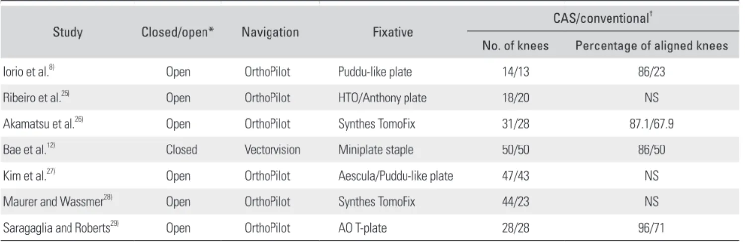
Table 1. Previous Comparative Studies of Computer-Assisted and Conventional HTOs
Study Closed/open* Navigation Fixative CAS/conventional†
No. of knees Percentage of aligned knees
Iorio et al.8) Open OrthoPilot Puddu-like plate 14/13 86/23
Ribeiro et al.25) Open OrthoPilot HTO/Anthony plate 18/20 NS
Akamatsu et al.26) Open OrthoPilot Synthes TomoFix 31/28 87.1/67.9
Bae et al.12) Closed Vectorvision Miniplate staple 50/50 86/50
Kim et al.27) Open OrthoPilot Aescula/Puddu-like plate 47/43 NS
Maurer and Wassmer28) Open OrthoPilot Synthes TomoFix 44/23 NS
Saragaglia and Roberts29) Open OrthoPilot AO T-plate 28/28 96/71
HTO: high tibial osteotomy, NS: not stated.
*Closed/open: closed-wedge high tibial osteotomy/open-wedge high tibial osteotomy. †CAS/conventional: computer-assisted high tibial osteotomy/conventional high tibial osteotomy.
angle for the cases with a lateral cortical or a lateral tibial plateau fracture. The change in the tibial posterior slope angle was small in the computer-assisted group (0.6° vs.
3.5°; p = 0.001) although they used the two-dimensional navigation system (OrthoPilot ver. 1.3). They explained that it could decrease the change in the tibial slope because the same maximum extension angle of the knee was kept before and after osteotomy.
A comparative study between computer-assisted and conventional closed-wedge HTOs was performed by Bae et al.12) In the study, the postoperative coronal alignment of the mechanical axis percentage was more accurate (mean, 59% vs. 47%; p < 0.001) and more precise (variability, 2.3°
vs. 3.7°; p = 0.012) in the computer-assisted group than in the conventional group. The tibial posterior slope angle was less changed in the computer-assisted group (mean, 2.0° vs. 4.0°; p < 0.001). Another retrospective compara- tive study for open-wedge HTO26) showed that the weight- bearing line passing through the tibial plateau was 62.3% ± 2.9% in the computer-assisted group and 58.7% ± 2.9% in the conventional group (p = 0.001). The mean Lysholm (85 vs. 83; p = 0.047) and Hospital for Special Surgery (84 vs.
79; p = 0.009) knee scores at the 1-year follow-up were also better in the computer-assisted group. Although there were two knees of delayed union and one knee of varus collapse in the computer-assisted group, all of these complica- tions were associated with the breakage of the lateral tibial hinge and varus collapse. They could have been avoided with a locking plate instead of the dual open-wedge plate (Aesculap, Seoul, Korea) that was used. Maurer and Was- smer28) reported the results of 67 open-wedge HTOs. They compared the first 23 knees of conventional HTO and the next 44 knees of computer-assisted HTO, and the computer-assisted group resulted in a higher accuracy of the postoperative mechanical axis within the stated target of 3° to 5° valgus without producing additional complica- tion. Saragaglia and Roberts29) performed a matched pair analysis between 28 computer-assisted open-wedge HTOs and his retrospective control group of 28 conventional pa- tients. The goal of final mechanical alignment of 184° ± 2°
was achieved in 96% of the computer-assisted group and in 71% of the conventional group (p < 0.01).
ADVANTAGES AND LITERATURE REVIEW
The most important advantage of computer-assisted HTO is to improve the accuracy and precision of the aiming alignment, and it has been consistently demonstrated in cadaveric and clinical studies.4,12,16,26-30) Computer-assisted HTO can also provide real-time intraoperative informa-
tion concerning coronal, sagittal and transverse axes, which can compensate for the shortcomings of preopera- tive radiographic planning. It can improve postoperative results with decreased radiation exposure.7,31)
Computer-assisted HTO can resolve changes in the mechanical axis with varus and valgus external force.18) It can also provide information on the medial or lateral soft tissue status.18) The evaluation of the postoperative mechanical axis under external force or pushing heels can also provide information on the opposite cortical hinge fracture, postoperative fixation stability, and leg axis dur- ing full-weight bearing.
The incidence of opposite cortical fracture, 9% to 80%, has been reported in both closed- and open-wedge HTOs.32-35) Kessler et al.36) reported that the maximum cor- rection angle prior to the medial cortical fracture was 6.5°
in closed-wedge HTO when the osteotomy was terminated 10 mm from the medial cortex and approximately 20 mm below the plateau. Additionally, the correction angle could be increased to 10° when the osteotomy was terminated in a 5-mm-diameter hole, drilled in an anteroposterior direction. Accurate control of the position of the cortical hinge using navigation guidance and the effort for plastic deformation of the opposite cortex during wedge closing or opening can help to avoid hinge fractures in HTO.
In addition, navigation can be used in laborato- ries and the operating room as a teaching tool for less- experienced surgeons to shorten the learning curve.10) Computer-assisted navigation may play a role in aiding more complex osteotomies, such as combined femoral and tibial osteotomies (double-level osteotomy) for severe genu varum and no tibia vara.37) Computer-assisted navigation can serve as a valuable research tool, facilitating precise measurements of overall limb alignment that normally require additional radiographic procedures and offering data previously confined to cadaver studies such as real- time knee kinematics.10)
DISADVANTAGES AND LITERATURE REVIEW
The current barriers to widespread use of computer- assisted navigation include increased costs, operating time, and inconvenience of surgery.10) Economic analyses indicate that these high-cost technologies may only be cost effective in high-volume hospitals.38) Clinical studies have shown that computer-assisted navigation improves the accuracy of lower-volume surgeons to a greater degree such that they can obtain similar results to those of high- volume surgeons.38) However, the cost of most navigation apparatuses may limit their use in low-volume hospitals.
Another disadvantage is the additional time re- quired for the registration step, ranging from approximate- ly 10 to 30 minutes.7,28) There are also technique-related disadvantages, such as the long learning curve, line of sight issues, registration failures, and mechanical or software malfunctions.1) Gebhard et al.16) reported on the influence of surgeon’s experience and perioperative complications in computer-assisted open-wedge HTO. Seven intraoperative complications were reported from a total of 59 patients (12%); they were all derived from the navigation system, and the majority occurred during one study center’s learn- ing phase. There was loosening of the DRB (three knees), system failure (two knees), loss of orientation after chang- ing the reference pins (one knee), and unavailability of the navigation instrument (one knee). The procedures can appear cumbersome compared with those of conventional techniques. Also, several procedures may be required before the surgeon feels comfortable with the navigation system.16)
Another complication of CAOS is the increased in- cidence of deep infection due to the longer operating time.
The use of DRB entails stab wounds in the distal femur and middle tibia, which consequently increases the risk of infection, fracture, and heterotopic ossification.8,39)
AUTHORS’ PERSPECTIVES
The main criticism of the application of the navigation system in HTO to determine the active weight-bearing alignment is that data are acquired in the supine position.
Therefore, future research should elucidate the relation- ship between the alignment assessed in the supine position in the operating room and the weight-bearing alignment in daily living activities.
Previous studies have suggested convincing evidence that computer-assisted navigation provides more accurate and precise postoperative alignment in HTO.8,10,12) Howev- er, no long-term clinical studies or randomized controlled trials have provided evidence that the navigation system either improves clinical results or lowers the conversion
rate to total knee arthroplasty. Questions remain whether reducing the outliers of alignment would outweigh the ini- tial cost for the navigation system. Future studies should have high methodological standards, including prospec- tive randomization with control of preoperative, intraop- erative, and postoperative variables and long-term follow- up to analyze the survival rate.
Biomechanical studies will also be required to define ideal alignments in the coronal, sagittal, and axial planes.
To produce sound evidence concerning the advantages and disadvantages of CAOS, it is necessary to ascertain the kinematic patterns of patients before and after surgery.
The software for navigation system is expected to evolve with greater convenience and greater accuracy. The navigation equipment should eventually become less ex- pensive, simpler, and easier to use.
CONCLUSIONS
The benefit of computer-assisted navigation lies in the improved accuracy and precision of postoperative coronal and sagittal alignments. In addition, the navigation system can allow adjustment of the hinge axis position and reduce the risk of opposite cortical hinge fracture.21) However, additional studies are necessary to determine whether the improvement in alignment and hinge axis influences the long-term results and survival rate to offset the increased surgical time and potential complication of CAOS.
The orthopedic surgeon’s experience, adaptability, and knowledge of technology regarding computer-assisted HTO are crucial to the surgical success. Only an orthope- dic surgeon who clearly understands the technology, goals, surgical technique, potential pitfalls, advantages, and limi- tations of the navigation system can apply the CAOS tech- nique appropriately for occasional cases of HTO.
CONFLICT OF INTEREST
No potential conflict of interest relevant to this article was reported.
REFERENCES
1. Picardo NE, Khan W, Johnstone D. Computer-assisted navi- gation in high tibial osteotomy: a systematic review of the literature. Open Orthop J. 2012;6:305-12.
2. Thompson SR, Zabtia N, Weening B, Zalzal P. Arthroscopic and computer-assisted high tibial osteotomy using standard total knee arthroplasty navigation software. Arthrosc Tech.
2013;2(2):e161-6.
3. Sharma L, Chmiel JS, Almagor O, et al. The role of varus and valgus alignment in the initial development of knee car- tilage damage by MRI: the MOST study. Ann Rheum Dis.
2013;72(2):235-40.
4. Lutzner J, Gross AF, Gunther KP, Kirschner S. Precision of
navigated and conventional open-wedge high tibial oste- otomy in a cadaver study. Eur J Med Res. 2010;15(3):117-20.
5. Miniaci A, Ballmer FT, Ballmer PM, Jakob RP. Proximal tibial osteotomy: a new fixation device. Clin Orthop Relat Res. 1989;(246):250-9.
6. Noyes FR, Barber SD, Simon R. High tibial osteotomy and ligament reconstruction in varus angulated, anterior cruci- ate ligament-deficient knees: a two- to seven-year follow-up study. Am J Sports Med. 1993;21(1):2-12.
7. Hankemeier S, Hufner T, Wang G, et al. Navigated open- wedge high tibial osteotomy: advantages and disadvantages compared to the conventional technique in a cadaver study.
Knee Surg Sports Traumatol Arthrosc. 2006;14(10):917-21.
8. Iorio R, Pagnottelli M, Vadala A, et al. Open-wedge high tibial osteotomy: comparison between manual and com- puter-assisted techniques. Knee Surg Sports Traumatol Ar- throsc. 2013;21(1):113-9.
9. Noyes FR, Goebel SX, West J. Opening wedge tibial oste- otomy: the 3-triangle method to correct axial alignment and tibial slope. Am J Sports Med. 2005;33(3):378-87.
10. Young SW, Safran MR, Clatworthy M. Applications of computer navigation in sports medicine knee surgery:
an evidence-based review. Curr Rev Musculoskelet Med.
2013;6(2):150-7.
11. Bae DK, Song SJ. Computer assisted navigation in knee ar- throplasty. Clin Orthop Surg. 2011;3(4):259-67.
12. Bae DK, Song SJ, Yoon KH. Closed-wedge high tibial osteot- omy using computer-assisted surgery compared to the con- ventional technique. J Bone Joint Surg Br. 2009;91(9):1164- 71.
13. Song EK, Seon JK, Park SJ. How to avoid unintended increase of posterior slope in navigation-assisted open- wedge high tibial osteotomy. Orthopedics. 2007;30(10 Suppl):S127-31.
14. Yim JH, Seon JK, Song EK. Posterior tibial slope in medial opening-wedge high tibial osteotomy: 2-D versus 3-D navi- gation. Orthopedics. 2012;35(10 Suppl):60-3.
15. Khadem R, Yeh CC, Sadeghi-Tehrani M, et al. Comparative tracking error analysis of five different optical tracking sys- tems. Comput Aided Surg. 2000;5(2):98-107.
16. Gebhard F, Krettek C, Hufner T, et al. Reliability of com- puter-assisted surgery as an intraoperative ruler in navi- gated high tibial osteotomy. Arch Orthop Trauma Surg.
2011;131(3):297-302.
17. Keppler P, Gebhard F, Grutzner PA, et al. Computer aided high tibial open wedge osteotomy. Injury. 2004;35 Suppl 1:S- A68-78.
18. Kendoff DO, Fragomen AT, Pearle AD, Citak M, Rozbruch SR. Computer navigation and fixator-assisted femoral oste- otomy for correction of malunion after periprosthetic femur fracture. J Arthroplasty. 2010;25(2):333.e13-9.
19. Yaffe MA, Koo SS, Stulberg SD. Radiographic and naviga- tion measurements of TKA limb alignment do not correlate.
Clin Orthop Relat Res. 2008;466(11):2736-44.
20. Kyung BS, Kim JG, Jang KM, et al. Are navigation systems accurate enough to predict the correction angle during high tibial osteotomy? Comparison of navigation systems with 3-dimensional computed tomography and standing radio- graphs. Am J Sports Med. 2013;41(10):2368-74.
21. Bae DK, Park CH, Kim EJ, Song SJ. Medial cortical fractures in computer-assisted closing-wedge high tibial osteotomy.
Knee. 2016;23(2):295-9.
22. Goleski P, Warkentine B, Lo D, Gyuricza C, Kendoff D, Pearle AD. Reliability of navigated lower limb alignment in high tibial osteotomies. Am J Sports Med. 2008;36(11):2179- 86.
23. Yamamoto Y, Ishibashi Y, Tsuda E, Tsukada H, Kimura Y, Toh S. Validation of computer-assisted open-wedge high tibial osteotomy using three-dimensional navigation. Or- thopedics. 2008;31(10 Suppl 1):68-71.
24. Saragaglia D, Chedal-Bornu B. Computer-assisted oste- otomy for valgus knees: medium-term results of 29 cases.
Orthop Traumatol Surg Res. 2014;100(5):527-30.
25. Ribeiro CH, Severino NR, Moraes de Barros Fucs PM.
Opening wedge high tibial osteotomy: navigation system compared to the conventional technique in a controlled clinical study. Int Orthop. 2014;38(8):1627-31.
26. Akamatsu Y, Mitsugi N, Mochida Y, et al. Navigated open- ing wedge high tibial osteotomy improves intraoperative correction angle compared with conventional method. Knee Surg Sports Traumatol Arthrosc. 2012;20(3):586-93.
27. Kim SJ, Koh YG, Chun YM, Kim YC, Park YS, Sung CH.
Medial opening wedge high-tibial osteotomy using a kine- matic navigation system versus a conventional method: a 1-year retrospective, comparative study. Knee Surg Sports Traumatol Arthrosc. 2009;17(2):128-34.
28. Maurer F, Wassmer G. High tibial osteotomy: does navigation improve results? Orthopedics. 2006;29(10 Suppl):S130-2.
29. Saragaglia D, Roberts J. Navigated osteotomies around the knee in 170 patients with osteoarthritis secondary to genu varum. Orthopedics. 2005;28(10 Suppl):s1269-74.
30. Iorio R, Vadala A, Giannetti S, et al. Computer-assisted high tibial osteotomy: preliminary results. Orthopedics.
2010;33(10 Suppl):82-6.
31. Na YG, Eom SH, Kim SJ, Chang MJ, Kim TK. The use of navigation in medial opening wedge high tibial osteotomy can improve tibial slope maintenance and reduce radiation exposure. Int Orthop. 2016;40(3):499-507.
32. Miller BS, Downie B, McDonough EB, Wojtys EM. Compli- cations after medial opening wedge high tibial osteotomy.
Arthroscopy. 2009;25(6):639-46.
33. Nelissen EM, van Langelaan EJ, Nelissen RG. Stability of medial opening wedge high tibial osteotomy: a failure analy- sis. Int Orthop. 2010;34(2):217-23.
34. Takeuchi R, Umemoto Y, Aratake M, et al. A mid term comparison of open wedge high tibial osteotomy vs uni- compartmental knee arthroplasty for medial compartment osteoarthritis of the knee. J Orthop Surg Res. 2010;5(1):65.
35. van Raaij TM, Brouwer RW, de Vlieger R, Reijman M, Ver-
haar JA. Opposite cortical fracture in high tibial osteotomy:
lateral closing compared to the medial opening-wedge tech- nique. Acta Orthop. 2008;79(4):508-14.
36. Kessler OC, Jacob HA, Romero J. Avoidance of medial cor- tical fracture in high tibial osteotomy: improved technique.
Clin Orthop Relat Res. 2002;(395):180-5.
37. Saragaglia D, Mercier N, Colle PE. Computer-assisted os- teotomies for genu varum deformity: which osteotomy for which varus? Int Orthop. 2010;34(2):185-90.
38. Goradia VK. Computer-assisted and robotic surgery in orthopedics: where we are in 2014. Sports Med Arthrosc.
2014;22(4):202-5.
39. Citak M, Kendoff D, O'Loughlin PF, Pearle AD. Heterotopic ossification post navigated high tibial osteotomy. Knee Surg Sports Traumatol Arthrosc. 2009;17(4):352-5.