26
26 THE EWHA MEDICAL JOURNALTHE EWHA MEDICAL JOURNAL
Acute Vulvar Ulcer (Lipschutz Ulcer) in a Sexually Inactive 11-Year- old Girl
Kyoung Won Cho, Shin Hye Kim, Ji Hyen Lee
1Department of Pediatrics, Myongji Hospital, Hanyang University College of Medicine, Goyang, 1Ewha Womans University College of Medicine, Seoul, Korea
Case Report
Ewha Med J 2021;44(1):26-29
https://doi.org/10.12771/emj.2021.44.1.26 eISSN 2234-2591
Non-sexually acquired genital ulceration, also known as Lipschutz ulcer, is a rare condi- tion that typically occurs in prepubertal and pubertal girls. It can be misdiagnosed as a sexually transmitted disease or even a sign of child abuse, causing great anxiety for patients and their families. It is often accompanied by systemic symptoms such as fever, myalgia, or lymphadenopathy. Several viruses such as Epstein-Barr virus, cytomegalovi- rus, and mumps virus have been associated with this entity. Furthermore, Mycoplasma pneumonia has rarely been linked to such ulcers in the literature. We present a case of Lipschutz ulcer in a sexually inactive 11-year-old girl. (Ewha Med J 2021;44(1):26-29)
Received August 27, 2020 Revised December 15, 2020 Accepted December 27, 2020 Corresponding author Ji Hyen Lee
Department of Pediatrics, Ewha Womans University College of Medicine, 260 Gonghang-daero, Gangseo-gu, Seoul 07804, Korea
Tel: 82-2-6986-1766, Fax: 82-2-6986-3024 E-mail: leejihyen@ewha.ac.kr
Key Words
Female; Genital diseases; Ulcer; Lipschutz ulcer
This is an Open Access article distributed under the terms of the Creative Commons Attribution Non-Commercial License (http://creativecommons.org/licenses/by-nc/4.0) which permits unrestricted non-commercial use, distribution, and reproduction in any medium, provided the original work is properly cited.
Introduction
The Austrian dermatologist Benjamin Lipschutz first de- scribed acute genital ulcers in adolescent girls in the absence of any evidence of sexually transmitted infections [1]. Lipschutz ulcer is an entity that presents as an acute ulcer in the labia mi- nora or majora, introitus, fourchette, or vestibule, accompanied by flu-like systemic symptoms such as fever, tonsillitis, and/or lymphadenopathy.
Lipschutz ulcer is a diagnosis of exclusion. It mimics a wide spectrum of diseases ranging from infective causes (syphilis, herpes genitalis, and chancroid), to inflammatory conditions (Behcet disease and Crohn disease), as well as trauma. After ruling out sexually transmitted infections, inflammatory condi- tions, and systemic illness, Lipschutz ulcer diagnosis is estab-
lished according to five major and one of two minor criteria (Table 1) [2].
The etiology of this disease remained obscure until the 1960s, when the first clues revealed the involvement of viruses or bac- teria. Studies have since then revealed that Lipschutz disease is associated with viruses such as Epstein-Barr virus (EBV), cyto- megalovirus (CMV), and mumps virus, as well as bacteria such as Salmonella, Mycoplasma pneumoniae, and M. fermentans [3].
Case
An 11-year-old girl was referred to our hospital because of fever, myalgia, and ulcer in her genital area. She had no history of recurrent oral or genital aphthous lesions. Four days before
27
THE EWHA MEDICAL JOURNAL Lipschutz Ulcer in a Sexually Inactive Korean Girl
admission, she started experiencing fever (max 39.7℃), sore throat, myalgia, and mild dry cough. Then she started experi- encing intense pain and burning sensation in her genital area on day 2 after the onset of fever.
On admission, she had fever, myalgia, and chills, but no diarrhea or abdominal pain. Physical examination revealed a painful ulcer with a single necrotic eschar (1.0 cm×1.5 cm) lo- cated inside the right side of the labia majora (Fig. 1). This ulcer was severely tender upon palpitation, and no oral ulcers or swollen lymph nodes were detected. She showed no symptoms related to gastrointestinal, neurologic, or orbital lesions, and furthermore, she was not sexually active and had no previous history of similar ulcers. She was initially treated with intra- venously administered antibiotics and local wound care with antiseptics.
Analytical test results showed elevated C-reactive protein (0.86 mg/dL; normal range, <0.5 mg/dL). Her complete blood count and liver enzyme values were normal at admission. C- reactive protein levels dropped to 0.57 mg/dL after starting treatment with azithromycin and ampicillin-sulbactam intra- venously. Her serum test was negative for herpes simplex virus
(HSV), CMV, EBV, parvovirus, mumps virus, varicella-zoster virus, hepatitis B, or hepatitis C. Furthermore, her serum anti- streptolysin O was normal, and human immunodeficiency virus and syphilis serologic tests were all negative. Her blood and urine cultures were negative. Antibodies against antineu- trophil cytoplasmic antibodies-proteinase, and antineutrophil cytoplasmic antibodies-myeloperoxidase were all negative.
Antinuclear antibody screening and titer were all negative. Her immunoglobulin G, A, and M were normal. Furthermore, her serum complement factor 3 and 4, and CH50 tests were normal. Her nasopharyngeal secretion was tested for a panel PCR for respiratory virus, and the results were all negative for adenovirus, respiratory syncytial virus A and B, influenza A and B, parainfluenza 1-4, metapneumovirus, rhinovirus A, B, and C, bocavirus, enterovirus, and human coronaviruses 229E, OC43, and NL63. The results of her stool PCR for gastroen- teritis related virus and bacteria were all negative. Her serum M. pneumoniae IgM test was with in borderline range (0.93;
borderline, 0.91 to 1.09). Four days after admission, her serum M. pneumoniae IgM was still borderline, although the titer was slightly elevated (0.96; borderline, 0.91 to 1.09). PCR test and culture of her vaginal discharge were negative for Chlamydia trachomatis, Neisseria gonorrhea, Trichomonas vaginalis, Treponemapallidum, HSV type I and II, and Candida albicans.
Only normal floras were checked in the wound culture. Soft tissue sonography of her genital ulcer revealed that there was no abscess pocket in the swollen region of the right major labia.
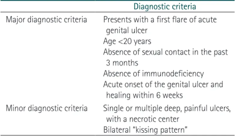
She became afebrile after 5 days of treatment starting with azithromycin and ampicillin-sulbactam. Treatments during hospitalization included supportive local care, pain and fever control with oral analgesics and topical steroid ointment. She was discharged from hospital in good condition after a total hospital stay of seven days. After a 2-week follow-up, her Table 1. Diagnostic criteria for Lipschutz ulcer
Diagnostic criteria Major diagnostic criteria Presents with a first flare of acute
genital ulcer Age <20 years
Absence of sexual contact in the past 3 months
Absence of immunodeficiency Acute onset of the genital ulcer and
healing within 6 weeks
Minor diagnostic criteria Single or multiple deep, painful ulcers, with a necrotic center
Bilateral “kissing pattern”
A B C D
Fig. 1. Single necrotic genital ulceration with eschar. (A) Admission day 1, (B) ad- mission day 3, (C) admission day 5, and (D) admission day 8. Informed consent for medical publication of the clinical images was obtained from the patient and her parent.
28 THE EWHA MEDICAL JOURNAL Cho KW, et al
genital necrotic eschar dropped off and showed re-epitheli- zation of the vulvar skin (Fig. 2). Six months later, she had no recurrence of the ulcer.
This study was a retrospective study, and it was approved by the institutional review board of the Myongji Hospital (MJH2020-01-008).
Discussion
A Lipschutz ulcer is defined as a vulvar ulcer with no identi- fiable etiology, based on clinical, histopathologic, serologic, and microbiologic findings. These ulcers are characterized by sud- den painful genital ulceration, occurring mostly in young vir- gin girls, accompanied with malaise, fever, and other systemic symptoms. These distressing symptoms are rare and may be presented to dermatologists, gynecologists, or pediatricians. It is often misdiagnosed as a sexually transmitted disease or even taken as a sign of child abuse. Some clinicians have noted that the presenting symptoms of a Lipschutz ulcer may be confused with sexual abuse, a terrible diagnosis, especially in children, and can lead to unnecessary investigations, treatments, and anxiety within the family [2]. Generally, the natural course is benign, with spontaneous regression occurring within just a few weeks.
Despite its long history, this condition is not well recognized,
and its root cause is still poorly understood. Over 70% of all reported patients were ultimately diagnosed with idiopathic vulvar ulcers. Some cases were reported to be preceded or ac- companied by systemic infections such as infectious mononu- cleosis (EBV), influenza, CMV infection, mumps, paratyphoid fever, or mycoplasma pneumonia. Other reported preceding illnesses include viral gastroenteritis, viral upper respiratory tract illness, and streptococcal pharyngitis. The pathogenesis of Lip- schutz ulcer is still an enigma. It could develop from a hema- togenous spread or autoinoculation, although one hypothesis suggests that it could arise from a hypersensitive reaction to a viral or bacterial infection, leading to the deposition of immune complexes in the dermal vessels, which in turn activates the complementary systems, resulting in micro-thrombi formation and subsequent tissue necrosis [4,5].
Extensive laboratory work is often conducted, creating a great deal of anxiety in both patients and parents, while also generating enormous and unnecessary medical expenses. A limited infectious evaluation, including serologies (EBV, CMV, influenza, and mycoplasma) may be considered in patients with systemic symptoms, including persistent fever, extreme fatigue, swollen lymph nodes, and/or persistent or severe sore throat. Furthermore, testing for human immunodeficiency virus, syphilis, HSV, and hepatitis B and C was negative in all reported patients. In addition, through careful physical exami- nation and history, it is imperative to rule out other causes of more frequent genital ulcers, particularly sexually transmitted diseases, and to discuss the main differential diagnoses, such as Behcet disease or cutaneous localization of Crohn disease.
Routine bacterial and fungal cultures reveal skin flora or non- pathogenic bacteria and do not offer any additional benefit [6].
Skin biopsies are also not recommended as a first-line investi- gation because they only indicate non-specific dermal infiltrate of mixed inflammatory cells in most patients. A skin biopsy from the edge of the ulcer is advised for ulcers lasting longer than 4 weeks [7].
Lipschutz ulcers are self-limiting and generally recover spon- taneously. Treatments are mainly aimed at relieving pain and healing of the ulcer. Therefore, patients in many case reports were treated symptomatically with analgesics, topical steroids, and antibiotics. In addition, treatment with a brief course of systemic corticosteroid (0.5 mg/kg of prednisolone for 1 to 2 weeks) may help in healing severely painful, multiple, or ne- Fig. 2. Ulcer was almost completely healed at 22 days without any
sequelae. Informed consent for medical publication of the clinical im- ages was obtained from the patient and her parent.
29
THE EWHA MEDICAL JOURNAL Lipschutz Ulcer in a Sexually Inactive Korean Girl
crotic ulcers [8,9]. Antiviral therapy can be chosen if HSV was thought, but patient’s ulcer lesions didn’t look like genital HSV (vesicles), we didn’t use antiviral agents [10]. Healing occurs within 6 weeks, without scarring. The mean healing time was reported to be16 to 21 days (range, 5 to 52 days) [11,12].
A case in South Korea, a sexually inactive 16-year-old girl with fever, oral ulcers, genital ulcers was diagnosed with Bechet disease. She had no history of oral or genital ulcers and Bechet disease. Serologic tests included VDRL, EBV, HSV IgM, HLA- 51 were negative. After 4 weeks, ulcers were disappeared. Dur- ing 4-month follow-up, there was no recurrence of ulcer [13].
Another case in South Korea, a 13-year-old previously healthy girl, with no personal history of recurrent oral or genital aph- thous lesions, presented with sudden onset of two painful vul- var ulcers and fever. Serologic studies were all negative. She was discharged from hospital in good condition without pain after a total hospital stay of 8 days. The 3-week follow-up showed total resolution and re-epithelization of the vulvar lesion. No recurrences occurredduring the following 6 months [14].
To conclude, we presented a case of Lipschutz ulcer in a sex- ually inactive 11-year-old girl in South Korea. Lipschutz vul- var ulcer is a rare clinical entity due to the deleterious, extremely painful ulcerations among sexually inactive peripubertal girls. It is often misdiagnosed, over-investigated, and under-reported.
Recognition of Lipschutz ulcer is important to the extent that patients receive appropriate and timely treatment,in addition to prognostic counseling. More than 70% of Lipschutz ulcers are idiopathic. No formal treatment guidelines for Lipschutz ulcer exist. Therefore, more studies are necessary to determine the organisms associated with Lipschutz ulcer and the relevant guidelines for confirming diagnosis.
References
1. Lipschutz B. Through a strange Geschwursform of the female genitalia (ulcer Vulvae Acutum). Arch Dermatol Syphilis (Ber- lin) 1913;114:363-395.
2. Garcia JG, Pavon BM, Martin LM, Martinez BF, Norniella CM, Caro FA. Lipschutz ulcer: a cause of misdiagnosis when suspect- ing child abuse. Am J Emerg Med 2016;34:1326.
3. Horie C, Kano Y, Mitomo T, Shiohara T. Possible involvement of mycoplasma fermentans in the development of nonsexually acquired genital ulceration (Lipschütz ulcers) in 3 young female patients. JAMA Dermatol 2015;151:1388-1389.
4. Leigh R, Nyirjesy P. Genitourinary manifestations of Epstein- Barr virus infections. Curr Infect Dis Rep 2009;11:449-456.
5. Portnoy J, Ahronheim GA, Ghibu F, Clecner B, Joncas JH. Re- covery of Epstein-Barr virus from genital ulcers. N Engl J Med 1984;311:966-968.
6. Bandow GD. Diagnosis and management of vulvar ulcers. Der- matol Clin 2010;28:753-763.
7. Huppert JS. Lipschutz ulcers: evaluation and management of acute genital ulcers in women. Dermatol Ther 2010;23:533-540.
8. Lampert A, Assier-Bonnet H, Chevallier B, Clerici T, Saiag P. Lip- schutz's genital ulceration: a manifestation of Epstein-Barr virus primary infection. Br J Dermatol 1996;135:663-665.
9. Taylor S, Drake SM, Dedicoat M, Wood MJ. Genital ulcers associ- ated with acute Epstein-Barr virus infection. Sex Transm Infect 1998;74:296-297.
10. Workowski KA, Bolan GA; Centers for Disease Control and Prevention. Sexually transmitted diseases treatment guidelines, 2015. MMWR Recomm Rep 2015;64:1-137.
11. Berlin C. The pathogenesis of the so-called ulcus vulvae acutum.
Acta Derm Venereol 1965;45:221-222.
12. Healy CM, Thornhill MH. An association between recurrent oro- genital ulceration and non-steroidal anti-inflammatory drugs. J Oral Pathol Med 1995;24:46-48.
13. Soh BW, Lee ES. Non-sexually related genital ulcer in a 16-year- old Girl (Lipschutz ulcer). Korean J Dermatol 2017;55:73-74.
14. Kim JK, Jeon JS, Park JW, Kang YD. Non-sexually related acute genital ulcers in a pubertal girl. Australas Med J 2017;10:628-631.