대한치주과학회지 : Vol. 37, No. 2(Suppl.), 2007
Identification of Matrix Mineralization-Related Genes in Human Periodontal Ligament Cells Using cDNA
Microarray
Jae-Hee Shin1, Jin-Woo Park1, Shin-Il Yeo1, Woo-Chang Noh1, Moon-Kyu Kim2, Jung-Chul Kim2, Jo-Young Suh1*
1. Department of Periodontology, College of dentistry, Kyungpook National University 2. Department of Immunology, School of Medicine, Kyungpook National University
I. INTRODUCTION
The periodontal ligament (PDL) is derived from the inner layer of the dental follicle shortly root development begins1). Indeed, development and maturation of the PDL are dependent on the formation of root dentin and the synthesis of its investing mineralized tissue, cementum.1)
The PDL is a highly vascular and cellular connective tissue situated between the tooth and the alveolar bone that provide suppor- tive, attachment, and sensory functions. The normal PDL includes osteoblasts and osteo- clasts on the bone side; fibroblasts, epithelial cell rests of Malassez, macrophages, un- differentiated mesenchymal cells, neural ele- ments,and endothelial cells in the body; and
cementoblasts on the root surface. Fibroblasts are the predominant cells in the PDL. Their specialized functions are considered to be re- sponsible for not only the formation and maintenance of the PDL, but also for the re- pair, remodeling and regeneration of the ad- jacent alveolar bone and cementum2-4). According to clinical and histological re- searches5,6), periodontal regeneration is in- duced by the cells which are derived from the remaining healthy portions of the perio- dontal connective tissues.
Several studies have also demonstrated that the PDL cells are not homogenous but may consist of different subpopulation with unique phenotypes and distinct functional activities7-9). The cementum increases in thickness with age under normal physiological condition. In
* It's work was supported by basic research grant of Korea Research Foundation(E00044)
* Correspondence: Jo-Young Suh, D.D.S. Ph.D, Department of Periodontology, college of Dentistry, Kyungpook National University, Jung-Gu, , Daegu, 702-412, KOREA (E-Mail: jysuh@knu.ac.kr)
addition, deposition of newly formed ce- mentum-like layers are observed on the hard dental tissues in pathological conditions fol- lowing injury of the periodontium10). The al- veolar bone has been to undergo continued remodeling in physiological conditions.
Therefore, the continued differentiation of osteoblasts is required for the alveolar bone formation. Studies focusing on the character- ization of the PDL cells in vitro indicate that these cells have osteoblasts like proper- ties including synthesis and expression of al- kaline phosphatase(ALP) and osteopontin (OPN), synthesis of 1, 25-dihydroxyvitamin D3-induced bone 'gla' protein, responsive- ness to parathyroid hormone(PTH), and syn- thesis of dexamethasone induced PTH- medi- ated cAMP11,12). Furthermore, the PDL fibro- blasts have the capacity to produce mineral- ized nodules in vitro under the mineraliza- tion media which include ascorbic acid, β -glycerophosphate and dexamethasone11-13)
Therefore, cells in the PDL either have beenknown as multipotential cells or are composed of heterogeneous cell populations which have the capacities to differentiate in- to either cementoblasts or osteoblasts, de- pending on needs and conditions.
In spite of these well-known osteoblast- like propertiesof the PDL cells, very little is known about the molecules involved in the formation of the mineralized nodules in the PDL cells. cDNA microarray is one of the important tool in the gene expression analysis among many modern techniques. Therefore, in the present study, using a culture system
that facilitates the formations of mineralized nodules in the human PDL cells, we ana- lysed gene-expression profiles during the mineralization process by means of a cDNA microarray consisting of 3063 genes.
II. MATERIALS AND METHODS
1. Cell Isolation and Culture
We were received a patient written con- sent from patients. After, the PDL tissues were obtained from the periodontal ligament of premolar teeth extracted for orthodontic reasons. After extration the teeth were placed in biopsy media(Dulbecco's modified Eagles media [DMEM] with 10% fetal bo- vine serum [FBS] 500 U/ml penicillin and 500ug/ml streptomycin). Only periodontal ligament attached to the middle third of the root was removed with a curette to avoid contamination with gingival and apical tissues.
The PDL tissues were cut into small pieces, rinsed with biopsy media, and placed in small culture dishes. The PDL tissues were incubated in biopsy medium at a humidified atmosphere of 95% air. The medium was re- placed with culture medium (DMEM with 10% FBS 100 U/ml penicillin and 100 μg/
ml streptomycin). After reaching confluence, cells were passaged with 0.25% trypsin/0.1%
ethylene diaminoterraacetic acid (EDTA).
2. Mineralization Protocol
The PDL cells were seeded at an initial
density of 1 ×10⁶ cells in 100 mm dishes.
After reaching confluence, culture dishes were divided into 2 groups and cells were cultured in DMEM containing 1) 10% FBS (control); 2) 10% FBS with mineralization supplement (50 μg/ml ascorbic acid, 10 mM β-glycerophosphate and 100 nM dex- amethasone for 21 days The media for two groups were changed every 2 days. To de- tect the formation of mineral-like nodules, the cells were fixed and stained using the alizarin red S method14).
3. Total and messenger RNA extraction
Total RNA was extracted from cultured cells byusing a modified acid phenol method. Briefly, the growth medium was re- moved and the cells were lysed with Trizol (Life Technologies). The lysate was cleared and extracted with 1/10 volume of 1-bro- mo-3-chloporopane. The aqueous layer was collected in a newtube and precipitated with isoporpanol. After 75% ethanol washing, the pellet was air-dried and resuspended in DEPC-treated water, and quantified by A260/A280 measurement by using UV spec- trometer (DU530, Beckman, USA). For the assays of the quality, 3-5 μg of total RNA was loaded onto denaturing 1.0% form- aldehyde agarose gels and electrophoresed.
mRNA was isolated from the total RNA with an oligotex mRNA midi kit (Qiagen, Chatsworth, CA, USA)
4. cDNA Microarrays
The KNU Human 3K DNA chip was con- sisted of 3,063 cDNA clones (1741 of known genes, 1290 of novel genes, and 32 of internal control) isolated from human der- mal papilla cell cDNA library. Known genes were classified according to function (Table. 1).
1) Preparations of plasmid samples cDNA microarrays were produced by spot- ting PCR products representing specific genes onto a glass slide. Typically, the PCR products were purified by precipitation to re- move unwanted salts, detergents, PCR primers. All insert cDNAs were amplified by PCR for 35 cycles with C6 amine-modi- fied T7 (5'-AATTAACCCTAACTAAAGG-3') and T7 (5'-GTAATACGACTCACTATAGGGC-3') universal primer (IDT Ins., USA). For each 96 well plate to be amplified with PCR ma- chine (MJR, USA), PCR mixtures containing 1×PCR buffer, 0.2 μM each primer, 2.5 U Taq DNA polymerase (Promega, USA), and 0.2 μmM dNTPs (Amersham Phamaciabiotech., USA) were prepared. Thermal cycling was done for 35 cycles with 94℃ for 60 sec- onds, 57℃ for 90 seconds, and 72℃for 120 seconds and additional extension period at 72℃ for 7 minutes. 3 ㎕ of each PCR re- action was analyzed by electrophoresis in 1% agarose gel. The remaining 97 ㎕ of PCR reaction was precipitated in the 96-well plates by addition of 1/10 volume of 3 M sodium acetate (pH 5.2) and 2.5 volume of pure ethanol. The 96-well plates were in-
cubated at -20℃ for overnight and the DNA was pelleted by centrifugation at 3,000 rpm at 4℃ for 1 hour. The precipitated DNA was washed once with 75% ethanol. The ethanol wash was then removed by suction and pelleted DNA was dried by air. The amino-modified DNA pellets were suspended in 3×standard saline citrate (SSC) and trans- ferred from the 96-well plates to 384-well plates (ABgene House, UK)
2) Microarray printing
Amino-silane coated slides (CMT-GAPSTM coated slides ; Corning Inc., USA) wereused for printing microarrays. Using a robotic transfer device (Cartesian PixSYS 5500, USA), the DNA was transferred from the 384-well plates onto glass slides. A total of 3,063 cDNAs were arrayed in 1.8-cm2 areas.
3) Post-processing of arrays
After printing amino-linked cDNA, micro- arrays were allowed to dry under vacuum for several days and immobilized the printed DNA. First, microarray was rehydrated by holding slides (array side down) over a bath of hot water (95~100℃) until a light vapor coating is observed across the slide, and then we snap-dry each array (DNA side up) on a 100℃ hot plate for approximately 5 to 10 seconds. Using a UV Srtaralinker (Stratagene, USA) (60 to 300 mJ), DNA was immobilized on a slides. Blocking proc- ess was done. Microarray was soaked in the succinic anhydride/sodium borate solution (5.0 g of succinic anhydride dissolved in
315 ml of n-methyl-pyrrilidinone) to reduce free aldehydes and washed in dH2O at 95~100℃ for 2 minutes. And then slides were transferred quickly in 95% ethanol for 1 minute and dried using a centrifuge.
Microarrays were stored in a desiccator at room temperature with light protection
4) Preparation of fluorescent DNA probe from mRNA
Probes were made as described15) with several modifications. The reverse tran- scriptase used here was Superscript Ⅱ RNase H (Life Technologies). The Cy3 dUTP and Cy5 dUTP were purchased from Amersham. Each reverse transcripion re- action contained 4.0~6.0 ㎍ of mRNA and 1
㎍of random hexamers. Following the re- verse transcription step, samples were treated with each 1.0 ㎕ of 1.5 M sodium hydrox- ide and 30 mM EDTA for 10 minutes at 6 5℃, then neutralized by adding 468 ㎕ of TE buffer (pH 7.4). By using a Microcon 30 (Millipore, USA), the probe was purified and concentrated. Cy3 and Cy5 fluorescently labelled probes were mixed in 3×SSC, 0.1%
SDS with 0.5 mg/ml poly A blocker (Amersham), and 0.5 mg/ml yeast tRNA (Life Technologies) to a final volume of 25 ㎕.
5) Microarray hybridization
For amino-silane coated slides (CMT- GAPSTM coated slides), prehybridization is required. Arrays were prehybridized in 3.5
×SSC, 0.1% SDS, 10 mg/ml BSA in a cop- ing jar for 20 minutes at 50℃, washed by
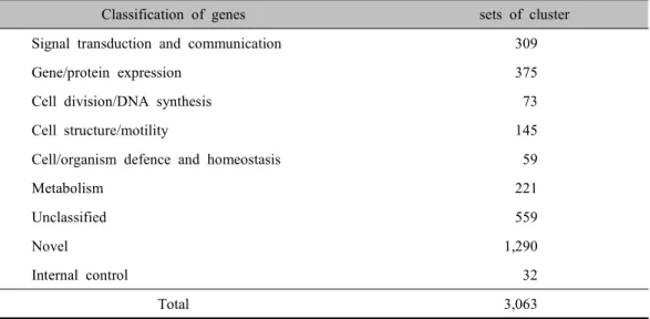
Classification of genes sets of cluster
Signal transduction and communication 309
Gene/protein expression 375
Cell division/DNA synthesis 73
Cell structure/motility 145
Cell/organism defence and homeostasis 59
Metabolism 221
Unclassified 559
Novel 1,290
Internal control 32
Total 3,063
Table 1. Classification of 3063 mesenchymal cell derived genes dipping in water and in isopropanol, and
dried by using centrifuge. The prepared probes was denatrured by heating at 95-10 0℃ for 2 minutes and added onto a array with cover slide. The hybridization was done in a CMT-Hybridization chamber (Corning) for 20 hours in a 50℃ waterbath. Arrays were washed for 5 minutes at room temper- ature in low stringency wash buffer (0.1×
SSC/0.1% SSC), and then twice for 5 mi- nutes in high stringency wash buffer (0.1×
SSC) and dried by using centrifuge.
6) Scanning and Image analysis Fluorescence intensities at immobilized targets were measured by using scanarray 4000 with a laser confocal micorscope (GSI Lumonics, USA). The two fluorescent im- ages (Cy3 and Cy5) were scanned separately from confocal microscope, and color images were formed by arbitrarily assigning differ- entiated cell intensity values into the red channel and control intensity into the green
channel and data were analyze by using Quantarray software (version 2.0.1, GSI Lumonics). Results were also analyzed by normalization between the images to adjust for the different efficiencies in labeling and detection with the two different fluors. This was achieved by matching of the detection sensitivities to bring a set of 32 internal control genes (β-actin and GAPDH) to nearly equal intensity.
For this analysis, we used a filter that in- cluded all gene exhibiting a minimum level of expression of intensify over 1,000 fluo- rescent units (on a scale of 0-65,535 fluo- rescent units) for both red and green chan- nels for each pair of experiments.
5. Northern blot analysis
Ten ㎍ of total RNAs were heated to 6 5℃ for 15 min in 50% formamide, 0.02%
formaldehyde, 40 mM MOPS (3-[-N-mor- pholino]propanesulfonic acid), 10 mM so-
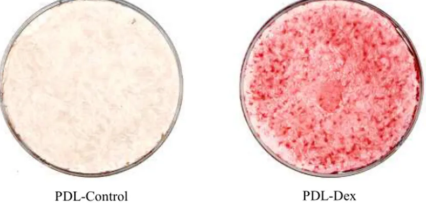
PDL-Control PDL-Dex
Figure 1. Alizarin red S staining of calcified matrix around PDL cells cultured for 21 days without (PDL-Control) and with mineralization supplements (50ug/ml ascorbic acid, 10mM β -glycerophosphate and 100nM dexamethasone) added to the culture medium(PDL-Dex).
Concentrations of each supplement and cell densities were described under Materials and Methods.
dium acetate, 1 mM EDTA, and 0.1 mg/ml ethodium bromide prior to gel electro- phoresis on 1% agarose, 55% formaldehyde, 40 mM MOPS, 10 mM sodium acetate, and 1 mM EDTA. The RNA was blotted onto a Zeta-Probe Blotting Membranes in 20×SSC.
The RNA was air-dried and then cross- linked by exposure to ultraviolet light. The probes were labelled with [α-P32]-dCTP by Megaprime DNA labelling system kit.
Prehybridization and hybridization were per- formed by using the Express Hyb solution.
After hybridization, the membrane was wash- ed in 2×SSC- 0.1% SDS at room temperature and then in 0.1×SSC /0.1% SDS at 55℃, and exposed to Agfa X-ray film at -70℃
with intensifying screens.
III. RESULTS
1. Morphology
Staining with alizarin red S distinguished
PDL cells cultured in the media with miner- alization supplements from those grown in the control media. The nodules of mineral- ized matrix were strongly stained with ali- zarin red S at day 21 (Figure 1). These data suggest that the media containing 50 μg/ml ascorbic acid, 10 mM β-glycerophosphate, and 100 nM dexamethasone are the essential supplementation for PDL cells to form ma- trix mineralization nodules, and that in- duction of these nodules in vitro requires about 3 weeks.
2. cDNA Microarray Analysis
We harvested PDL cells from each of the two different culture media on 21 day and isolated mRNAs for analysis on the cDNA microarray. After normalization of the re- sults, we selected genes that were up- or down-regulated on 21 day in cells cultured both in the media of the mineralization sup- plements and in the control media.
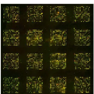
Figure 2. Image of microarray used to analyze gene expression level in PDL cells cultured with mineralization supplements and with 10% FBS. Total RNA was extracted from both cultures and each RNA sample was used as a template for synthesis of cDNA probes.
The probe for PDL cells cultured with 10%
FBS was labeled with Cy3(green) while the probe for PDL cells cultured with minera- lization supplements was labeled with Cy5 (red). The probes were mixed and hybridized to a microarray slide. The slides were scanned in a dual-laser scanning confocal microscope (GSI Lumonics,USA) and imageswere analyzed using a Quantarray software (Version 2.0.1, GSI Lumonics).
Green dots represented genes whose expression is decreased in PDL cells cultured with mineralization supplements while red dots presented genes whose expression is increased. Yellow dots indicate no change in gene expression.
Among 3,063 genes analyzed, 35 were up-regulated more than two-fold at one or more time points in cells that developed ma- trix mineralization nodules, and 38 were down-regulated to less than half their normal level of expression. The selected genes and their known or suspected functions are listed in Tables 2, 3, 4, 5, 6, 7, and 8.
In accord with the morphological change we observed, several genes related to cal- cium-related or mineral metabolism were in- duced in PDL cells during osteogenesis, such as insulin-like growth factor-II(IGF-II) and insulin-like growth factor-binding protein 2(IGFBP-2). Proteogycan 1, fibulin-5, keratin 5, β-actin, α-smooth muscle actin and cap- ping protein, and cytoskeleton and ex- tracellular matrix proteins were up-regulated during mineralization. Several genes encod- ing proteins related to apoptosis were ex- pressed in the PDL cells. Dickkopf-1
(Dkk-1) and Ninteen kD interacting protein-3 (Nip3, E1B 19K/Bcl-2-binding protein) were up-regulated, and Btf (Bcl-2 associated tran- scriptional factor) and Tax1 binding protein 1(TAX1BP1) were down-regulated during mineralization. Also periostin and S100 cal- cium-binding protein A4 were down-regu- lated during mineralization.
3. Verification of Expression of Selected Genes
To verify the microarray results, we per- formed Northern blots. Probes for each tested gene were PCR products amplified from plas- mid DNAs containing appropriate cDNAs.
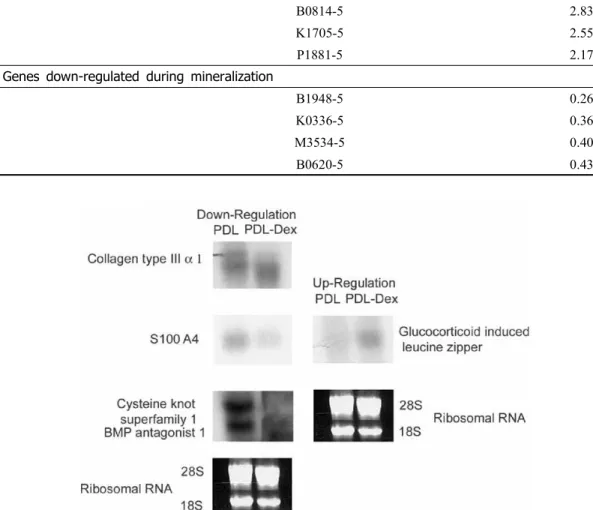
Among up-regulated genes, we selected gluco- corticoid-induced leucine zipper(GILZ) (Figure 3). From the list of down-regulated genes, we selected genes encoding collagen type III α1, S100A4, and cystein knot superfamily 1/BMP
Gen bank Name Ratio Genes up-regulated during mineralization
NM_012242 Dickkopf-1(hdkk-1)/Wnt antagonist 4.75
MAP 3.26
NM_000597 IGFBP 2 3.22
IGF-II 2.89
NM_018407 Putative integral membrane transporter(LC27) 2.20
NM_006367 Adenyl cyclase-associated protein(CAP) 2.14
NM_000227 Laminin-related protein(LamA3, epiligrin alpha 3 subunit) 2.11 Genes down-regulated during mineralization
NM_000598 IGFBP 3 0.15
OSF-1 0.16
NM_003299 Tumor rejection antigen (gp96) 1(TRA1) 0.22
NM_013372 Cysteine knot superfamily 1/BMP antagonist 1 0.27 NM_001257 H-cadherin(calcium dependent cell adhesion protein) 0.44 NM_006209 Ectonucleotide pyrophosphatase/phosphodiesterase 2(Autotayin) 0.44
NM_006475 OSF-2(Periostin) 0.45
NM_000484 Amyloid A4 protein 0.47
NM_021137 Tumor necrosis factor, alpha-induced protein 0.50
NM_019554 S100 calcium-binding protein A4 0.50
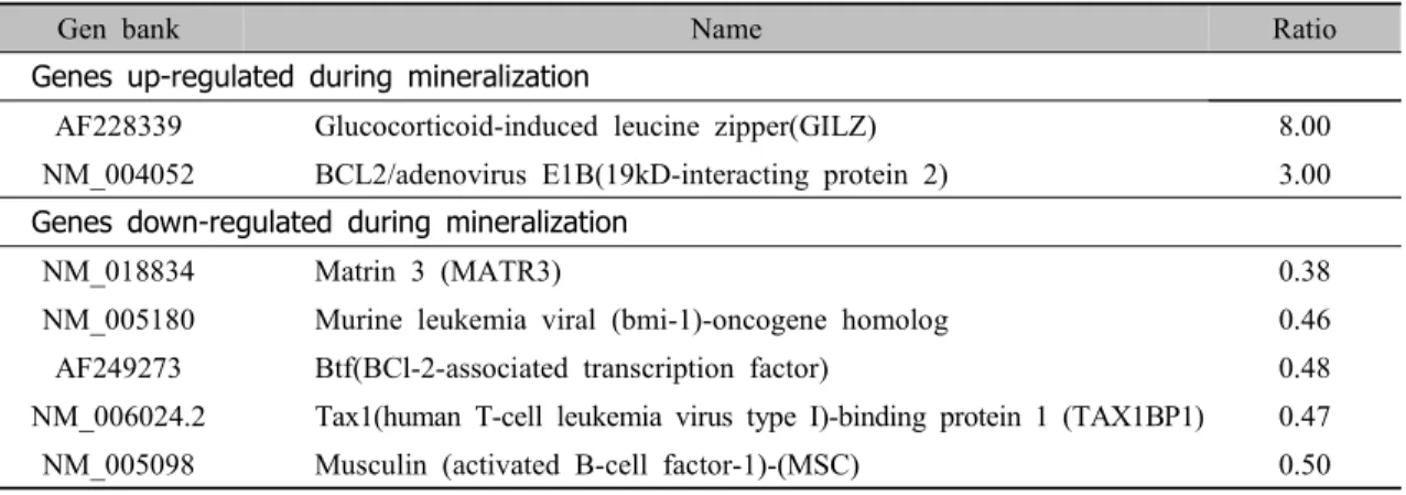
Table 3. Differently expressed genes related with gene and protein expression.
Gen bank Name Ratio
Genes up-regulated during mineralization
AF228339 Glucocorticoid-induced leucine zipper(GILZ) 8.00
NM_004052 BCL2/adenovirus E1B(19kD-interacting protein 2) 3.00 Genes down-regulated during mineralization
NM_018834 Matrin 3 (MATR3) 0.38
NM_005180 Murine leukemia viral (bmi-1)-oncogene homolog 0.46
AF249273 Btf(BCl-2-associated transcription factor) 0.48
NM_006024.2 Tax1(human T-cell leukemia virus type I)-binding protein 1 (TAX1BP1) 0.47
NM_005098 Musculin (activated B-cell factor-1)-(MSC) 0.50
Table 4. Differently expressed genes related with cell division and DNA synthesis.
Gen bank Name Ratio
Genes up-regulated during mineralization
NM_001753 Caveolin 1, caveolae protein, 22kD(CAV1) 2.40
NM_002106 Histone H2A.Z 2.18
Genes down-regulated during mineralization
NM_003589 Cullin 4A (CUL4A) 0.50
NM_001758 Cyclin D 1 (PRAD1: parathyroid adenomatosis 1) 0.50 Table 2. Differently expressed genes related with cell signal and communication
Gen bank Name Ratio Genes up-regulated during mineralization
NM_002727 Proteoglycan 1, secretory granule(PRG1) 2.48
NM_006329 Fibulin-5 2.27
Keratin 5(KRT5) 2.24
NM_001101 Actin, beta 2.16
NM_001613.1 Actin, alpha 2, smooth muscle, aorta(ACTA2) 2.09 NM_001747.1 Capping protein(actin filament),gelsolin-like 2.00 Genes down-regulated during mineralization
NM_000090.1 Collagen, type III, alpha 1 0.28
NM_004859.1 Clathrin, heavy polypeptide (Hc) (CLTC) 0.39
Table 6. Differently expressed genes related with metabolism.
Gen bank Name Ratio
Genes up-regulated during mineralization
NM-006793 Peroxiredoxin 3(PRDX3) 2.56
NM_020300 Glutathione S-transferase, microsomal 2.25
NM_001428 Enolase, alpha 2.23
NM_003945 ATPase, H+ transporting, lysosomal-(vacuolar proton pump) 2.12
NM_006169 Nicotinamide N-methyltransferase(NNMT) 2.11
NM_000291 Phosphoglycerate kinase 2.09
NM_003713 Phosphatidic acid phosphatase type 2b(PPAP2B) mRNA 2.00 Genes down-regulated during mineralization
NM_000918 Procollagen-proline, 2-oxoglutarate 4-dioxygenase 0.36
NM_024843 Hypothetical protein FLJ23462 0.38
NM_004786 Thioredoxin isolog 0.47
NM_005313 Glucose regulated protein, 58kD(thioredoxin) 0.50
Table 7. Differently expressed genes related with unclassified genes.
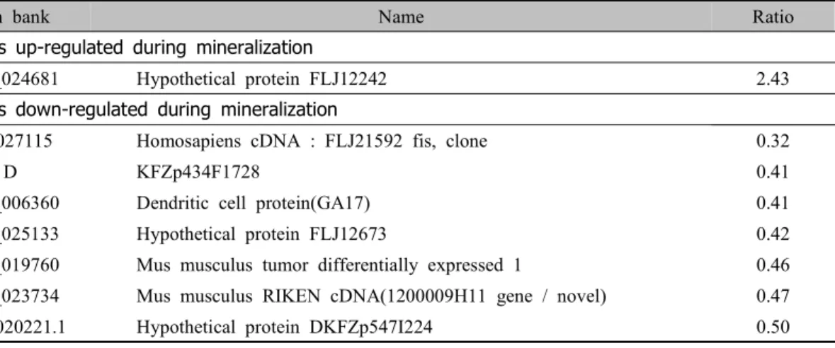
Gen bank Name Ratio
Genes up-regulated during mineralization
NM_024681 Hypothetical protein FLJ12242 2.43
Genes down-regulated during mineralization
AK027115 Homosapiens cDNA : FLJ21592 fis, clone 0.32
D KFZp434F1728 0.41
NM_006360 Dendritic cell protein(GA17) 0.41
NM_025133 Hypothetical protein FLJ12673 0.42
NM_019760 Mus musculus tumor differentially expressed 1 0.46 NM_023734 Mus musculus RIKEN cDNA(1200009H11 gene / novel) 0.47
NM_020221.1 Hypothetical protein DKFZp547I224 0.50
Table 5. Differently expressed genes related with cytoskeleton, adhesion and extracellular matrix.
Gen bank Name Ratio Genes up-regulated during mineralization
M0594-5 3.88
B0814-5 2.83
K1705-5 2.55
P1881-5 2.17
Genes down-regulated during mineralization
B1948-5 0.26
K0336-5 0.36
M3534-5 0.40
B0620-5 0.43
Table 8. Differently expressed genes related with novel genes.
Figure 3. Northern blot analysis of gene expression during mineralization of PDL cells. To verify microarray results on selected genes, we performed Northern blot analysis.
antagonist 1 to be verified(Figure 3). A ri- bosomal RNA (28S and 18S) was chosen to serve as a control(Figure 3). Our Northern blot results demonstrated identical expression pattern for all selected genes as revealed by microarray analysis. The results indicated that our microarray data were highly re- producible and reliable.
IV. DISCUSSION
To identify genes involved in the mineral- ization of extracellular matrix during the dif- ferentiation of PDL cells, we performed cDNA microarray analysis by using PDL cells that could be induced to differentiate into osteoblastic lineages16,17).
Among many modern techniques, one of
the most important tool in gene expression analysis is the development of DNA micro- array15,18). DNA microarrays provide a for- mat for the simultaneous measurement of the expression level of thousands of genes in a single hybridization asay. And it will be more efficient and resonable to have a tissue-specific DNA chip for specific area of research, as it already proved in a breast tis- sue DNA chip19), and a melanocyte-specific DNA chip20). We have generated a dermal papilla cDNA chip consisted of 3,063 cDNA clones isolated from human hairy mesen- chymal cell derived genes cDNA library.
Although this chip is not specific to discov- er mineralization-related genes, it is adequate to analysis the characterization of mesen- chymal-derived PDL cells.
In experiments using two different culture conditions, we clearly demonstrated that a me- dium containing mineralization supplements (50 μg/ml ascorbic acid, 10 mM β -glycerophosphate and 100 nM dexamethasone) could induce matrix mineralization, which was detected by virtue of strong staining with alizarin red S, but a medium without mineralization supplements could not (Figure 1).
Using acomputer analysis, we selected genes that showed an altered expression in the cells cultured in a medium with miner- alizationsupplements compared to in the cell cultured in the media with only 10% FBS . We identified 35 genes that were up-regu- lated more than two-fold and 38 that were down-regulated by more than 50% during the mineralization process. Among the se-
lected genes, several had already been noted for roles in osteogenesis. For example IGFBP2 binds prolong the half-life of the IGFs, inhibit or stimulate the growth pro- moting effects of the IGFs on cell culture, and alter the interaction of IGFs with their cell surface receptors21). IGF-II is a potent pleiotypic stimulator of both replication and differenciation of cells in the oteoblast line- age22) and increase proliferation and alkaline phosphatase activity in the human osteo- sarcoma cells23).
During the mineralization, S100 cal- cium-binding protein A4, a calcium-binding one with minerals in extracellular matrix, and OSF-2(periostin), a potential bone adhe- sion protein, were down-regulated. S100 cal- cium-binding proteins are small, acidic pro- teins which interact with target molecules and control various cellular processes such as cell cycle progression, differentiation, and cell metabolism after binding to Ca2+24). Interestingly Strutz et al25) have isolated one S100 calcium-binding protein known as S100A4 as a fibroblast-specific protein and demonstrated its high gene expression in the periodontal mesenchyme during early stages of tooth morphogenesis in an in situ hybrid- ization study. S100A4 might be a useful marker for distinguishing cells of the PDL from the gingiva26). S100A4 has an ex- tracellular role as a mineralization inhibitor, which is responsible for maintaining the pe- riodontal ligament space27). We found that the PDL cells culture medium with 10%
FBS at 21 day retained their characteristic
phenotypes during the culture period.
OSF-2(periostin)was originally identified as a secreted factor in a screen of a mouse os- teoblastic library, and is thought to function in bone adhesion, based on its sequence similarity to the insect adhesion protein fas- ciclin-128,29) because it is expressed in speci- alized connective tissues which form and support mineralized tissues30). Not all of the ALP positive cells in bone express OSF-2 (periostin), only those ALP positive cells in the periosteum. OSF-2(periostin)also appears to be secreted into the surrounding ex- tracellular matrix. Because strong expression of OSF-2(periostin)is observed in periosteal osteoblasts and not in the osteoblasts lining the trabecular bone where bone remodeling is actively taking place, it is assumed that OSF-2(periostin)is not directly but indirectly involoved in osteogenesis or in the process of bone formation or repair30). OSF-2(peri- ostin) expression in osteocytes was nearly undetectable, suggesting that osteoblasts cease the production of OSF-2(periostin) during the process of differentiation. A os- teocyte cell line, MLO-Y4, lacks detectable OSF-2(periostin) transcripts31), which further supports this observation. Osteocytes are likely derived by the terminal differentiation of osteoblasts which become trapped within the forming osteoid tissue during bone formation.
We identified the genes noted for roles in apoptosis during mineralization. Apoptosis during osteogenesis has not been well stud- ied but there are clear indications that it
must play an important role in the normal bone formation and remodeling. A major fraction of osteoblasts at bone remodeling sites cannot be accounted for and probably undergo apoptosis32)Dkk-1 and Nip3 were up-regulated, but Btf and TAX1BP1 were down-regulated during mineralization. Dkk-1 is a secreted protein that specially inhibits Wnt/β-catenin signaling by interacting with the co-receptor Lrp-633,34). Dkk-1 is origi- nally identified as a strong head inducer in Xenopus due to its potent anti-Wnt effect35). Dkk-1 promotes Bmp-triggered apoptosis and this might suggest that antagonism of Wnt/
β-catenin signals by Dkk-1 is necessary for Bmp-triggered apotosis36). Nip3 is an apopto- sis-inducing dimeric mitochondrial protein that can overcome Bcl-2 suppression37). Bcl-2 family members bear COOH-terminal transmembrane domains that allow their as- socition with the outer mitochondrial mem- brane38) and this mitochondrial localization is important for the suppressive function of Bcl-239,40). There is growing evidence that mitochondrial function is disturbed early in the apoptotic response and may be important in mediating apoptosis41-43). This is often seen as the loss of mitochondrial membrane potential41,42) and the release of cytochromec43), which has been implicated in the activation of caspases43-45). Bcl-2 can suppress the re- lease of cytochrome c from mitochondria and prevent caspase activation44,45). Btf is a novel death-promoting transcriptional repress- or that interacts with Bcl-2-related proteins and inhibits the apoptosis46). TAX1BP1 is a
novel substrate for caspase family members.
Thus TAX1BP1 appears to be a novel A20-binding protein which might mediate the anti-apoptotic activity of A20, and which can be processed by specific caspases47).
Our identification of a set of genes that may be associated with the mineralization of bone matrix provides important information toward a better understanding of the capacity to differentiate PDL cells into either ce- mentoblasts or osteoblasts, depending on needs and conditions as well as the molec- ular mechanism about the regeneration of periodontal tissues lost by the periodontal diseases.
V. SUMMARY
Periodontal ligament (PDL) cells have been known as multipotential cells, and as playing an important rolesin periodontal regeneration. The PDL cells are composed of heterogeneous cell populations which have the capacity to differentiate into either cementoblasts or osteoblasts, depending on needs and conditions. Therefore, PDL cells have the capacity to produce mineralized nod- ules in vitro in mineralization medium which include ascorbic acid, β-glycerophosphate and dexamethasone.
In spite of these well-known osteoblast like properties of PDL cells, very little is known about the molecules involved in the formation of the mineralized nodules in the PDL cells. In the present study, we analysed gene-expression profiles during the minerali-
zation process of cultured PDL cells by means of a cDNA microarray consisting of 3063 genes.
Nodules of mineralized matrix were strongly stained with alizarin red S on the PDL cells cultured in the media with miner- alization supplements.
Among 3,063 genes analyzed, 35 were up-regulated more than two-fold at one or more time points in cells that developed ma- trix mineralization nodules, and 38 were down-regulated to less than half their normal level of expression. In accord with the mor- phological change we observed, several genes related to calcium-related or mineral metabolism were induced in PDL cells dur- ing osteogenesis, such as IGF-II and IGFBP-2. Proteogycan 1, fibulin-5, keratin 5, β-actin, α-smooth muscle actin and capping protein, and cytoskeleton and extracellular matrix proteins were up-regulated during mineralization. Several genes encoding pro- teins related to apoptosis weredifferentially expressed in PDL cells cultured in the me- dium containing mineralization supplements.
Dkk-1 and Nip3, which are apoptosis-induc- ing agents, were up-regulated, and Btf and TAX1BP1, which have an anti-apoptosis ac- tivity, were down-regulated during minera- lization. Also periostin and S100 calcium- binding protein A4 were down-regulated dur- ing mineralization.
VI. REFERENCES
1. Ten Cate AR, Oral histology, 4th ed.,
Mosby, St. Louis, MO, pp. 1994;56-77 2. Hou LT, and Yaeger JA: Cloning and
characterization of human gingival and pe- riodontal ligament fibroblasts, J Periodontol 1993;64:1209-1218
3. Kuru L, Parkar MH, Griffiths GS, Newman HN, and Olsen I: Flow cy- tometry analysis of gingival and perio- dontal ligament cells, J Dent Res 1998;77:
555-564
4. Lackler KP, Cochran DL, Hoang AM, Takacs V, and Oates TW: Development of an in vitro wound healing model for periodontal cells, J Periodontol 2000;71:
226-237
5. Bowers GM, Granet M, Stevens M, Emerson J, Corio R, Mellonig J, Lewis SB, Peltzman B, Romberg E, and Risom L: Histological evaluation of new attach- ment in humans : A preliminary report, J periodontol 1985;56:381-396
6. Pontonero R, Lindhe JM, Nyman S, Karring T, Rosenberg E, and Sanavi F:
Guided tissue regeneration in degree II furcation involved mandibular molars : A clinical study, J Clin Periodontol 1988;15:
249-254
7. Carnes DL, Maeder CL, and Graves DT:
Cells with osteoblastic phenotypes can be explanted from human gingiva and perio- dontal ligament, J Periodontol 1997;68:
701-707
8. Brandsten C, Lundmark C, Christersson C, Hammarstrom L, and Wurtz T:
Expression of collagen alpha1(I) mRNA variants during tooth and bone formation in the rat, J Dent Res 1999;78:11-19 9. Chien HH, Lin WL, and Cho MI:
Expression of TGF-beta isoforms and their receptors during mineralized nodule formation by rat periodontal ligament cells in vitro, J Periodontal Res 1999;34:301-309
10. Beertsen W and Everts V: Formation of acellular root cementum in relation to dental and non-dental hard tissues in the rat, J Dent Res 1990;69:1669-1673 11. Somerman MJ, Archer SY, Imm GR, and
Foster RA: A comparative study of hu- man periodontal ligament cells and gin- gival fibroblasts in vitro, J Dent Res 1998;67:66-70
12. Yamaguchi M, Shimizu N, Shibata Y, and Abiko Y: Effects of different magni- tudes of tension-force on alkaline phos- phatase activity in periodontal ligament cells, J Dent Res 1996;75:889-894 13. Ramakrishnan PR, Lin WN, Sodek J, and
Cho M-I: Synthesis of non-collagenous extracelluar matrix proteins during devel- opment of mineralized nodules by rat pe- riodontal ligament cells in vitro, Calcif Tissue Int 1995;57:52-59
14. Dahl, LK: A simple and sensitive histo- chemical method for calcium, Proc. Soc.
Exp. Biol. Med. 1952;80:474-479
15. Schna M, Shalon D, Heller R, Chai A, Brown PO, and Davis RW: Parallel hu- man genome analysis: Microarray-based expression monitoring of 1000 genes, Proc. Natl Acad Sci USA 1996;93:10614 -10619
16. Bruder SP, Jaiswal N, and Haynesworth SE: Growth kinetics, self-renewal, and the osteogenic potential of purified human mesenchymal stem cells during extensive sub cultivation and following cry- opreservation, J Cell Biochem 1997;64:278- 294
17. Pittenger MF, Mackay AM, Beck SC, Jaiswal RK, Douglas R, Mosca JD, Moorman MA, Simonetti DW, Craig, S, and Marshak DR: Multilineage potential of adult human mesenchymal stem cells, Science 1999;284:143-147
18. Brown PO, and Bostein D: Exploring the new world of the genome with DNA mi- croarray, Nat Genet 1999;21:33-37 19. Perou CM, Jeffrey SS, Van de Rijn M,
Rees CA, Eisen MB, Ross DT, Pergamenschikou A, Wolliams CF, Zhu SX, Lee JC, Lashkari D, Shalon D, Brown Po, and Botstein D: Distinctive gene expression patterns in human mam- mary epithelial cell and breast cancers, Pro Natl Acad Sci 1999;96:9212-9217 20. Loftus SK, Chen Y, Gooden G, Ryan JF,
Birznieks G, Hilliard M, Baxevanis AD, Bittner M, Meltzer P, Trent J, and Pavan W: Informatic selection at a neural crest melanocyte cDNA set for microarray anal- ysis, Proc. Natl Acad Sci 1999;96:9277- 9288
21. Warg E, Warg J, Chin E, Zhan J, and Bondy LA: Cellular patterns of in- sulin-like growth factor system gene ex- pression in murine chondrogenesis and os- teogenesis, Endocrinology 1995;136:2741- 2751
22. Cahalis E, McCarthy R, and Centrella M:
Growth factors & cytokines in bone cell metabolism, Hnnu Rev Med 1991;42:17-24 23. Knutsen R, Honda Y, Strong DD, Sumpath TK, Boylink DJ, and Mohan S:
Regulation of insulin-like growth factor system components by osteogenic pro- tein-1 in human bone cells, Endocrinology 1995;136:857-865
24. Schafer BW, and Heizmann CW: The s100 family of EF-hand calcium-binding proteins :functions and pathology(review), Trends Biochem Sci 1996;21:134-140 25. Stratz F, Kada H, Lo CW, Danoff T,
Carare RL, and Tomaszewsk JE :Identification and characterization of a fi- broblast marker:FSP1, J Cell Biol 1995;
130:393-405
26. Duarte WR, Limura T, Takenaga K, Ohya K, and Ishikawa I: cDNA cloning of S100 calcium-binding proteins from bo- vine periodontal ligament and their ex- pression in oral tisues, J Dent Res 1998;77:1694-1699
27. Durate WR, Limura T, Takenaga K, Ohya K, Ishikawa I, and Kasugai S:
Extracellular role of S100A4 cal- cium-binding protein in the periodontal ligament. Biochem. Biophys Res Commun 1999;255:416-420
28. Takeshita S, Kikum R, Tezuka K, and Amann E: Osteoblast-specific factor 2:
Cloning of a putative bone adhesion pro- tein with homology with the insect protein fasciclin ?, Biochem J 1993;27:278-294 29. Sugiura T, Takamatsu H, Kudo A, and
Amann E: Expression and characterization of murine osteoblast-specific factor2 (OSF-2) in a baculovirus expression sys- tem, Protein Expr Purif 1995;6:305-311 30. Horiuchi K, Amizuka N, Takeshita S,
Takamatsu H, Katsuura M, Ozawa H, Toyama Y, Bonewald LF, and Kudo A:
Identification and characterization of a novel protein, periostin with restricted ex- pression to periosteum and periodontal ligament and increased expression by transforming growth factor beta, J Bone Miner Res 1993;14:1239-1249
31. Kato Y, Windle JJ, Koop BA, Mundy GR, and Bonewald LF: Establishment of an osteocyte-like cell line, MLO-Y4, J Bone Miner Res 1997;12:2014-2033 32. Tilka R, Weinstein RS, Bellido T, Parfitt
AM, and Manolagas S: Osteoblast pro- grammed cell death(Apoptosis):Modulation by growth factors and cytokines, J Bone Miner Res 1998;13:793-802
33. Bao B, Wu W, Li Y Stannek P, Glinka A, and Niehrs C: LDL-receptor-related
protein 6 is a receptor for Dickkopf pro- teins, Nature 2001;411:321-325
34. Zorn AM: Wnt signaling: antagonistic Dikkopfs, Curr Biol 2001;11:R592-R595 35. Glinka A, Wu W, Delius H, Monaghan
AP, Blumenstock C, and Niehr C:
Dickkopf-1 is a member of a new family of secreted proteins and functions in head induction, Nature 1998;391:357-362 36. Grotewold L and Ruther U: The Wnt an-
tagonist Dickkopf-1 is regulated by BMP signaling and c-Jun and modulates pro- grammed cell death, EMBO J 2002;21:
966-975
37. Chen G, Ray R, Doubit D, Shi L, Cizeau J, Bleackly C, Saxena S, Gietz RD, and Greenberg AH: The E1B 19k/Bcl-2-bind- ing protein Nip3 is a dimeric mitochon- drial protein that activates apoptosis, J Exp Med 1997;12:1975-1983
38. Krajewski S, Tanaka S, Takayama S, Schibler MJ, Fenton W, and Reed JC :Investigation of the subcellular dis- tribution of the Bcl-2 oncoprotein : Residence in the nuclear envelope, endo- plasmic reticulum, and outer mitochondrial membranes, Cancer Res 1993;53:4701-4714 39. Tanaka S, Saito K, and Reed JC:
Structure-function analysis of the Bcl-2 oncoprotein. Addition of a heterologous transmembrane domain to portions of the Bcl-2 beta protein restores function as a regulator of cell survival, J Biol Chem 1993;268:10920-10926
40. Zhu W, Cowie A, Wasfy GW, Penn LZ, Leber B, and DA Andrews: Bcl-2 mutants with restricted subcellular location reveal spatially distinct pathways for apoptosis in different cell types, EMBO J 1996;15:
4130-4141
41. Zamzami N, Susin SA, Marchetti P, Hirsch T, Gomez-Monterrey I, Castedo M, and Kroemer G: Mitochondrial coordi- nating control of nuclear apoptosis, J Exp Med 1996;183:1533-1544
42. Marchetti P, Castedo M, Susin SA, Zamzami N, Hirsch T, Macho A, Haeffiner A, Hirsch F, Geuskens M, and Kroemer G: Mitochondrial permeability transition is a central coordinating event of apoptosis, J Exp Med 1996;184:1155- 1160
43. Liu X, Kim C, Yang J, Jemmerson R, and Wang X: Induction of apoptotic pro- gram in cell-free extracts: Requirement for dATP and cytochrome c, Cell 1996;86:
147-157
44. Yang J, Liu XS, Bhalla K, Kim CN, Ibrado AM, Cai JY, Peng TI, Jones DP, and Wang XD: Prevention of apoptosis by Bcl-2: Release of cytochrome c from mitochondria blocked, Science 1997;275:
1129-1132
45. Kluck RM, Bossy-Wetzel E, Green DR, and Newmeyer DD : The release of cyto- chrome c from mitochondria: A primary site for Bcl-2 regulation of apoptosis, Science 1997;275:1132-1136
46. Kasof GM, Goyal L, and White E: Btf, a novel death-promoting Transcriptional re- pressor hat interacts with Bcl-2-related proteins, Mol Cell Biol 1999;19:4390-4404 47. Devalck D, Jim DY, Heyninck K, Van de Craen M, Contresras R, Fieb W, Jearg KT, and Beyaert R: The zinc finger pro- tein A20 interacts with a novel anti-apop- totic protein which is cleared by specific caspases, Oncogen 1999;18:4182-4190
-국문초록-
cDNA microarray에 의한 치주인대세포의 광물화 결절형성에 관여하는 유전자들의 분석
신재희1, 박진우1, 여신일1, 노우창1, 김문규2, 김정철2, 서조영1
1. 경북대학교 치의학 전문대학원 치주과학교실 2. 경북대학교 의학 전문대학원 면역학교실
치주인대세포는 시험관적 실험에서 광물화 결절형성을 유도할 수 있으므로 광물화 결 절형성에 관여하는 유전자들을 특이하게 발현할 것으로 여겨진다. 이에 본 실험은 cDNA microarray를 이용한 동시 유전자분석을 시행하여 치주인대세포의 분화에 의한 광물화 결 절형성시 나타나는 유전자의 특징적 발현 양상을 알아보고자 하였다.
교정치료를 목적으로 경북대학교병원에 내원한 환자의 제일소구치를 발치하여 통상적
방법으로 치주인대세포를 분리, 배양하였고, 3세대의 치주인대세포를 사용하여 실험을 시
행하였다. 치주인대세포를 100 mm 배양접시에 넣고 배양하여 매 2일 마다 배지를 교환해 주고, 10% FBS 만을 투여한 군을 대조군으로, ascorbic acid (50 ㎍/㎖), β-glycerophosphate (10 mM) 및 100 nM dexamethasone을 투여한 군을 실험군으로 하였다.
배양된 치주인대세포에 ascorbic acid, β-glycerophosphate, 그리고 dexamethasone을 투여 한 실험군에서 21일째 광물화된 결정을 관찰할 수 있었으나 대조군에서는 관찰할 수 없 었다.
3063개의 유전자를 분석한 결과 35개 유전자가 대조군에 비해 2배이상 발현이 증가하 였고, 38개 유전자는 2배이상 발현이 감소하였다. 형태학적 검사에서 보여준 바와 같이 광물화 형성과정시 관여하는 IGF-II과 IGFBP2와 같은 유전자가 실험군에서 증가하였으 며, 세포골격과 세포외기질 형성에 관여하는 proteogycan 1, fibulin-5, keratin 5, β-actin, α -smooth muscle actin, capping protein 등도 발현이 실험군에서 증가하였다. 한편 periostin and S100 calcium-binding protein A4는 대조군에서 오히려 높게 나타나므로 이는 배양된 치주인대세포가 그 자체의 표현형을 유지하고 있음을 보여 주고 있다. 그 외 apoptosis를 유발시키는데 관여하는 Dkk-1와 Nip3는 실험군에서 높게 발현되었고, apoptosis를 억제시
키는데 관여하는 Btf와 TAX1BP1는 오히려 낮게 발현됨을 알 수 있으므로 이는 실험군에
서 치주인대세포가 골아세포로의 분화되었음을 나타낸다.2)
Key worlds: Periodontal ligament cells, cDNA Microarray, Mineralization, Apoptosis