대한치주과학회지 : Vol. 36, No. 4, 2006
리포터유전자를 이용한 조골세포 분화정도에 관한 연구
김경화
1,2, 박윤정
3, 이용무
1, 한중석
4, 이동수
5, 이승진
6, 정종평
1, 설양조
1,*1. 서울대학교 치과대학 치주과학교실, 2. 서울대학교 BK21 치의학생명과학사업단, 3. 서울대학교 치과대학 두개악안면재건과학교실, 4. 서울대학교 치과대학 보철학교실, 5. 서울대학교 의과대학 핵의학과, 6. 이화여자대학교 약학대학 물리약학교실
I. 서론
조골세포의 분화에 대한 과정은 아직 완벽하게 밝 혀지지는 않았지만 ALPase, 교원질, proteoglycan, bone sialoprotein, osteopontin, ostecalcin 같은 세포외기질을 발현시키는 유전자의 순차적인 활성 과, 비타민 D3 와 parathormone receptor 같은 호 르몬 수용체들에 의해서 분화가 표지될 수 있다고 알려져 있다1). 이러한 단백질들은 세포의 분화에 따 라 조골세포에서 발현되기도 하지만 다른 종류의 세 포에서 관찰되기도 하므로 조직특이성이 부족하다고 할 수 있다. 그러나 osteocalin의 경우 분화된 조골 세포나 odontoblast에서만 높게 발현되는 것으로 밝 혀져 골에 관한 연구 시 매우 적절한 조직특이적인 분화 표지자가 될 수 있다2). Osteocalcin은 46-50 개의 아미노산으로 이루어진 4.9 kD의 비교원성단 백질로서 골의 세포외기질(ECM)의 약 10%를 차지한 다. Osteocacin은 세포들이 석회화가 시작되면서 나 타나며, 골의 세포외기질 안에 축적되거나3) 새로 합 성된 osteocalcin의 일부가 혈중으로 방출되기도 한
다. 이러한 특성을 이용하여 ostoecalcin의 혈중 농도에 의해 골형성의 정도를 예측하기도 한다4). Osteocalcin 은 칼슘이나 수산화인회석 (hydroxyapatite)와 결합하 는 γ-carboxyglutamic acid (Gla)를 포함하고 있기 때문에 bone gla protein (BGP)라고 불려지기도 한 다. 다른 종류의 세포들의 세포외기질에서는 관찰되 지 않기 때문에 조골세포 특이 유전자의 발현을 조 절하는 메커니즘에 관한 연구에서 많이 이용되고 있 다5). 마우스의 조골세포와 odontoblast에서 osteo- clain을 발현시키는 유전자는 두 개이며, osteo- calcin gene 1 (OG1) 과 osteocalcin gene 2 (OG2) 로 알려져 있다. 이들은 거의 같은 단백질을 코딩하 면서 같은 발현양상을 보여주고, 또한 그들의 pro- moter 부위가 93%정도가 일치한다6). OG2 pro- moter에 관한 연구에서 160bp 크기의 promoter 부 위가 조골세포에 필수적이면서 조골세포에서만 특이 하게 발현이 되도록 조절할 수 있는 곳으로 알려져 있다1).1)
Tissue engineering을 통한 골조직 재생에 관한 연구에서 종종 골형성이 가능한 세포들을 생체재료
본 연구는 과학기술부 특정연구개발사업 (M10528010004-06N2801-00410)의 지원에 의하여 이루어진 것임
* 교신저자 : 설양조, 서울시 종로구 연건동 28 서울대학교 치과대학 치주과학교실, 110-748 (전자우편 : [email protected])
에 접종한 후 접종된 세포의 성장 및 분화를 관찰하 거나 체내에 이식한 후 조직 형성을 관찰한다. 세포 의 성장 및 생존은 세포의 상태 및 재료의 bio- compatibility에 따라 결정되어지기 때문에 오랜 시 간 동안의 관찰이 필요하며 연구의 편이를 위해 간 편하고 경제적이면서 객관적인 데이터 분석을 할 수 있는 연구방법이 필요하다. 최근에는 이러한 목적을 위하여 luciferase나 green fluorescence protein (GFP) 같은 리포터단백질에서 발생되는 bio- luminescence를 이용하는 방법들이 많이 연구되고 있다. luciferase는 bioluminescence를 발생시킬 수 있는 대표적인 효소로 ATP와 반응하여 그것의 기질 인 D-luciferin을 oxoluciferin으로 전환될 수 있게 촉매작용을 하면서 빛을 발생시킨다. 이 종류의 빛 들은 생체 내부의 장기들에서 발광이 되어도 조직을 투과할 수 있기 때문에 체외에서도 검출이 가능하다
7,8). 이런 점을 이용하여 luciferase를 발현시키는 유전자를 체내에 위치시킨 후 리포터단백질이 발현 되는 곳의 위치, 발현정도, 발현기간 등을 동물들을 희생시키지 않고도 외부에서 관찰이 가능하게 만들 수 있다9). 암세포를 luciferase 발현 유전자로 형질 전환시킨 후 동물에 이식하여 암을 유발시킨 뒤 이 세포들로 인해 암이 발생한 부위에 치료제를 주사하 여 암세포의 사멸을 관찰하는 방법은 이미 암치료제 를 연구하는 곳에서 널리 사용되고 있다10). 특정유전 자가 특정 조직에서만 발현되는 점이나 시간특이적 인 발현을 한다는 점을 이용하면 골조직의 생성과정 을 추적해 낼 수 있을 것이며 특히 리포터단백질로 luciferase를 이용하면 생체내부에서의 발현을 동물 의 희생 없이 외부에서 관찰할 수 있을 것이다.
본 연구의 목적은 골모세포에서만 발현되는 os- teocalcin 유전자의 promoter를 luciferase 유전자 의 promoter로 사용하여 골모세포에서만 luciferase 가 발현되도록 하여 골모세포의 분화 정도를 관찰할 수 있도록 하는 것이다.
II. 연구재료 및 방법
1. 재조합 DNA 제조
Luciferase발현을 위한 promoter는 osteocalcin promoter 중 OG2의 proximal promoter 상의 -147 과 -34에 위치한 곳을 사용하였다. 이 곳은 osteo- calcin이 골세포에서 특이하게 발현되도록 조절하는 데 필수적인 promoter region으로 알려져 있다1). 마우스의 세포에서 DNA를 분리하여 마우스의 OG2 promoter (mOGP)의 PCR을 위한 주형으로 사용하 였다. 제한효소인 Kpn I (NEB, USA) 및 Hind III (NEB, USA)의 인식 사이트가 부착된, 서열에 적합 한 primer를 제작하여 PCR을 수행하여 mOGP promoter 서열을 증폭시켰다. 증폭된 mOGP 서열을 Gel extraction kit (QIAGEN, USA)를 사용하여 분 리정제하였다. mOGP 서열을 Kpn I 및 Hind III로 자른 후 promoter가 없는 pGL4 plasmid vector (Promega, USA)에 삽입하여 pGL4/mOGP를 제작 하였다. 재조합 된 DNA를 대장균에 transduction 시킨 후 배양하여 재조합 DNA를 증폭시켰다. 재조 합 DNA의 서열을 분석하여 mOGP의 서열이 plas- mid DNA에 존재하는 지 유무와 방향을 확인하였다.
2. Luciferase 발현 평가
mOGP의 promoter로서의 기능 및 발현양상을 확 인하기 위하여 마우스의 조골세포주인 MC3T3-E1세 포에 ExGen 500 (Fermentas, USA)를 이용하여 pGL4 plasmid vector, pGL4/mOGP 를 전이시켰다.
어떠한 vector도 전이되지 않은 세포를 대조군으로 하였다. 형질전환된 세포들을 1% 항생제, 15% fetal bovine serum (FBS), 5×10⁻⁷ M Dexamethason, 0.5% ascorbic acid, 10uM ß-glycerolphosphate 가 첨가된 Dulbecco’s modified Eagle’s medium (DMEM), 즉 분화유도용 배지 또는 1% 항생제, 10%
FBS만 첨가된 DMEM, 즉 세포성장용 배지에서 2주 동안 배양하였다. Luciferase activity 정량을 위하
Cells cultured in mineralization medium
Cells cultured in growth medium
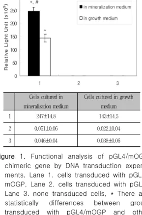
1 247±14.8 143±14.5
2 0.051±0.06 0.022±0.04
3 0.046±0.04 0.038±0.06
Figure 1. Functional analysis of pGL4/mOGP chimeric gene by DNA transduction experi- ments, Lane 1. cells transduced with pGL4/
mOGP, Lane 2. cells transduced with pGL4, Lane 3. none transduced cells, * There are statistically differences between group transduced with pGL4/mOGP and other groups(p<0.05). # There is statistically dif- ference between cells cultured in minerali- zation medium and in growth medium (p<0.05). Error bars represent standard deviation. Values represent the mean±SD of three samples.
여 2주일 후에 배지를 제거하고 luciferase reporter assay kit (Promega, USA)를 이용하여 luciferase activity를 luminometer (BMG LABTECH, Germany)와 cooled charged coupled device(CCCD) camera (Fusifilm, Japan)에서 확인하였다. 세포에 서의 발현양상을 확인하기 위하여 형질전환 2주 후 에 luciferin (Promega, USA)을 첨가하고 Confocal laser scanning microscope (CLSM)(Carl Zeiss, Germany)으로 관찰하였다.
3. Osteocalcin 생성 평가
Osteocalcin의 발현과 pGL4/mOGP와의 상관성을 확인하기 위하여 MC3T3-E1세포에 pGL4/mOGP vector를 전이시켰다. 다음날에 100 ng/ml 농도의 rhBMP-2 (R&D systems, USA)를 첨가하고 1% 항 생제, 10% FBS가 첨가된 DMEM에서 배양하였다. 1, 3, 7, 14일 후 배양액을 모아서 Mouse osteocalcin ELISA kit (BTI, USA)를 이용하여 세포에서 분비된 osteocalcin의 양을 측정하였다. 남아있는 세포들은 cell lysis buffer로 용해 시킨 후 luciferase re- porter assay kit를 이용하여 luciferase activity를 확인하였다.
4. Ti disc에 이식된 세포에서의 luciferase 발현 평가
pGL4/mOGP에 의해 발현된 luciferase가 생체재 료에 이식된 세포의 분화를 시각적으로 보여줄 수 있는 지를 확인하기 위하여 다음과 같이 시행하였다.
직경 5 mm의 Ti disc를 96well plate에 넣은 후 MC3TC-E1세포를 1×10⁴의 수로 접종하고 다음 날 pGL4/mOGP를 세포에 전이시켰다. 다음 날에 100 ng/ml 농도의 rhBMP-2를 첨가하고 1% 항생제, 10% FBS가 첨가된 DMEM에서 배양하였다. 1일, 3 일 뒤 각 well에 cell lysis buffer를 첨가하여 세포 를 용해 시킨 후 luciferase 활성도를 luminometer 및 CCCD camera 에서 확인하였다.
5. 통계학적 분석
모든 실험은 3회 실험하였으며, 실험에서 얻어진 수치의 통계학적 분석을 위하여 one-way ANOVA 와 사후 검정에 Duncan 법을 시행하였다. α=0.05 에서 통계학적 유의성을 확인하였다.
III. 결과
1. Luciferase 유전자 발현 평가
제작된 pGL4/mOGP 유전자가 본 연구의 목적에 맞게 발현되는 지를 확인하기 위하여 조골세포에 유
Figure 2. Images of MC3TC-E1 cells at 14days after transduction with pGL4/mOGP by CLSM, (a)none transduction cells, (b)cells transduced with pGL4/mOGP, (c)cells transduced with pGL4
Figure 3. Images of MC3TC-E1 cells at 14days after transduction with pGL4/mOGP by CCCD camera, (a)none transduced cells, (b)cells transduced with pGL4/mOGP, (c)cells transduced with pGL4, (d)pseudo- color reference represents light intensity (blue least intense and red most intense), A: cells cultured in growth medium, B:
cells culutured in mineralization medium
Figure 4. Production of osteocalcin from cells and luciferase activity of cells, (a)production of osteocalcin from cell at 1, 3, 7 and 14days after transduction with pGL4/mOGP, (b)lucifer- ase activity of cells at 1, 3, 7 and 14days after transduction with pGL4/mOGP. Error bars represent standard deviation. Values represent the mean±SD of three samples. * There is statistically difference between BMP treated and untreated cells (p<0.05)
Figure 5. Images of MC3TC-E1 cells on Ti disc at 1 and 3days after transducing with pGL4/mOGP, (a)cells on Ti disc at 1day after tranduction with pGL4/mOGP, (b)cells on Ti disc at 3days after trans- duction with pGL4/mOGP, (c)pseudo- color reference represents light intensity (blue least intense and red most in- tense), A: none transduced cells, B:
cells transduced with pGL4/mOGP
전자를 전이시킨 후 luciferase activity를 측정하였 다. 조골모세포인 MC3T3-E1세포에 유전자를 전이 시키고 각각 일반배지와 분화유도용 배지에서 14일 동안 배양한 결과 pGL4에 의해 형질전환된 세포나 대조군에 비해 pGL4/mOGP에 의해 형질전환된 세 포들에서 luciferase의 활성이 통계학적으로 유의성 있는 차이를 보이며 높게 나타났다. 또한 pGL4/
mOGP에 의해 형질전환된 세포들 중 분화유도용 배 지에서 배양된 세포에서의 luciferase의 활성이 일반 배지에서 배양된 세포에서 보다 유의성 있게 높게 나타났다(figure 1).
또한 형질 전환된 세포들을 CLSM에서 관찰한 결과 pGL4/mOGP가 전이된 세포에서만 형광에 의한 형태를 관찰할 수 있었으며 다른 군들에서는 형광에 의한 세포 들의 모습은 관찰할 수 없었다 (figure 2). CCCD cam- era에서 luminescence를 관찰한 결과 pGL4/mOGP에 의해서 형질 전환된 모든 세포에서 luminescence를 관 찰할 수 있었으며, 일반배지에서 성장한 세포들에서 보 다 분화유도용 배지에서 성장한 세포들에서 lumines- cence의 강도가 더 높게 나타났다(figure 3).
2. Osteocalcin 생성 평가
Osteocalcin의 생성과 osteocalcin promoter에 의한 luciferase의 발현과의 관련성을 평가하기 위하 여 세포들을 pGL4/mOGP로 형질전환시킨 후 rhBMP-2를 첨가하여 골세포로의 분화를 유도하고, 세포에서 분비된 osteocalcin의 양과 luciferase의 활성을 관찰하였다. 시간이 지남에 따라 osteocalcin 의 생성은 점차 증가되었으며, 7일째에 rhBMP-2 처리군과 처리되지 않은 군 간의 osteocalcin의 생 성양이 차이가 나기 시작했으며, 14일째에 두 군간 의 통계학적으로 유의성 있는 차이를 나타내었다 (figure 4, a). 이에 비해 luciferase의 활성은 3일 부터 7일까지 두 군간의 차이가 통계학적으로 유의 성 있게 나타났으며, 7일째부터는 활성이 3일 째에 비해 두 군 모두에서 감소하는 양상을 보여주었다 (figure 4, b). 그러나 rhBMP-2 처리군과 처리되지
않은 군간의 활성양의 차이는 계속 나타났다.
3. Ti disc 에서 luciferase 발현 평가
생체재료에서의 세포의 분화를 실시간으로 관찰할 수 있는 지 확인하기 위하여 세포를 Ti disc에 접종 한 후 pGL4/mOGP로 형질전환시켰다. 1일과 3일 후에 luminescence를 CCCD camera에서 관찰한 결 과 1일과 3일 모두에서 luminescence를 관찰할 수 있었다. 1일 보다는 3일째에 luminescence의 강도 가 더 높게 나타났으며, 3일째에 luminescence의 강도가 모든 곳에서 고르게 나타나지 않고 분화된 양상에 따라서 다르게 나타났다(figure 5).
IV. 고안
조골세포에서 골생성까지의 기전은 재료 자체로의 접근방법이 용이하지 않기 때문에 아직까지 완전하 게 알려져 있지 않고, 일부만이 알려져 있다. 현재까 지 연구에서는 ALPase, 교원질, proteoglycan, bone sialoprotein, osteopontin, ostecalcin 등이 골세포로의 분화 과정 중에 나타나는 중요한 표지인 자들로 알려져 있다11). ALPase나 osteopontin 같은 경우 골에서뿐만 아니라 콩팥12), 태반12), 유선13) 등 에서도 발현되는 단백질들로 조직특이성이 부족하 다. 그러나 osteocalcin은 분화된 조골세포나 odon- toblast에서만 높게 발현되는 조직특이성을 가지고 있어 조골세포의 분화를 잘 나타낼 수 있는 표지인 자라고 할 수 있으며 이러한 osteocalcin의 전사를 조절하는 promoter작동을 관찰함으로써 세포의 분 화를 관찰할 수 있을 것이다14). 이 연구에서는 표지 인자중의 하나인 osteocalcin의 발현을 조절하는 promoter의 일정부위를 선택하여 리포터유전자로 잘 알려져 있는 luciferase의 발현을 조절하도록 하 여 골세포로의 분화를 간편하게 단시간 내에 실시간 으로 관찰할 수 있는 방법을 개발하고자 하였다. 이 를 위해 promoter가 없는 플라스미드 벡터에 os- teocalcin promoter 중 조골세포에서만 관찰되는 특
정부위를 삽입한 후 promoter 조절에 의한 lucifer- ase의 활성을 관찰하였다. Luminometer와 CLSM에 의한 관찰에서 조골세포에 전이된 osteocalcin pro- moter 유전자는 luciferase를 정상적으로 발현시켰 으며, 일반 배지에서 보다 골분화유도 배지에서 lu- ciferase의 활성이 더 높게 나타났다(figure 1, 2).
CCCD camera는 적은 양의 photon도 검출할 수 있 는 장치로 생체내에서나 세포에서 luciferase나 GFP 같은 리포터 단백질들에서 방출되는 photon을 감지 함으로써 실시간으로 세포들의 반응이나 위치를 관 찰할 수 있다15). 본 연구에서도 CCCD camera에 의 해서 세포들에서 발현되고 있는 luciferase를 관찰할 수 있었으며, 골분화유도용배지에 의한 세포의 분화 정도에 따라 luminescence의 강도가 다르게 나타남 을 확인할 수 있었다(figure 3). 또한 간단히 세포에 assay 시약을 넣어줌으로써 실시간으로 간편하게 luminescence를 관찰할 수 있었다. 이 결과들은 osteocalcin promoter가 luciferase의 발현을 조절 할 수 있으며, 분화의 정도에 따라 luciferase의 발 현양이 조절됨을 보여줌으로써 osteocalcin pro- moter에 의한 리포터단백질의 발현조절이 조골세포 의 분화를 관찰할 수 있는 방법이 될 수 있음을 시 사한다고 할 수 있다. 이러한 결과는 인간의 osteo- calcin promoter의 전체적인 서열을 이용하여 골형 성과정을 추적한 연구에서도 보여지고 있다. 이 연구 에서도 인간의 osteocalcin promoter조절에 따른 lu- ciferase의 발현을 관찰함으로써 충분히 골형성 단계 를 확인할 수 있다고 보고 하고 있다16). Ostoecalcin promoter에 의한 luciferase의 발현과 실제적인 osteocalcin생성과의 관련성을 관찰하기 위하여 골 유도능이 우수한 BMP-2를 14일 동안 배지에 첨가 하여 골조직분화를 유도한 후 osteocalcin 생성 및 luciferase 활성도를 동시에 측정하였다. BMP-2에 의한 osteocalcin 생성은 분화초기 단계인 3일 까지 는 대조군과 별다른 차이가 없었으나 7일부터는 두 군간의 차이가 나타나기 시작했으며 14일에는 통계 학적으로 유의성 있는 차이가 나타났다(figure 4).
이에 비해 luciferase 활성은 3일째 부터 두 군간의
유의성 있는 차이를 보였으며, 14일까지 두 군간의 차이를 보여주었다. 다른 연구 결과에서도 이와 비 슷하게 osteocalcin promoter에 의한 luciferase 활 성이 세포의 분화에 따라 차이가 나타나기까지는 4 일 정도가 필요하며 골분화유도인자에 의해서 세포 들이 osteocalcin을 분비하기까지는 7일 정도의 시 간이 필요한 것으로 보고 하고 있다17,18). 이러한 결 과들에서와 마찬가지로 osteocalcin promoter에 의 한 luciferase의 활성을 3일 만에 관찰할 수 있는 것 처럼 좀 더 빠른 시간에 세포의 분화를 확인할 수 있다는 것은 다양한 골재생 관련 연구에서 이 방법 들이 좀 더 편리하고 경제적인 것이 될 수 있음을 의미한다. 한편, 두 군간의 luciferase의 발현량의 차이가 14일 까지 계속적으로 나타나는 반면 7일째 에 3일째보다 luciferase의 활성이 감소됐으며 14일 까지 계속 감소하는 경향을 보이는 결과는 플라스미 드 벡터를 세포에 전이시킨 후 바로 사용했기 때문에 나타나는 현상으로 보이며 stable cell line을 제조하 거나 형질전환마우스를 제조하여 형질 전환된 세포를 분리하면 문제를 해결할 수 있을 것으로 보인다.
골조직 재생에 관한 연구는 현재 활발하게 진행되 고 있는 분야로, 다양한 재료 및 방법들이 개발되고 있다. 이러한 연구들에서 줄기세포나 골모세포의 골 세포로의 분화는 중요한 목표이면서 도구가 된다.
골세포로의 분화를 확인하기 위해서는 다양한 표지 인자들이 연구되고 있으며, 관찰방법도 매우 다양하 다. 그러나 대부분의 방법들이 관찰결과를 얻기까지 시간과 비용이 많이 소요되고 연구방법을 시행하면 서 여러 가지 변수들이나 오차가 발생할 수도 있다.
본 연구에서도 생체재료에 대한 세포의 반응을 관찰 하기 위한 도구로 pGL4/mOGP를 이용할 수 있는 지의 여부를 확인하기 위하여 치과에서 많이 사용되 고 있는 Ti에 세포를 부착시키고, pGL4/mOGP를 전 이시킨 다음 CCCD camera에서 luciferase의 활성 을 관찰하였다. Ti disc에 부착된 세포에서 lucifer- ase의 활성은 CCCD camera에 의해서 실시간으로 관찰이 가능했으며, 1일 보다는 3일 째에 더 높은 활 성을 보였다. 또한 분화된 세포의 위치에 따라 활성
이 다르게 나타나는 것을 알 수 있었다(figure 5).
이 결과들은 pGL4/mOGP가 세포의 분화정도를 반 영해서 실시간으로 보여줄 수 있으며 세포가 분화되 는 위치를 시각적으로 알려줄 수 있는 도구가 될 가 능성을 보여준다고 할 수 있다. 또한 3차원적인 구 조의 생체재료에 대한 세포의 반응관찰에서 재료의 구조 때문에 쉽게 관찰하기 어려운 세포의 분화를 본 연구의 방법을 사용하면 좀더 용이하게 확인할 수 있을 것으로 보여진다.
V. 결론 및 요약
이 연구에서 pGL4/mOGP를 이용하여 세포의 분 화를 CCCD camera로 실시간 관찰할 수 있었으며, 매우 간편하고 빠르게 세포의 분화를 보여줄 수 있 는 도구로서의 가능성을 보여주었다. 또한 생체재료 에서 pGL4/mOGP에 의한 luciferase 활성을 CCCD camera로 관찰이 가능하다는 결과는 생체재료에서 의 세포분화를 용이하게 실시간으로 관찰할 수 있는 가능성을 보여주었다고 할 수 있으며, 이 방법으로 시간과 경비를 절약할 수 있을 것으로 보여진다. 그 러나 세포에서의 발현되는 기간이 짧아지는 문제점 을 보여주고 있어 장기간의 세포의 분화관찰에는 아 직까지 사용이 가능하지 않으며, 이러한 문제점은 stable cell line 이나 형질전환마우스를 제작하여 세포를 분리해서 사용하면 해결할 수 있을 것으로 생각된다. 또한 앞으로 리포터 단백질로서 GFP 같 은 다른 형광물질을 사용하여 좀 더 간편하게 세포 의 분화를 관찰할 수 있는 방법들이나19,20) 시기가 다 르게 발현되는 분화표지인자에 2 종류 이상의 리포터 단백질을 적용함으로써21) 분화를 시기 별로 관찰하는 방법 등에 대한 추가 연구가 필요할 것으로 보인다.
VI. 참고문헌
1. Ducy P, Karsenty G. Two distinct osteo- blast-specific cis-acting elements control expression of a mouse osteocalcin gene.
Mol Cell Biol. 1995;15:1858-1869.
2. Price PA. Role of vitamin-K-dependent proteins in bone metabolism. Annu Rev Nutr. 1988;8:565-583.
3. Gallop PM, Lian JB, Hauschka PV.
Carboxylated calcium-binding proteins and vitamin-k. N Engl J Med. 1980;302:1460-1466.
4. Lian JB, Gundberg CM. Osteocalcin - bio- chemical considerations and clinical- applications. Clin Orthop Relat Res. 1988;
226:267-291.
5. Frendo JL, Xiao GZ, Fuchs S, Franceschi RT, Karsenty G, Ducy P. Functional hier- archy between two OSE2 elements in the control of osteocalcin gene expression in vivo. J Biol Chem. 1998;273:30509-30516.
6. Desbois C, Hogue DA, Karsenty G. The mouse osteocalcin gene-cluster contains 3 genes with 2 separate spatial and temporal patterns of expression. J Biol Chem.
1994;269:1183-1190.
7. Honigman A, Zeira E, Ohana P, et al.
Imaging transgene expression in live animals. Mol Ther. 2001;4:239-249.
8. Wu JC, Sundaresan G, Iyer M, Gambhir SS.
Noninvasive optical imaging of firefly luci- ferase reporter gene expression in skeletal muscles of living mice. Mol Ther. 2001;4:
297-306.
9. de Boer J, van Blitterswijk C, Lowik C.
Bioluminescent imaging: emerging technol- ogy for non-invasive imaging of bone tis- sue engineering. Biomaterials. 2006;27:
1851-1858.
10. Hoffman RM. Real-time subcellular imag- ing in live animals: new visible targets for cancer drug discovery. IDrugs. 2006;9:632-635.
11. Aubin JE, Liu F, Malaval L, Gupta AK.
Osteoblast and chondroblast differentiation.
Bone. 1995;17(2 Suppl):77S-83S.
12. Nomura S, Wills AJ, Edwards DR, Heath JK, Hogan BLM. Developmental expression of ear (osteopontin) and sparc (osteonectin) rna as revealed by in-situ hybridization. J Cell Biol. 1988;106:441-450.
13. Rittling SR, Novick KE. Osteopontin ex- pression in mammary gland development and tumorigenesis. Cell Growth Differ.
1997;8:1061-1069.
14. Lian JB, Stein GS, Stein JL, van Wijnen AJ. Osteocalcin gene promoter: unlocking the secrets for regulation of osteoblast growth and differentiation. J Cell Biochem.
1998;30-31:62-72.
15. Zhang W, Feng JQ, Harris SE, Contag PR, Stevenson DK, Contag CH. Rapid in vivo functional analysis of transgenes in mice using whole body imaging of luciferase expression. Transgenic Res. 2001;10:423 -434.
16. Iris B, Zilberman Y, Zeira E, et al.
Molecular imaging of the skeleton: quanti- tative real-time bioluminescence monitor- ing gene expression in bone repair and development. J Bone Miner Res. 2003;18:
570-578.
17. Xiao G, Cui Y, Ducy P, Karsenty G, Franceschi RT. Ascorbic acid-dependent activation of the osteocalcin promoter in MC3T3-E1 preosteoblasts: requirement for collagen matrix synthesis and the presence of an intact OSE2 sequence. Mol Endocrinol 1997;11:1103-1113.
18. Clemens TL, Tang H, Maeda S, et al.
Analysis of osteocalcin expression in transgenic mice reveals a species difference in vitamin D regulation of mouse and hu- man osteocalcin genes. J Bone Miner Res.
1997;12:1570-1576.
19. Bilic-Curcic I, Kronenberg M, Jiang X, et al. Visualizing levels of osteoblast differ- entiation by a two-color promoter-GFP strategy: Type I collagen-GFPcyan and os- teocalcin-GFPtpz. Genesis. 2005;43:87-98.
20. Yang M, Baranov E, Moossa AR, Penman S, Hoffman RM. Visualizing gene expression by whole-body fluorescence imaging.
PNAS. 2000;97:12278-12282.
21. Yamamoto N, Jiang P, Yang M, et al.
Cellular dynamics visualized in live cells in vitro and in vivo by differential dual-color nuclear-cytoplasmic fluorescent-protein expression. Cancer Res. 2004;64:4251-4256.
- Abstract -
A study on the osteoblast differentiation using osteo- calcin gene promoter controlling luciferase expression
Kyoung-Hwa Kim
1,2, Yoon-Jeong Park
3, Yong-Moo Lee
1, Jung-Suk Han
4, Dong-Soo Lee
5, Seung-Jin Lee
6, Chong-Pyoung Chung
1, Yang-Jo Seol
11. Dept. of Periodontology, 2. BK21 Craniomaxillofacial life science, 3. Craniomaxillofacial Reconstructive Science,
4. Dept. of Prosthodontics, School of Dentistry,
5. Dept. of Nuclear Medicine, College of Medicine, Seoul National University, 6. College of Pharmacy, Ewha Womans University
The aim of this study is to monitor reporter gene expression under osteocalcin gene promoter, us- ing a real-time molecular imaging system, as tool to investigate osteoblast differentiation.
The promoter region of mouse osteocalcin gene 2 (mOG2), the best-characterized osteo- blast-specific gene, was inserted in promoterless luciferase reporter vector. Expression of reporter gene was confirmed and relationship between the reporter gene expression and osteoblastic differ- entiation was evaluated. Gene expression according to osteoblstic differentiation on biomaterials, utilizing a real-time molecular imaging system, was monitored. Luciferase was expressed at the only cells transduced with pGL4/mOGP and the level of expression was statistically higher at cells cul- tured in mineralization medium than cells in growth medium. CCCD camera detected the luciferase expression and was visible differentiation-dependent intensity of luminescence. The cells produced osteocalcin with time-dependent increment in BMP-2 treated cells and there was difference between BMP-2 treated cells and untreated cells at 14days. There was difference at the level of luciferase expression under pGL4/mOGP between BMP-2 treated cells and untreated cells at 3days. CCCD cam- era detected the luciferase expression at cells transduced with pGL4/mOGP on Ti disc and was visible differentiation-dependent intensity of luminescence
This study shows that 1) expression of luciferase is regulated by the mouse OC promoter, 2) the CCCD detection system is a reliable quantitative gene detection tool for the osteoblast differ- entiation, 3) the dynamics of mouse OC promoter regulation during osteoblast differentiation is ach- ieved in real time and quantitatively on biomaterial. The present system is a very reliable system for monitoring of osteoblast differentiation in real time and may be used for monitoring the effects of growth factors, drug, cytokines and biomaterials on osteoblast differentiation in animal.2)
Key words : osteocalcin promoter, OG2, bone differentiation, luciferase, osteoblast