• 검색 결과가 없습니다.

Suppressive Effect of Der p 2 on Constitutive Neutrophil Apoptosis by Cytokine Secretion of Normal and Allergic Lymphocytes

N/A
N/A
Protected

Academic year: 2021

Share "Suppressive Effect of Der p 2 on Constitutive Neutrophil Apoptosis by Cytokine Secretion of Normal and Allergic Lymphocytes"

Copied!
7
0
0

로드 중.... (전체 텍스트 보기)

전체 글

(1)

pISSN 1738-3544 eISSN 2288-1662

Suppressive Effect of Der p 2 on Constitutive Neutrophil Apoptosis by Cytokine Secretion of Normal and Allergic Lymphocytes

In Sik Kim 1,2 , Na Rae Lee 1 , and Ji-Sook Lee 3

1

Department of Biomedical Laboratory Science, School of Medicine, Eulji University, Daejeon 34824, Korea

2

Department of Senior Healthcare, BK21 Plus Program, Graduate School, Eulji University, Daejeon 34824, Korea

3

Department of Clinical Laboratory Science, Wonkwang Health Science University, Iksan 54538, Korea

정상인과 알레르기 환자의 림프구에서 Der p 2에 의한 사이토카인 분비가 호중구의 세포고사 억제에 미치는 효과

김인식 1,2 , 이나래 1 , 이지숙 3

1

을지대학교 대전캠퍼스 임상병리학과,

2

을지대학교 대전캠퍼스 BK21 플러스 프로그램 시니어 헬스케어학과,

3

원광보건대학교 임상병리과

Der p 2 is the major allergen of the house dust mite (HDM) associated with the development of allergic diseases. The pathogenic mechanism of the allergy is related to cytokine release of lymphocytes and constitutive apoptosis of neutrophils. In the present study, we examined whether Der p 2 induces cytokine release of lymphocytes, which is involved in regulation of neutrophil apoptosis. In normal and allergic subjects, Der p 2 enhanced the secretion of IL-6, IL-8, MCP-1, and GM-CSF in a time-dependent manner. Although Der p 2 was weakly effective against neutrophil apoptosis, conditioned media collected from normal and allergic lymphocytes after Der p 2 treatment inhibited the apoptosis of normal and allergic neutrophils. Der p 2 showed stronger inhibition of apoptosis of allergic neutrophils cocultured with allergic lymphocytes than normal neutrophils cocultured with normal lymphocytes. These findings improve our understanding of the role of Der p 2 in regulation of lymphocytes and neutrophils and will enable elucidation of allergy pathogenesis.

Keywords: Der p 2, Cytokine, Lymphocyte, Neutrophil apoptosis, Allergy

Corresponding author: Ji-Sook Lee Department of Clinical Laboratory Science, Wonkwang Health Science University, 501, Iksan-daero, Iksan 54538, Korea Tel: 82-63-840-1216 Fax: 82-63-840-1219 E-mail: [email protected]

This is an Open Access article distributed under the terms of the Creative Commons Attribution Non-Commercial License (http://creativecommons.org/licenses/by-nc/4.0) which permits unrestricted non-commercial use, distribution, and reproduction in any medium, provided the original work is properly cited.

Copyright © 2016 The Korean Society for Clinical Laboratory Science. All rights reserved.

Received: April 26, 2016 Revised: May 16, 2016 Accepted: May 30, 2016

Introduction

Allergic diseases including allergic rhinitis, asthma, and allergic dermatitis are caused by environmental, genetic, and immunological elements [1-4]. House dust mites (HDMs), which primarily consist of Dermatophagoides pteronissinus and Dermatophagoides farinae , are related to allergy

pathogenesis [1,5-7]. Der p 2, a group II allergen from Dermatophagoides pteronissinus , is the major allergen of HDM. Der p 2-specific immunoglobin E (IgE) is positively related to 87.8% of asthmatic subjects [8]. Der p 2 triggers an inflammatory process through TLR4-dependent signaling [9].

Der p 2 also induces IL-4 expression in T helper type 2 (Th2)

cells and activation of inflammasome in monocytes [10]. In

(2)

addition, allergic diseases, specifically asthma, are aggravated by inflammatory responses induced by inhibiting neutrophil apoptosis [11].

Lymphocytes mainly contain B cells and T cells, and release inflammatory cytokines associated with allergic diseases [12].

Overexpression of cytokines is caused by the pathogenesis of allergic diseases. The secretion of IL-6, IL-8/CXCL8, MCP-1/CCL2 and GM-CSF is involved in inhibition of neutrophil apoptosis, and these cytokines are induced by HDM in monocytes [13,14]. HDM induces the secretion of Th2 cytokines such as IL-4 and IL-5 in lymphocytes [15].

In this study, we studied the role of Der p 2 in cytokine production of normal and allergic lymphocytes, as well as constitutive neutrophil apoptosis of normal and allergic subjects due to cytokines secreted by Der p 2.

Materials and Methods

1. Normal and allergic subjects

Allergy patients (n=17), including allergic asthma and allergic rhinitis subjects, were recruited from Eulji University Hospital. Allergic patients had mild to severe symptoms of the disease. Allergic status was based on the presence of positive results of a skin prick test (≥2+), multiple allergen simul- taneous test (MAST) (≥class 2), or evaluation of specific HDM IgE using the Pharmacia Unicap 100 system for common allergens (≥100 IU/mL). The normal subjects (n=16) had no history of asthma or allergic rhinitis. This study was approved by the Institutional Review Board of Eulji University for normal volunteers and the Institutional Review Board of Eulji University Hospital for allergic patients (EU 14∼33). All participants in this study gave their written informed consent.

2. Isolation of lymphocytes and neutrophils and cell culture

Human lymphocytes and neutrophils were isolated from the peripheral blood of normal and allergic subjects using Ficoll-Hypaque (Amersham Phamacia biotechnology, Buc- kinghamshire, UK) gradient centrifugation. CD16 microbeads magnetic cell sorting kit and a monocyte isolation kit II (Miltenyi Biotec, Bergisch Gladbach, Germany) were used for

neutrophil and lymphocyte isolation, respectively. The cells were washed after hypotonic lysis to remove erythrocytes.

Neutrophils and lymphocytes were resuspended at 3×10 6 /mL and 2×10 6 /mL in RPMI 1640 medium with 1%

penicillin-streptomycin and 10% FBS (Life Technologies Inc., Gaithersburg, MD, USA). This method routinely yielded greater than 97% neutrophil purity and more than 99%

lymphocyte purity.

3. 3-(4,5-dimethylthiazol-2-yl)-2,5-diphenyl tetrazollum bromide (MTT) assay

MTT assay was performed to determine cell viability using the cell proliferation kit (Roche, Penzberg, Germany).

Lymphocytes in 100 L of the culture medium were plated into a 96-well culture plate. Der p 2 (INDOOR biotechnologies, Charlottesville, VA, USA) was added to each well. The plate was then incubated for 24 hr at 37 o C. 10 L of MTT solution was added in each well. After incubation of the plate at 37 o C for 4 hr, 100 L of solubilization solution was added to each well. After 24 hr incubation, the absorbance was measured using an ELISA reader (Bio-Tek Instruments, Winooski, VT, USA) at 550 nm.

4. Enzyme-linked immunosorbent assay (ELISA)

After treatment with Der p 2 for 12 hr, 24 hr, and 48 hr in normal and allergic lymphocytes, the concentrations of IL-6, IL-8, GM-CSF, and MCP-1 in a cell supernatant were measured with a sandwich enzyme-linked immunosorbent assay (ELISA) using OptEIA TM Set human IL-6, IL-8, GM-CSF, and MCP-1 (BD Biosciences, San Diego, CA, USA) according to the manufacturer's instructions. The concentration was calculated using a linear-regression equation obtained from the standard absorbance values. All assays were performed in at least three independent experiments.

5. Detection of apoptosis

An annexin V–fluorescein isothiocyanate (FITC) apoptosis

detection kit (BD Biosciences, San Diego, CA, USA) was used

for the detection of neutrophil apoptosis. Isolated neutrophils

were treated with DP, and then incubated with FITC-labeled

annexin V and propidium iodide (PI) for 15 min at room

(3)

Fig. 1. Derp 2 has no cytotoxicity in lymphocytes. Lymphocytes isolated from normal (n=7) (A) and allergic subjects (n=5) (B) were incubated in the absence or presence of Der p 2 at the indicated concentrations for 24 hr, after which the survival rate was measured by MTT assay.

temperature. Apoptotic neutrophils were analyzed using a FACSCalibur flow cytometer with the CellQuest software (BD bioscience) and reported as the percentage of cells showing annexin V+/PI- and annexin V+/PI+. For the morphological estimation of neutrophil apoptosis, neutrophils were cytocentrifuged and stained with Wright staining solution.

6. Statistical analysis

Data were expressed as the means±S.E.M. Statistical differences were analyzed using a paired t -test for two-group comparisons and one-way ANOVA for comparison of more than two groups. All analyses were conducted using the SPSS statistical software version 10.0 (SPSS Inc., Chicago, IL,USA), and a p- value<0.05 was considered to indicate statistical significance.

Results

1. Der p 2 increases the release of IL-6, IL-8, MCP-1, and GM-CSF in normal and allergic lymphocytes

We investigated, for the first time, the cytotoxic effect of Der p 2 in normal and allergic lymphocytes before examining cytokine secretion. Der p 2 at the concentrations of 2 g/mL, 5 g/mL and 10 g/mL has no cytotoxicity on normal and allergic lymphocytes (Fig. 1). Der p 2 increased the production of IL-6, IL-8, MCP-1 and GM-CSF of normal and allergic lymphocytes in a time-dependent manner (Fig. 2A and B). As shown in Fig. 2C, cytokine secretion induced by Der p 2 in allergic subjects was higher than in normal subjects. These results indicate that the effects of Der p 2 on cytokine release influence both normal and allergic lymphocytes.

2. Cytokine release due to Der p 2 suppresses spontaneous apoptosis of normal and allergic neutrophils

Because Der p 2 increases cytokines associated with neutrophil survival, we examined whether the molecules released by Der p 2 inhibits constitutive neutrophil apoptosis.

We first collected supernatant after Der p 2 treatment in normal and allergic lymphocytes, then treated normal and allergic neutrophils with this supernatant. As shown in Fig. 3A and B, Der p 2 was weakly effective on normal and allergic neutrophil apoptosis without statistical significance. The supernatant treated with Der p 2 in normal and allergic lymphocytes significantly suppressed the spontaneous apoptosis of normal and allergic neutrophils relative to the control supernatant ( p <0.05). Control supernatants of normal and allergic neutrophils had significant effects on apoptosis of allergic neutrophils ( p <0.05). As shown in Fig.

3C, the anti-apoptotic effect of Der p 2-stimulated supernatants on allergic neutrophils was stronger than on normal neutrophils. These results indicate that the cytokines after exposure to Der p 2 are associated with suppression of neutrophil apoptosis.

3. Der p 2 has anti-apoptotic effects on neutrophils in coculture with lymphocytes

Because secretory molecules of lymphocytes after Der p 2 stimulation are involved in neutrophil apoptosis, we investigated whether the coculture of neutrophils with lymphocytes affects neutrophil apoptosis. As shown in Fig.

4A and B, neutrophil apoptosis was weakly inhibited by the

(4)

Fig. 2. Der p 2 induces the secretion of IL-6, IL-8, MCP-1, and GM-CSF in a time-dependent course in normal and allergic lymphocytes.

Normal (7<n<13) (A) and allergic (5<n<9) (B) lymphocytes were incubated with 10 g/mL Der p 2 for the indicated time. The supernatant

was collected and analyzed by ELISA. Data are expressed as the means±S.E.M. (C) Data from A and B were reconstituted to compare

normal with allergic subjects. * p <0.05 indicates a significant difference between the control and Der p 2-treated groups or between

the normal and allergic groups.

(5)

Fig. 3. Cytokine secretion due to Der p 2 suppresses constitutive apoptosis of normal and allergic neutrophils.

Normal (6<n<13) and allergic (6<n

<8) lymphocytes were incubated with and without 10 g/mL of Der p 2 for 24 hr. The supernatant (Sup) was collected and added to fresh neu- trophils isolated from of normal (A) and allergic subjects (B). Neutrophils apoptosis was analyzed by measuring the binding of annexin V-FITC and PI.

Data are presented relative to the control, which was set at 100% of the means±S.E.M. (C) Data from A and B were reconstituted to compare normal with allergic subjects. * p <0.05 and

** p <0.01 indicate a significant dif- ference between the media and su- pernatant-treated groups or between the normal and allergic groups.

Fig. 4. Der p 2 has anti-apoptotic effect on neutrophils in coculture with lymphocytes. (A) Normal neutrophils or neutrophils and lymphocytes (n=3) (1:1 ratio) were incubated for 24 h in the absence and presence of Der p 2 (10 g/mL). (B) Allergic neutrophils or neutrophils and lymphocytes (n=3) (1:1 ratio) were incubated for 24 hr in the absence and presence of Der p 2 (10 g/mL). Apoptosis was analy- zed by measuring the binding of annexin V-FITC and PI. Data are expressed as the means±S.E.M and are presented relative to the control, which was set at 100%. (C) Data from A and B were reconstituted to compare normal with allergic subjects. * p <0.05 indicates a significant difference bet- ween the neutrophils and neutrophils/

lymphocytes groups or between the

normal and allergic groups.

(6)

coculture of neutrophil with lymphocytes. Der p 2 suppressed apoptosis of normal and allergic neutrophils in the coculture with lymphocytes. The anti-apoptotic effect of Der p 2 on allergic neutrophils in the presence of lymphocytes was stronger than on normal neutrophils ( p <0.05) (Fig. 4C).

Discussion

HDMs include a variety of allergen proteins, which function as cysteine and serine proteases, MD-mimic molecule, alpha amylase, chitinase, and arginine kinase [2,3,11,15]. Der p 2 is an essential allergen, which binds to serum IgE of allergy patients. We previously demonstrated that allergic disease is associated with regulation of neutrophil apoptosis and HDM, and that HDM inhibits the spontaneous apoptosis of neutrophils by cytokine secretion of lymphocytes via PAR2 [16,17]. In this study, we examined that Der p 2 regulates neutrophil apoptosis by inducing cytokine production in lymphocytes.

As shown in Figs. 2 and 3, Der p 2 induced the secretion of IL-6, IL-8, MCP-1, and GM-CSF in normal and allergic lymphocytes, and the cytokines produced by Der p 2 suppressed neutrophil apoptosis. Dysregulation of cytokine secretion is an essential step in the pathogenesis of immune-related disorders, particularly allergic diseases. IL-6 is a pleiotropic cytokine and increases neutrophil survival [13]. It also may trigger the shift from acute to chronic phase in atopic dermatitis [18,19]. IL-8 and GM-CSF induce the differentiation, chemotaxis and proliferation of neutrophils [20]. MCP-1 plays as a chemotactic factor attracting monocytes as well as a regulator of neutrophil apoptosis [14,21]. The increased cytokines may regulate various immune responses including regulation of neutrophil survival. Effects of Der p 2 on lymphocytes and neutrophils were different between normal and allergic subjects. Der p 2 more strongly increased IL-6, IL-8, MCP-1, and GM-CSF in normal lymlphocytes than in allergic lymphocytes (Fig. 2C). The anti-apoptotic effects of Der p 2-stimulated normal and allergy supernatants on allergic neutrophils were stronger than on normal neutrophils (Fig. 3C). The anti-apoptotic effect of Der p 2 on allergic neutrophils cocultured with

lymphocytes was stronger than on normal neutrophils (Fig.

4C). These results indicate that primary cells isolated from normal and allergic patients are different, and Der p 2 differently shows its effect depending on normal and allergic status.

More than 50% of known major allergens are lipid-binding proteins. Der p 2 is a MD2-like protein [9]. Der p 2 promotes TLR4 aggregation, which is required for receptor activation.

Der p 2 induces airway smooth muscle cells in a TLR2/MyD88-dependent course to promote an inflammatory response [22]. However, Der p 2 stimulates apoptosis of bronchial epithelial BEAS-2B cells via TLR2 [23]. This study indicates that Der p 2 is an important allergen in regulation of neutrophil apoptosis mediated by cytokines of lymphocytes.

The exact mechanism due to Der p 2 remains to be elucidated.

Further study is needed to find the Der p 2-mediated signaling associated with cytokine production and anti-apoptotic signaling.

요 약

Der p 2는 알레르기 질환과 관련이 있는 집먼지 진드기의 주요 알러젠이다. 알레르기 질환의 병인기전은 림프구의 사이토카인 분 비와 호중구의 세포고사와 관련이 있다. 본 연구에서는 Der p 2가 림프구의 사이토카인 분비를 유도하고, 유도된 사이토카인이 호중 구의 세포고사 조절에 효과가 있는 지를 확인하였다. Der p 2는 정 상인과 알레르기 질환의 림프구에서 IL-6, IL-8, MCP-1, GM- CSF의 분비를 증가시켰다. Der p 2는 호중구의 세포고사에 어떠한 효과도 없었지만, Der p 2로 림프구를 자극한 뒤 모은 상층액이 호 중구의 자발적 세포고사를 억제시켰다. Der p 2는 정상인의 림프 구와 호중구를 공동배양에서 자극한 것 보다 알레르기 질환의 림프 구와 호중구의 공동배양에서 자극했을 때 호중구의 세포고사를 더 크게 억제시켰다. Der p 2의 림프구와 호중구의 조절기전 규명은 알레르기 질환의 병인기전을 규명하는데 유용한 결과가 될 것이다.

Acknowledgements: This paper was supported by Wonkwang Health Science University in 2016.

Funding: None

Conflict of interest: None

(7)

References

1. Hogate ST. Pathogenesis of asthma. Clin Exp Allergy. 2008;

38:872-897

2. Rondón C, Campo P, Togias A, Fokkens WJ, Durham SR, Powe DG, et al. Local allergic rhinitis: concept, pathophysiology, and management. J Allergy Clin Immunol. 2012;129:1460-1467.

3. Kim IS, Lee JS. Suppressive effect of arazyme on neutrophil apoptosis in normal and allergic subjects. Biomed Sci Lett.

2014;20:244-249.

4. Kim IS, Lee NR, Lee JS. Der p 1 inhibits spontaneous neutrophil apoptosis by cytokine secretion of normal and allergic lymphocytes. Korean J Clin Lab Sci. 2015;47(4):230-236.

5. Gaffin, JM, Phipatanakul W. The role of indoor allergens in the development of asthma. Curr Opin Allergy Clin Immunol.

2009;9:128-135.

6. Kim IS, Kim EH, Kim DH, Kim JS, Lee JS. Effect of house dust mite and CCL2 on S100A8 and S100A9 expression in human monocytes. Biomed Sci Lett. 2013;19:344-347.

7. Kang BK, Kim MA, Park SH, Lee EJ, Kim JS, Kim EJ, et al. The house dust mite allergen, Dermatophagoides pteronyssinus regulates the constitutive apoptosis and cytokine secretion of human eosinophils. Biomed Sci Lett. 2014;20:39-42.

8. Tsai JJ1, Shen HD, Chua KY. Purification of group 2 Dermatophagoides pteronyssinus allergen and prevalence of its specific IgE in asthmatics. Int Arch Allergy Immunol. 2000;

121(3):205-210.

9. Gregory LG1, Lloyd CM. Orchestrating house dust mite-asso- ciated allergy in the lung. Trends Immunol. 2011;32:402-411.

10. Yu SJ, Liao EC, Sheu ML, Chang DT, Tsai JJ. Cell-penetrating peptide derived from human eosinophil cationic protein in- hibits mite allergen Der p 2 induced inflammasome activation.

PLoS One. 2015;10:e0129187.

11. Lee JS, Chooi E, Yang EJ, Lee NR, Baek SY, Kim EJ, et al.

Induction of the neutrophil migration in normal subjects due to asthmatic bronchoalveolar lavage fluid (BALF). Biomed Sci Lett.

2014;20:1-6.

12. Kudo M, Melton AC, Chen C, Engler MB, Huang KE, Ren X, et al.

IL-17A produced by  T cells drives airway hyper-responsive- ness in mice and enhances mouse and human airway smooth muscle contraction. Nat Med. 2012;18:547-554.

13. Lee JS, Kim IS, Ryu JS, Yun CY. House dust mite, dermatopha- goides pteronissinus increases expression of MCP-1, IL-6, and IL-8 in human monocytic THP-1 cells. Cytokine. 2008;42:

365-371.

14. Yang EJ, Choi E, Ko J, Kim DH, Lee JS. Differential effect of CCL2 on constitutive neutrophil apoptosis between normal and asthmatic subjects. J Cell Physiol. 2012;227:2567-2577.

15. Thomas WR, Hales BJ, Smith WA. House dust mite allergens in asthma and allergy. Trends Mol Med. 2010;16:321-328.

16. Kim EH, Lee JS, Lee NR, Baek SY, Kim EJ, Lee SJ, et al.

Regulation of constitutive neutrophil apoptosis due to house dust mite allergen in normal and allergic rhinitis subjects. PLoS One. 2014;9:e105814.

17. Lee NR, Baek SY, Gu A, Kim DH, Kim SY, Lee JS, et al. House dust mite allergen suppresses neutrophil apoptosis by cytokine release via PAR2 in normal and allergic lymphocytes. Immunol Res. 2016;64:123-132.

18. Bellanti JA. Cytokines and allergic diseases: clinical aspects.

Allergy Asthma Proc. 1998;19:337-341.

19. Gabay C. Interleukin-6 and chronic inflammation. Arthritis Res Ther. 2006;8(Suppl 2):S3.

20. Kim DH, Choi E, Lee JS, Lee NR, Baek SY, Gu A, et al. House dust mite allergen regulates constitutive apoptosis of normal and asthmatic neutrophils via toll-like receptor 4. PLoS One.

2015;10:e0125983.

21. Rossi D, Zlotnik A. The biology of chemokines and their receptors. Annu Rev Immunol. 2000;18:217-242.

22. Chiou YL, Lin CY. Der p2 activates airway smooth muscle cells in a TLR2/MyD88-dependent manner to induce an in- flammatory response. J Cell Physiol. 2009;20(2):311-318.

23. Lin CH, Hong YC, Kao SH. Aeroallergen Der p 2 induces apop-

tosis of bronchial epithelial BEAS-2B cells via activation of both

intrinsic and extrinsic pathway. Cell Biosci. 2015;5:71-81.

수치

Fig.  2.  Der  p  2  induces  the  secretion  of  IL-6,  IL-8,  MCP-1,  and  GM-CSF  in  a  time-dependent  course  in  normal  and  allergic  lymphocytes
Fig.  3.  Cytokine  secretion  due  to  Der  p  2  suppresses  constitutive  apoptosis  of  normal  and  allergic  neutrophils

참조

관련 문서

Clustering Study on Constitutive Equations for using Integral Effect Test Data to Improve Accuracy of a Reactor Safety Analysis Code. ChoHwan Oh, DohHyeon Kim, Jaehyeong Sim,

Effects of Anthriscus sylvestris Hoffmann aqueous layer (ASAL) on cell apoptosis and PARP activity in NIH/3T3 fibroblast and KB, FaDu oral cancer cells by

In other words, pomegranate extract has an effect of inhibiting the progression of alveolar bone loss due to periodontal inflammation by reducing the expression of COX-1 and

‹ The science of Geophysics is the application of physics to investigations of the Earth, Moon and Planets3. × In

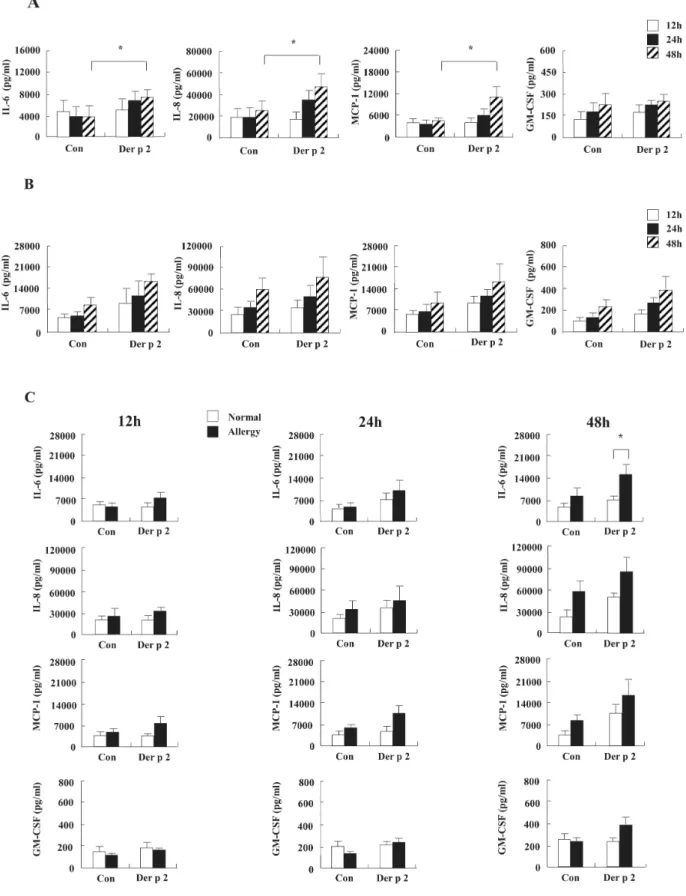
Pro-allergic cytokines were important mediators of allergic inflammation, cell recruitment and allergenic response decided to further investigate the

PI3K inhibition decreased antioxidants/GD-induced apoptosis in A549 cells, and PI3K inhibitor LY294002 had inhibitory effect on antioxidants/GD-induced caspase-3

Observed and simulated means, mean bias of modeled against observed PM 10 , correlation coefficients of Asian dust cases in 2011 springtime at sites located in Erdene

Effect of plate thickness on normal overall spectral transmittance of soda-lime glass (includes surface reflection) at 298 K. Transmittance