J Korean Ophthalmol Soc 2010;51(12):1584-1589 pISSN: 0378-6471
eISSN: 2092-9374
DOI : 10.3341/jkos.2010.51.12.1584
= 증례보고 =
삼체형과 일체형 비구면 인공수정체의 안정성 비교
손성완1 서정원1 신승주1 정성근2
삼육서울병원 안과1, 가톨릭대학교 의과대학 안과 및 시과학교실2
목적: 삼체형과 일체형 비구면 인공수정체의 중심이탈, 경사도, 전방깊이 및 굴절력을 검사하여 안정성을 비교하였다.
대상과 방법: 101안의 백내장수술 환자 중 삼체형인 TecnisⓇ ZA9003 삽입군(54안)과 일체형인 AcrySofⓇ IQ 삽입군(47안)을 대상으로 술 후 1일, 1달, 2달에 전안부분석기(EAS-1000, Nidek, Japan)로 중심이탈, 경사도, 전방깊이, 굴절력을 검사하였다.
결과: 중심이탈은 술 후 1일(p = 0.04)에 차이를 보였으나 1달(p = 0.15), 2달(p = 0.13)에는 유의한 차이는 없었다. 경사도는 술 후 1일, 1달, 2달에 유의한 차이가 없었고 전방깊이는 술 후 1일, 1달, 2달에서 모두 유의한 차이가 없었다. 굴절력은 술 후 1일(p = 0.03)에 차이를 보였으나 1달(p = 0.07), 2달(p = 0.07)에는 유의한 차이가 없었다.
결론: 삼체형과 일체형 비구면 인공수정체의 중심이탈, 경사도, 전방깊이 및 굴절력은 술 후 2달까지 유의한 차이가 없었다. 따라서 두 인공수정체의 2달까지 낭내 안정성은 차이가 없는 것으로 생각된다.
<대한안과학회지 2010;51(12):1584-1589>
접 수 일: 2010년 6월 4일 심사통과일: 2010년 11월 10일
책 임 저 자: 정 성 근
서울시 영등포구 여의도동 62 가톨릭대학교 성모병원 안과
Tel: 02-3779-1150, Fax: 02-761-6869, E-mail: [email protected]
* 본 논문의 요지는 2009년 대한안과학회 제102회 학술대회에서 포스터로 일부 발표되었음.
백내장수술 후 만족할 만한 시력을 얻지 못하는 원인 중 하나로 예측한 굴절력과의 차이가 원인인 경우가 많았 다.1-4굴절 오차의 원인으로 인공수정체 도수계산의 오차, 백내장수술 방법, 수술 중의 절개 방법, 수술 유발 난시 등 이 보고되었다.4-8또한 안축장의 측정 방법 차이와 인공수 정체의 종류, 인공수정체가 삽입된 위치의 차이에 따른 굴 절력의 변화에 대해서도 많은 연구가 있었다.9-14
백내장수술 기법 발달과 더불어 인공수정체의 발달은 백 내장수술 후의 시력에 획기적인 향상을 가져왔다. 최근에는 수술 후 단순 시력 교정을 넘어서 시력의 질적 향상을 위해 기존의 구면 인공수정체 이외에 비구면 인공수정체를 개발 하여 사용하게 되었다.15-17기존의 구면 인공수정체는 백내 장수술 후 양의 각막 구면수차 값을 더욱 증가시켜 대비감 도 등이 감소하여 환자의 만족도는 상대적으로 떨어지는 결과를 보였다.18-21따라서 이러한 문제점을 보완한 비구면 인공수정체의 사용이 증가하고 있다.
백내장수술에서 사용되는 비구면 인공수정체는 TecnisⓇ Z9003 (AMO, Santa Ana, CA, USA), AcrySofⓇIQ (Alcon,
Fort Worth, TX, USA) 등이 있으며, 두 인공수정체는 재 질과 크기는 동일하나 구조가 삼체형과 일체형으로 다르고 굴절률도 차이가 있다. 그러나 이러한 차이가 두 인공수정 체 간의 중심이탈, 경사도, 전방깊이와 굴절률에 영향을 주 어 안정성에 어떤 영향을 미치지는지에 대한 연구는 없었다.
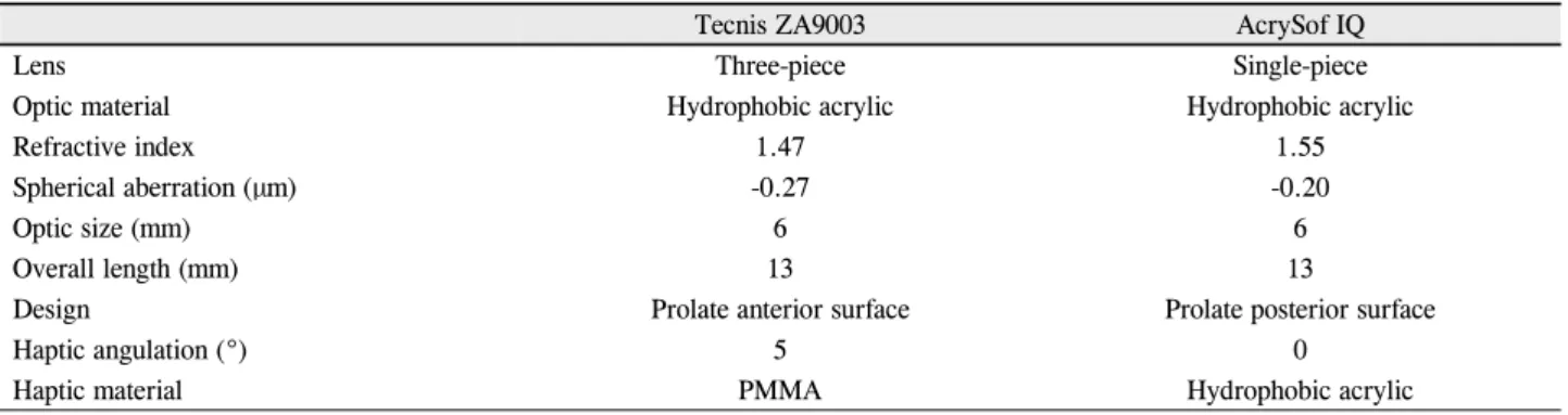
TecnisⓇZ9003은 굴절계수가 1.47로 광학부가 소수성 아 크릴 재질이며 지지부가 polymethylmetacrylate mono- filament로 이루어진 삼체형의 인공수정체이다. 전체 직경 은 13 mm, 광학부의 직경은 6 mm로 인공수정체의 전면이 비구면이며, -0.27 µm의 구면수차를 가지고, 광학부의 가 장자리의 경사가 완만하게 만들어진 구조로 광학부가 자외 선 차단 기능이 있다(Table 1).22
AcrySofⓇIQ는 광학부 재질이 동일한 일체형 비구면 인 공수정체로 굴절계수가 1.55이다. 전체직경은 13 mm로 광 학부 직경이 6 mm로서 광학부의 후면이 비구면인 형태이 며, -0.20 µm의 구면수차를 가진다. 또한 광학부 자체가 자 외선과 가시광선의 청색범위 파장을 차단하는 기능이 있고, 광학부 후면이 비구면으로 인공수정체의 두께가 비교적 얇 게 만들어진 특징이 있다.20,22
비구면 인공수정체를 삽입한 백내장수술에서는 구면수 차의 역할이 중요하고 중심이탈이나 경사도가 구면수차에 영향을 주어 술 후 낮은 시력의 질의 원인이 되며, 전방깊 이는 술 후 굴절력에 영향을 준다고 알려져 있다.20-23하지 만 이런 중요성에 비해 삼체형과 일체형 비구면 인공수정 체 삽입 후 중심이탈, 경사도및 전방깊이, 굴절력의 변화에 대한 연구는 없었다. 따라서 본 연구에서는 여러 비구면 인
Figure 1. The decentration, tilt, anterior chamber depth of
IOL are measured at 90° (top), 180° (bottom) direction using the Scheimpflug camera. Vertical green line denotes visual ax- is, and yellow line denotes the axis of intraocular lens.Decentration of intraocular lens is measured by calculating the distance between the vertex point of the intraocular lens and the visual axis. Anterior chamber depth is calculated by meas- uring the average distance between the corneal surface to the anterior surface of the intraocular lens at 90° and 180° angle from the Scheimpflug image.
Table 1. Characteristics of the three-piece and single-piece aspheric intraocular lens
Tecnis ZA9003 AcrySof IQ
Lens Three-piece Single-piece
Optic material Hydrophobic acrylic Hydrophobic acrylic
Refractive index 1.47 1.55
Spherical aberration (µm) -0.27 -0.20
Optic size (mm) 6 6
Overall length (mm) 13 13
Design Prolate anterior surface Prolate posterior surface
Haptic angulation (°) 5 0
Haptic material PMMA Hydrophobic acrylic
공수정체 중 삼체형 인공수정체인 TecnisⓇ ZA9003과 일 체형 인공수정체인 AcrySofⓇ IQ를 삽입한 환자에서 백내 장수술 후 두 비구면 인공수정체간의 중심이탈, 경사도 및 전방깊이, 굴절력의 변화를 검사하여 두 인공수정체의 안정 성의 차이를 알아보고자 하였다.
대상과 방법
2007년 12월부터 2010년 2월까지 노인백내장으로 내원 한 환자 89명 101안을 대상으로 하였으며, 그 중 이전에 안 과수술을 받은 병력이 있거나 시력에 영향을 줄 만한 각막 혼탁, 약시, 녹내장, 시신경병증, 망막이상 등의 질환을 가 진 환자와 수술 중의 후낭파열, 인공수정체 이탈 등이 발생 한 환자는 제외하였다.
환자군의 평균나이는 68.12 ± 12.28세였으며, 남자가 46 명,여자가 55명이었고, 54안에는 TecnisⓇZA9003을, 47 안에는 AcrySofⓇ IQ를 삽입하였다. 인공수정체의 도수는 수술 전 각 눈의 안축장과 각막곡률반경을 측정하여 SRK-
Ⅱ 공식에 대입하였고, 안축장이 22.0 mm 이하이거나 25.0 mm 이상인 경우는 제외하였다. 수술 후 목표 굴절력은 -0.5D에 가깝도록 인공수정체의 도수를 결정하였다. 모든 수술과정은 3 mm 너비의 귀쪽 투명 각막절개를 만들고 5 mm의 수정체낭원형절개술을 시행하였으며 중심이탈에 영 향을 미칠 수 있는 원형절개술이 치우치거나 확장된 경우 는 제외하였다. 수력분리술과 수력분층술을 시행 후 절개 부위를 통해 수정체유화술을 시행하고 인공수정체를 수정 체낭내에 삽입하였다. 모든 수술은 동일한 술자에 의해 시 행되었으며 수술 후 경과관찰은 1일, 1달째, 2달째에 시행 하였고 동일한 검사자에 의해 3회 측정 후 평균값으로 하였 다. 산동 후 전안부 분석기(Scheimpflug camera: EAS- 1,000, Nidek, Japan)를 이용하여 두 인공수정체의 중심이탈, 경사도 및 전방깊이를 검사하였다. 인공수정체의 Scheimpflug 영상은 90°와 180°에서 CCD 카메라로 촬영 후, 컴퓨터에 내장된 프로그램을 통해 분석함으로써 인공수정체와 주위
구조물과의 기하학적인 상관관계를 나타낸다.24 이때 내장 된 프로그램은 광학부 정점에서 시축까지의 거리를 중심이 탈 값으로 나타내게 되고, 경사도는 인공수정체의 광축과 시 축과의 기울어진 정도를 측정하여 자동으로 그 값을 측정하 게 된다.24전방깊이는 90°와 180°에서 촬영한 Scheimpflug 영상의 각막후면에서 인공수정체의 전면까지의 거리의 평 균으로 계산하였다(Fig. 2).20수술 후 1일, 1달째, 2달째에 자동굴절검사기를 이용하여 구면렌즈대응치를 수술 후 굴 절력으로 하였다.
두 군의 중심이탈, 경사도, 전방깊이의 변화, 굴절력은 SPSS 14.0 for windows (SPSS Inc., Chicago, Illinois, U.S.A.)를 이용하여 독립표본 t-test를 시행하여 유의확률
A B
(A) The three points of the corneal surface (B) The anterior surface of the intraocular lens
C
Figure 2. The tilt is measured by connecting the three points of
the corneal surface and the anterior and posterior surface of the intraocular lens and measuring the angle between the axis of in- traocular lens and visual axis and used the automatic image analysis program for calculation.(C) Posterior surface of the intraocular lens
Table 2. Postoperative changes in the decentration length, tilt, anterior chamber depth
IOL groups Postoperative day
1 day 1 mon 2 months
Decentration (mm) Tecnis ZA9003 0.071 ± 0.051 0.096 ± 0.063 0.062 ± 0.039
AcrySof IQ 0.112 ± 0.087 0.176 ± 0.414 0.052 ± 0.062
p value* 0.04 0.15 0.13
Tilt (°) Tecnis ZA9003 2.47 ± 0.94 2.01 ± 0.81 1.71 ± 0.78
AcrySof IQ 2.69 ± 0.84 2.35 ± 1.01 2.18 ± 0.85
p value* 0.14 0.16 0.14
Anterior chamber depth (mm) Tecnis ZA9003 0.354 ± 0.041 0.353 ± 0.031 0.352 ± 0.029
AcrySof IQ 0.272 ± 0.058 0.198 ± 0.051 0.184 ± 0.041
p value* 0.05 0.07 0.08
*Independent samples t-test.
0.05 미만(p-value<0.05)인 경우를 통계적 유의성이 있 다고 판단하였다.
결 과
중심이탈은 TecnisⓇZA9003에서 술 후 1일에 0.071 ± 0.051 mm, 1달에 0.096 ± 0.063 mm, 2달에는 0.062 ±
0.039 mm이었으며, AcrySofⓇIQ는 술 후 1일에 0.112 ± 0.087 mm, 술 후 1달에 0.176 ± 0.414 mm, 술 후 2달에 는 0.052 ± 0.062 mm이었다. 중심이탈의 정도는 술 후 1 일(p= 0.04)에는 통계적으로 유의한 차이를 보였으나 술 후 1달(p= 0.15), 술 후 2달(p= 0.13)에는 유의한 차이를 보이지 않았다(Table 2).
경사도는 TecnisⓇ ZA9003에서 술 후 1일에 2.47 ±
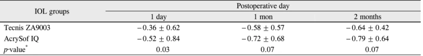
Table 3. Postoperative changes in the mean spherical equivalent (D)
IOL groups Postoperative day
1 day 1 mon 2 months
Tecnis ZA9003 0.36 ± 0.62 0.58 ± 0.57 0.64 ± 0.42
AcrySof IQ 0.52 ± 0.84 0.72 ± 0.68 0.79 ± 0.64
p value* 0.03 0.07 0.07
*Independent samples t-test.
0.94°, 술 후 1달에 2.01 ± 0.81°, 술 후 2달에는 1.71 ± 0.78°이었으며, AcrySofⓇIQ는 술 후 1일에 2.69 ± 0.84°, 술 후 1달에 2.35 ± 1.01°, 술 후 2달에는 2.18 ± 0.85°이 었다. 경사도는 술 후 1일(p= 0.14), 술 후 1달(p= 0.16), 술 후 2달(p= 0.14) 모두 유의한 차이를 보이지 않았다 (Table 2).
수술 전 측정한 평균 전방깊이는 TecnisⓇZA9003에서 3.26 ± 0.49 mm, AcrySofⓇ IQ는 3.18 ± 0.51 mm였으며 두 군간의 통계학적 차이는 없었다(p= 0.214). 전방깊이 의 변화값은 수술 전의 전방깊이에 비하여 전방깊이가 깊어 지는 경우는 양수를, 얕아지는 경우는 음수로 기록하였다. 전 방깊이의 변화는 TecnisⓇ ZA9003에서 술 후 1일에 0.354
± 0.041 mm, 1달에 0.353 ± 0.031 mm, 술 후 2달에는 0.352 ± 0.029 mm이었으며, AcrySofⓇ IQ는 술 후 1일 에 0.272 ± 0.058 mm, 1달에 0.198 ± 0.051 mm, 2달에는 0.184 ± 0.041 mm이었고, 술 후 1일(p= 0.05), 술 후 1달(p
= 0.07), 술 후 2달(p= 0.08) 모두 통계적으로 유의한 차 이를 보이지 않았다(Table 2).
술 후 각각의 목표 굴절력은 TecnisⓇZA9003는 -0.47
± 0.20D, AcrySofⓇIQ는 -0.49 ± 0.18D이었다. 술 후 굴 절력은 TecnisⓇZA9003에서 술 후 1일에 -0.36 ± 0.62D, 술 후 1달에 -0.58 ± 0.57D, 술 후 2달에는 -0.64 ± 0.42D 이었으며, AcrySofⓇIQ는 술 후 1일에 -0.52 ± 0.84D, 술 후 1달에 -0.72 ± 0.68D, 술 후 2달에는 -0.79 ± 0.64D이 었고, 술 후 1일에(p= 0.03)는 통계적으로 유의한 차이를 보였으나 술 후 1달(p= 0.07), 술 후 2달(p= 0.07)에는 통계적으로 유의한 차이를 보이지 않았다(Table 3).
고 찰
현재 백내장수술은 인공수정체의 발달과 더불어 인공수 정체 도수계산공식의 발전으로 적절한 인공수정체를 삽입 하여 수술 후 굴절력을 정확하게 예측할 수 있게 되었다.18 그러나 예측한 굴절력과 실제의 굴절력과의 오차를 보이는 경우가 있어 원인 규명에 대한 여러 연구가 있었고, 이로 인해굴절력의 오차는 감소하였으며 나아가서 시력의 질적 인 향상에 관심이 증가하게 되었다.18-22현재 주로 사용되
는 구면 인공수정체는 양의 구면수차 값을 갖게 되어 백내 장수술 후 높은 구면수차 값을 갖게 되므로 눈부심 등의 증 상을 일으켜 시력의 질이 떨어지는 문제점이 있었다.20 따 라서 음의 구면수차 값을 갖는 비구면 인공수정체를 개발 하여 이러한 단점을 보완하게 되었다.21
백내장수술 후 구면수차에 영향을 주는 것은 인공수정체 의 구면수차 이외에 동공의 직경, 각막의 비구면도 등이 있 고, 수술 후에 인공수정체의 중심이탈이나 경사도 등이 구 면수차에 영향을 준다. 이런 이유로 비구면 인공수정체는 인공수정체의 위치이상으로 시축에서 벗어났거나, 각막 중 심이 달라진 원추 각막 환자의 경우 구면수차 교정 효과가 떨어지는 것으로 알려져 있다.25 따라서 비구면 인공수정체 에서는 낭내 위치가 더욱 중요한 역할을 하므로 비구면 인 공수정체간의 중심이탈이나 경사도가 비구면 인공수정체의 광학적 효과를 나타내는 데 중요할 것으로 생각된다.20 이 런 비구면 인공수정체의 장점이 있어 여러 방면에서 비구 면 인공수정체에 관한 연구 보고가 나오고 있다. 백내장수 술에서 삽입된 비구면 인공수정체의 이전 연구로 Ahn et al21의비구면 인공수정체와 구면 인공수정체 삽입 후 수차 및 시기능 비교연구, Kang et al22의 서로 다른 비구면 인공 수정체 삽입안에서 술 후 시기능 비교연구, Lee et al20의 일체형 구면 인공수정체와 비구면 인공 수정체에서의 중심 이탈, 경사도 및 전방깊이의 비교 등의 연구가 있었으나 삼 체형과 일체형 비구면 인공수정체의 안정성을 비교한 연구 는 없었다.
Wirtitsch et al23와 Mutlu et al26은 구면 인공수정체간에 지지부 각도 차이로 A-상수 값이 서로 다르지만 술 후 중 심이탈과 경사도와는 무관하다고 보고하였고, Cha et al27 은 A-상수 값이 같은 삼체형과 일체형 구면 인공수정체의 술 후 2달까지의 중심이탈과 경사도 역시 유의한 차이를 보 이지 않았음을 보고하였다. 본 연구의 결과에서도 중심이탈 은 술 후 1일에 통계적으로 유의한 차이가 있었지만 술 후 1달, 2달째에는 유의한 차이가 없었다. 그러나 TecnisⓇ ZA9003이 술 후 1일에서 2달에 이르기까지 AcrySofⓇIQ 에 비해 중심이탈의 변화량이 적었다. 경사도는 술 후 1일, 술 후 1달, 술 후 2달째 모두 유의한 차이가 없어 비구면 인공수정체의 삼체형과 일체형 구조에 따른 인공수정체의
안정성 차이는 없는 것으로 나타났다.
전방깊이는 술 후 1일, 술 후 1달째, 술 후 2달째 모두 유 의한 차이가 없었고 굴절력은 술 후 1일에 통계적으로 유의 한 차이가 있었지만 술 후 1달째, 술 후 2달째에서 통계적 으로 유의한 차이를 보이지 않았다. 그러나 통계적으로 유 의한 차이는 없지만 술 후 1일째, 술 후 1달째, 술 후 2달째 에 전방깊이의 변화량이 TecnisⓇZA9003이 AcrySofⓇIQ 에 비해 더 깊어졌으며, TecnisⓇZA9003은 술 후 1일부터 술 후 2달까지 전방깊이의 큰 변화를 보이지 않았지만 AcrySofⓇIQ는 술 후 깊어졌다가 술 후 2달까지 지속적으 로 전방깊이가 얕아지는 결과를 보였다. 이는 Chae et al18 의 연구에서 일체형 구면 인공수정체가 술 후 지속적으로 전방깊이가 얕아지고, 삼체형은 일정한 양상을 보이는 결과 와 일치하였다. 술 후 굴절력은 삼체형과 일체형 비구면 인 공 수정체 모두에서 근시로의 이행을 보였으나 술 후 2달에 는 유의한 차이를 보이지 않았다. 수술 후 측정한 굴절력과 목표 굴절력과의 차이를 비교해보면, TecnisⓇ ZA9003이 수술 후 1일째 목표 굴절력에 비해 부족 교정 양상을 보였 으나 술 후 1달, 2달에는 약간 근시로 이행하였다. AcrySof
ⓇIQ는 술 후 1일째부터 목표 굴절력보다 근시상태이었으 며 술 후 2달까지 지속적으로 근시로 이행하는 결과를 보였 다. TecnisⓇZA9003와 AcrySofⓇIQ가 술 후 1달부터 2달 까지 굴절력의 차이가 통계적인 유의성은 없지만 AcrySofⓇ IQ가 더 근시로 이행되었고 이는 AcrySofⓇIQ가 더 앞쪽으 로 위치해 술 후 2달까지 전방깊이가 앝아지는 것으로 생각 된다. 그러나 TecnisⓇZA9003는 수술 후 1일부터 2달까지 지속적으로 근시로 이행하였으나 전방깊이는 거의 변화가 없어 이들과의 상관관계는 알 수 없어 다른 요소가 관여할 것으로 생각된다. 또한 전방깊이와 굴절력은 통계적인 유의 성은 없었지만 p값이 0.05에 근접하는 값을 가졌고 이는 더 많은 대상과 장기간의 연구가 필요하다고 생각된다.
이 연구의 한계점으로는 연구기간이 짧아 백내장수술 후 발생하는 후낭혼탁 등의 합병증 연구를 하지 못한 점과, 인 공수정체의 종류가 삼체형과 일체형 비구면 인공수정체를 1개씩만 사용한 점, 백내장수술 후 후 굴절력은 전방깊이 뿐 아니라 각막두께와 같은 다른 인자에도 연관이 있지만 이에 대한 연구가 없었다는 것이다. 이러한 문제점을 보완 하기 위해 향후 장기적인 연구와 더 많은 종류의 인공 수정 체, 각막두께와 같은 다른 인자도 고려된 연구가 필요할 것 으로 생각된다.
결론적으로, 삼체형과 일체형 비구면 인공수정체 삽입 후 중심이탈, 경사도 및 전방깊이, 굴절력의 변화를 연구한 결과 술 후 2달까지는 두 인공수정체간에 낭내 안정성은 차 이가 없다고 생각된다.
참고문헌
1) Brandser R, Haaskjold E, Drolsum L. Accuracy of IOL calculation in cataract surgery. Acta Ophthalmol Scand 1997;75:162-5.
2) Zaldivar R, Shultz MC, Davidorf JM, Holladay JT. Intraocular lens power calculation in patients with extreme myopia. J Cataract Refract Surg 2000;26:668-4.
3) Olsen T, Corydon L, Gimbel H. Intraocular lens power calculation with an improved anterior chamber prediction algorithm. J Cataract Refract Surg 1995;21:313-9.
4) Minassian DC, Rosen P, Dart JK, et al. Extracapsular cataract ex- traction compared with small incision surgery by phacoemulsifica- tion: a randomized trial. Br J Ophthalmol 2001;85:822-9.
5) Kondrot EC. Keratometric cylinder and visual recovery following Phacoemulsification and intraocular lens implantation using a self-sealing cataract incision. J Cataract Refract Surg 1991;17:
731-3.
6) Merriam JC, Zheng L, Merriam JE, et al. The effect of incisions for cataract curvature. Ophthalmology 2003;110:1807-13.
7) Long DA, Monica ML. A prospective evaluation of corneal curva- ture changes with 3.0 to 3.5 mm corneal tunnel phacoemulsification.
Ophthalmology 1996;103:226-32.
8) Ali JL, Rodfiguez-Prats JL, Gala A, Ramzy M. Outcomes of mi- croincision cataract surgery versus coaxial phacoemulsification.
Ophthalmology 2005;112:1997-2003.
9) Drews RC. Five year study of astigmatic stability after cataract sur- gery with intraocular lens implantation: comparison of wound sizes. J Cataract Refract Surg 2000;26:250-3.
10) Nemeth J, Fekete O, Pesztenlehrer N. Optical and ultrasound measurement of axial length and anterior chamber depth for intra- ocular lens power calculation. J Cataract Refract Surg 2003;29:85-8.
11) Olsen T, Nielsen PJ. Immersion versus contact technique in the measurement of axial length by ultrasound. Acta Ophthalmol (Copenh) 1989;67:101-2.
12) Watson A , Armstrong R. Contact or immersion technique for axial length measurement. Aust N Z J Ophthalmol 1999;27:49-51.
13) Rose LT, Moshegov CN. Comparison of Zeiss IOL master and ap- plication A-scan ultrasound: biometry for intraocular lens calculation.
Clin Experiment Ophthalmol 2003;31:121-4.
14) Brint SF, Ostrick DM, Bryan JE. Keratometric cylinder and visual performance following phacoemulsification and implantation with silicon small incision or poly(methylmethacrylate) intraocular lenses. J Cataract Refract Surg 1991;17:32-6.
15) Guirao A, Redondo M, Geraghty E, et al. Corneal optical aberra- tions and retinal image quality in patients in whom monofocal in- traocular lenses were implanted. Arch Ophthalmol 2002;120:
1143-51.
16) Werner L, Mamalis N. Wavefront corrections of intraocular lenses.
Ophthalmol Clin North Am 2004;17:233-45.
17) Chalita MR, Krueger RR. Correlation of aberrations with visual acuity and symptoms. Ophthalmol Clin North Am 2004;17:135-42.
18) Chae JK, Jang JW, Choi TH, Lee HB. Changes in refraction and an- terior chamber depth according to type of intraocular lenes. J Korean Ophthalmol Soc 2006;47:1935-42.
19) Kim HS, Kim SW, Ha BJ, et al. Ocular aberrations and contrast sensitivity in eyes implanted with aspheric and spherical intra- ocular lenses. J Korean Ophthalmol Soc 2008;49:1256-62.
=ABSTRACT=
Comparison of the Stability Between Three-piece and Single-piece Aspheric Intraocular Lenses
Sung Wan Son, MD1, Jung Won Seo, MD1, Seong Joo Shin, MD1, Sung Kun Chung, MD, PhD2
Department of Ophthalmology, SahmYook Seoul Hospital1, Seoul, Korea
Department of Ophthalmology, St, Mary’s Hospital, The Catholic University of Korea School of Medicine2, Seoul, Korea
Purpose: The goal of the present study was to compare the stability of the 3-piece (TecnisⓇ ZA9003) and single-piece (AcrySofⓇ IQ) aspheric intraocular lenses (IOL) by testing decentration, tilt, anterior chamber depth, and refraction.
Methods: The subjects of this study consisted of 101 eyes who had undergone cataract surgeries with a 3-piece aspheric IOL (54 eyes) and with a single-piece aspheric IOL (47 eyes). The decentration, tilt, anterior chamber depth, and refraction were measured on postoperative day 1, 1 month, and 2 months, using an anterior eye segment analysis system (EAS-1000, Nidek, Japan).
Results: There was a statistically significant difference in the decentration on the postoperative day 1 (p = 0.04). However, there was no statistically significant difference on postoperative 1 month (p = 0.15) and 2 months (p = 0.13). There was no statistically significant difference in the tilt on postoperative day 1, 1 month, and 2 months. There was no statistically sig- nificant difference in the anterior chamber depth on postoperative day 1, 1 month, and 2 months. There was a statistically significant difference in the refraction on postoperative day 1 (p = 0.03). However, there was no statistically significant dif- ference on postoperative 1 month (p = 0.07) and 2 months (p = 0.07).
Conclusions: There was no statistically significant difference in the decentration, anterior chamber depth, and refraction between the 3-piece and single-piece aspheric IOL. Therefore, there is no difference between the 3-piece and single-piece aspheric IOL in the capsular bag stability until 2 months postoperatively.
J Korean Ophthalmol Soc 2010;51(12):1584-1589
Key Words: Anterior chamber depth, Aspheric IOL, Decentration, Refraction, Tilt
Address reprint requests to Sung Kun Chung, MD, PhD
Department of Ophthalmology, St. Mary’s Hospital, The Catholic University of Korea School of Medicine
#62 Yeouido-dong, Yeongdeungpo-gu, Seoul 150-713, Korea
Tel: 82-2-3779-1150, Fax: 82-2-761-6869, E-mail: [email protected] 20) Lee JY, Lee SH, Chung SK. Decentration, tilt and anterior chamber
depth: aspheric vs spheric acrylic intraocular lens. J Korean Ophthalmol Soc 2009;50:852-7.
21) Ahn HS, Kim SW, Kim EK, Kim TI. Wavefront and visual analysis after aspherical and spherical intraocular lenses implantation. J Korean Ophthalmol Soc 2008;49:1248-55.
22) Kang IS, You IC, Pack YG, Yoon KC. Comparision of visual func- tion among aspheric intraocular lenes. J Korean Ophthalmol Soc 2009;50:691-7.
23) Wirtitsch MG, Findl O, Menapace R, et al. Effect of haptic design on change in axial lens position after cataract surgery. J Cataract Refract Surg 2004;30:45-51.
24) Kim JS, Shyn KH. Periodic Biometry in Three Types of Posteriorly Implanted IOLs: PMMA, Silicone, and Acrylic Soft, by EAS- 1000 Scheimpflug Photography. J Korean Ophthalmol Soc 2000;
41:2205-10.
25) Dietze HH, Cox MJ. Limitations of correcting spherical aberration with aspheric intraocular lenses. J Refract Surg 2005;31:574 85.
26) Mutlu FM, Erdurman C, Sobaci G, Bayraktar MZ. Comparision of tilt and decentration of 1-piece and 3-piece hydrophobic acrylic in- traocular lenses. J Cataract Refract Surg 2005;31:343-7.
27) Cha YD, Oh SH, Lee DH. Comparative assessment of clinical re- sults in various acrylate IOLs. J Korean Ophthalmol Soc 2006;47:
740-7.