pISSN: 0378-6471 eISSN: 2092-9374 http://dx.doi.org/10.3341/jkos.2013.54.8.1293
= 증례보고 =
유리체 출혈을 동반한 망막색소변성 1예
정인영⋅허현도⋅김성재⋅한용섭⋅서성욱⋅박종문 경상대학교 의학전문대학원 안과학교실, 경상대학교 건강과학연구원
목적: 초기 양안 유리체염을 보이다가 신생혈관이 생기고 우안 유리체 출혈로 유리체 절제술을 시행하여 시력호전을 보인 망막색소변 성 환아를 경험하였기에 보고하고자 한다.
증례요약: 8세 남아가 야맹증을 주소로 내원하였다. 초기 양안의 유리체 염증과 시신경 신생혈관으로 치료도중 우안의 유리체 출혈이 발생하여 유리체 절제술을 시행하였고 좌안에 녹내장 발생하여 녹내장 수술을 시행하였으며 현재 양얀 시력 0.6으로 호전되었다.
결론: 망막색소변성의 합병증 중 유리체 출혈은 만성 유리체 염증과 관련이 있을 것으로 생각되며 아주 드물게 발생한다. 유리체 절제 술로 치료후 안정적으로 유지되고 있어 보고하고자 한다.
<대한안과학회지 2013;54(8):1293-1297>
■Received: 2013. 1. 26. ■ Revised: 2013. 4. 13.
■Accepted: 2013. 6. 27.
■Address reprint requests to Jong Moon Park, MD, PhD Department of Ophthalmology, Gyeongsang National University Hospital, #79 Gangnam-ro, Jinju 660-702, Korea Tel: 82-55-750-8167, Fax: 82-55-758-4158
E-mail: [email protected]
망막 색소 변성은 전세계적으로 4,000명에 한 명꼴로 발 생하는 실명의 주요한 원인질환으로 여러 다양한 유전자들 의 변이에 의해 발생하는 것으로 알려졌다.1,2 유전자 변이 가 어떠한 생물학적 과정을 거쳐 광수용체세포의 괴사로 진행되는지는 아직도 알려지지 않았으며 최근 보고에서는 염증성 반응이 병인의 주요한 인자이며 유리체강내 염증세 포의 침윤이 초기 징후일 수 있다고 하였다.3-5
망막 색소 변성의 합병증으로 백내장, 녹내장, 낭포성 황 반부종, 황반전막등이 있으며 드물게 유리체 출혈이 동반되 기도 하나 아직 국내에 보고된 적은 없다.3,6,7
이에 저자들은 초기에 양안 유리체염증을 보이다가 양안 시신경의 신생혈관을 동반한 후 우안 유리체 출혈로 유리 체 절제술을 시행한 환자를 경험하였기에 관련 문헌 고찰 과 함께 이를 보고하고자 한다.
증례보고
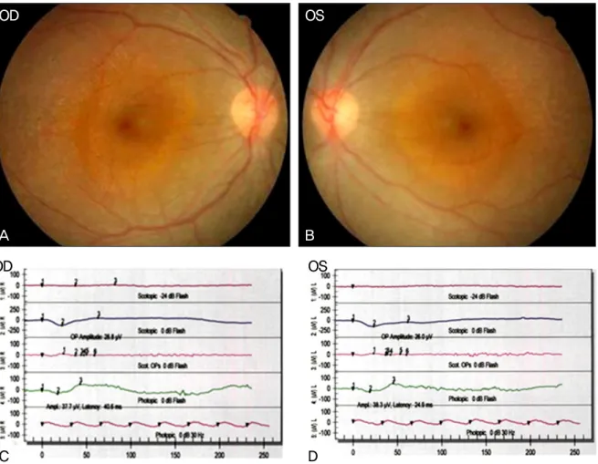
12세된 남아가 양안 야맹증을 주소로 내원하였다. 내원 당시 최대교정시력은 양안 1.0 이었고 안압은 정상이었으 며 안저검사상 황반부를 제외한 망막 대부분에 색소 변성 을 보이고 망막전위도 검사에서도 양안 모두 암순응과 명
순응에서 a파와 b파가 거의 소실되어 망막색소변성으로 진 단하였다(Fig. 1).
2년 뒤 양안의 시력저하를 주소로 내원하였는데 당시 교 정시력은 우안 0.4, 좌안 0.3이었고 전방에 염증소견 없었 으나 유리체강 내 염증소견 보이고, Stratus OCT (Carl Zeiss Meditec, USA)에서 양안 황반 부종이 있었다. 형광 안저혈관조영술에서 양안의 시신경주위에 신생혈관이 관찰 되었다(Fig. 2). 양안에 유리체강 내 항혈관내피성장인자인 Bevacizumab (Avastin®, Genetech Inc., San Francisco, CA, USA) 1.5 mg/0.05 cc를 주사하였으나 황반부종은 호 전되지 않았다. 2개월 뒤 유리체강내 Bevacizumab 1.5 mg/0.05 cc와 트리암시놀론(Triamcinolone acetonide, Dongkwang, Seoul, Korea) 2 mg/0.05 cc를 함께 주사하였 고 황반부종의 호전과 함께 양안교정시력이 0.8로 향상되 었다. 하지만 이 때부터 양안의 안압이 우안 24 mmHg, 좌안 23 mmHg로 상승되어 안압하강제 0.15% brimonidine tar- trate (Alphagan-P®, Allergan, USA), 0.005% Latanoprost (Xalatan®, Pfizer, USA), Brinzolamide/Timolol (Elazop®, Alcon, USA)을 양안에 사용하였고 안압은 정상으로 유지 되었다.
4개월 후에 우안 시력은 안전수동으로 교정되지 않았으 며, 좌안 교정 시력은 0.4로 저하되어 시행한 검사에서 양 안의 백내장과 함께 우안에 유리체 출혈과 좌안의 황반부 종이 동반되었다. 이에 우안에는 유리체 절제술, 초음파 유 화술 및 인공수정체 삽입술을 함께 시행하였고, 좌안에는 유리체강내 Bevacizumab 1.5 mg/0.05 cc과 트리암시놀론 2 mg/0.05 cc를 함께 주사하였다(Fig. 3). 우안 교정 시력
A B
C D
OD OS
OD OS
Figure 1. (A, B) Fundus photographs show diffuse retinal degeneration with sparing of the macula in both eyes. (C, D)
Electroretinograms showing nearly reduced ‘a’ and ‘b’ waves in the both eyes.OD OS
A B
Figure 2. (A, B) Fluorescein angiogram of the both eyes show dye leakage from new vessels around the optic disc margin.
0.6으로 호전되었으나 좌안은 안압하강제(Alphagan-P®, Elazop®, Xalatan®)의 사용에도 지속적으로 안압이 30 mmHg 이상 상승되어 0.3 mg/ml 농도의 mitomycin-C를 사용한
섬유주절제술을 시행하였으며, 이 후 좌안에도 백내장이 진 행하여 초음파유화술 및 인공수정체 삽입술을 시행하였다.
현재 술 후 약 1년간 경과관찰 중이며 재출혈 등의 합병
A B
OD OS
Figure 3. Fundus photographs show a complete resolusion of the vitreous hemorrhage. (A) Preoperative fundus
photograph. (B) Postoperative fundus photograph at 1 month after vitrectomy.A B
OD OS
Figure 4. Humphrey visual field test. The test shows peripheral field constriction of the both eyes.
증은 발생하지 않았고, 양안 교정시력 0.6으로 유지되고 있 으며 안압은 안압하강제 Elazop®을 양안에 점안하면서 15 mmHg로 조절되며, 양안 시야검사상 10도 이내의 위축된 관모양 시야장애를 보이고 있다(Fig. 4).
고 찰
망막 색소 변성의 합병증으로 널리 알려진 백내장, 녹내 장, 낭포성 황반부종에 비해 유리체 출혈은 드물게 발생하 며 현재까지 전세계적으로 수예가 증례보고되었으나 국내 에 보고된 적은 없다.6,7
망막 색소 변성은 40여종 이상의 여러 유전자의 변이와
관련되어 있다는 사실을 알고 있으나 그 정확한 기전이나 병인에 대해서는 아직도 모르는 부분이 많고 효과적인 치 료법이 아직 없어서 많은 환자들이 실명으로 진행되고 있 다. 이 질환의 일차적인 원인은 유전자의 결함으로 알려졌 으나 최근에는 염증반응이 중요한 요인으로 작용하리라는 연구결과들이 있다. 이러한 염증성 반응이 변성의 진행에 있어서 일차적인 역할을 하는지, 아니면 이차적이고 수동적 인 역할을 하는지에 관하여는 명확히 알 수 없으나 일부 연 구자들은 이런 유리체강내 염증변화가 망막색소변성의 가 장 빠른 징후일 수 있다고 하였다.3-5Yosida et al5은 지속 적이고 만성적인 유리체강내 염증성 반응이 망막색소변성 의 병인의 기저에 자리잡고 있으며, 이러한 염증반응에 대
한 개입이 환자의 치료에 필요할 수 있다고 보고하면서 특 히 젊은 나이의 환자에서 나이든 환자에 비해 더 강한 염증 반응이 나타난다고 하였다. 또한 Henkind8는 만성 염증성 질환에서 망막의 무혈관부위가 없어도 신생혈관이 나타날 수 있다고 하였다.
본 증례는 비교적 어린 나이에 양안 야맹증을 주소로 내 원하였으며 처음 나타난 징후가 유리체 염증이었고 형광안 저촬영으로 시신경의 신생혈관을 확인할수 있었다. 함께 동반된 수포성 황반부종을 치료하는 중에 우안에 유리체 출혈이 발생하였다. 본 환아의 형광 안저소견에서 주변부 망막의 무혈관부위는 발견할 수 없었으므로 유리체 출혈의 원인으로는 만성 염증과 관련된 시신경 신생혈관으로 인한 것으로 생각한다. 저자들은 신생혈관 및 염증 반응, 황반부 종에 대한 치료로 항 혈관내피생성인자를 유리체강내 주입 하였으나 큰 효과를 보지 못하였고 다시 트리암시놀론을 병합하여 유리체강내 주사하였고 스테로이드 병합 사용으 로 일시적인 염증 호전 및 황반부종의 호전을 경험하였다.
하지만 다시 발생한 우안의 심한 유리체 출혈로 인하여 유 리체 절제술을 시행하였고 현재 1년 이상 재발이나 후유증 없이 유지되고 있다. 좌안은 황반부종이 재발하여 병합약물 을 유리체강내 재주사하였으나 조절되지 않는 고안압이 발 생하여 녹내장수술을 시행하였고 현재 안압 및 시력이 안 정적으로 유지되고 있다.
저자들은 아직 국내에 보고된 적 없는 어린 환아에서 발 생한 유리체 출혈을 동반한 망막색소변성을 경험하였기에 이를 관련 문헌 고찰과 함께 보고하고자 한다.
REFERENCES
1) Wong P, Borst DE, Farber D, et al. Increased TRPM-2/clusterin mRNA levels during the time of retinal degeneration in mouse models of retinitis pigmentosa. Biochem Cell Biol 1994;72(9-10):
439-46.
2) Cotran PR, Bruns GA, Berson EL, Dryja TP. Genetic analysis of patients with retinitis pigmentosa using a cloned cDNA probe for the human gamma subunit of cyclic GMP phosphodiesterase. Exp Eye Res 1991;53:557-64.
3) Uliss AE, Gregor ZJ, Bird AC. Retinitis pigmentosa and retinal neovascularization. Ophthalmology 1986;93:1599-603.
4) Newsome DA, Michels RG. Detection of lymphocytes in the vitre- ous gel of patients with retinitis pigmentosa. Am J Ophthalmol 1988;105:596-602.
5) Yoshida N, Ikeda Y, Notomi S, et al. Clinical evidence of sustained chronic inflammatory reaction in retinitis pigmentosa. Ophthalmology 2013;120:100-5.
6) Nao-i N, Fukiyama J, Sawada A. Retinitis pigmentosa with re- current vitreous hemorrhage. Acta Ophthalmol Scand 1996;74:509-12.
7) Bressler NM, Graqoudas ES. Neovascularization of the optic disk associated with atypical retinitis pigmentosa. Am J Ophthalmol 1985;100:431-3.
8) Henkind P. Ocular neovascularization. The Krill memorial lecture.
Am J Ophthalmol 1978;85:287-301.
=ABSTRACT=
Retinitis Pigmentosa Complicated by Vitreous Hemorrhage in a Young Patient: A Case Report
In Young Chung, MD, PhD, Hyoun Do Huh, MD, Seong Jae Kim, MD, Yong Seop Han, MD, PhD, Seong Wook Seo, MD, PhD, Jong Moon Park, MD, PhD
Department of Ophthalmology, Gyeongsang National University School of Medicine, Jinju, Korea Institute of Health Science, Gyeongsang National University, Jinju, Korea
Purpose: To report a case of a young male patient with retinitis pigmentosa (RP) accompanied by vitritis and neo- vascularization of the optic disk in both eyes who underwent unilateral vitrectomy for the treatment of vitreous hemorrhage in the right eye.
Case summary: An 8-year-old boy visited our clinic with a complaint of night blindness. Both eyes showed inflammatory cells in the anterior vitreous and neovascularization of the optic disk confirmed by fluorescein angiography. Extensive vitreous hemorrhage developed in his right eye and he underwent unilateral vitrectomy. His final visual acuity was 0.6 in both eyes.
Conclusions: Vitreous hemorrhage may be related to chronic inflammation in the vitreous and is a very rare RP complication. Vitrectomy can be an effective treatment option for RP complicated by vitreous hemorrhage.
J Korean Ophthalmol Soc 2013;54(8):1293-1297
Key Words: Retinitis pigmentosa (RP), Vitrectomy, Vitreous hemorrhage
Address reprint requests to Jong Moon Park, MD, PhD
Department of Ophthalmology, Gyeongsang National University Hospital
#79 Gangnam-ro, Jinju 660-702, Korea
Tel: 82-55-750-8167, Fax: 82-55-758-4158, E-mail: [email protected]