CASE REPORT
방선균증이 동반된 대장의 미만성 거대 B-세포 림프종 증례
이상훈, 남승주, 이성준, 박성철, 강창돈, 최대희, 박진명, 이승구1
강원대학교 의학전문대학원 내과학교실, 해부병리학교실1
Colonic Diffuse Large B-cell Lymphoma Hidden in Actinomycosis
Sang Hoon Lee, Seung-Joo Nam, Sung Joon Lee, Sung Chul Park, Chang Don Kang, Dae Hee Choi, Jin Myung Park and Seung Koo Lee1
Departments of Internal Medicine and Anatomic Pathology1, Kangwon National University School of Medicine, Chuncheon, Korea
Actinomycosis can mask malignant diseases. This paper reports a case of colonic diffuse large B-cell lymphoma (DLBCL), which was misdiagnosed as abdominal actinomycosis. A 76-year-old woman presented with right flank pain and weight loss. Abdominal CT and colonoscopy revealed a huge ascending colon mass. Despite the initial impression of a malignancy, a colonoscopic biopsy revealed no malignant cells, but sulfur granules and a filamentous organism suggesting actinomycosis. Intravenous penicillin G was ad- ministered under the impression of abdominal actinomycosis but her condition deteriorated rapidly. Follow up CT showed markedly increased colon mass and new multiple nodular lesions around the ascending colon. Sono-guided percutaneous biopsy of the nodular lesion was performed. The pathological result was DLBCL. The patient was scheduled to undergo chemotherapy but the patient expired due to cancer progression. The diagnosis of gastrointestinal infiltrating tumors is often difficult because a superficial biopsy usually does not provide a confirmative diagnosis. This case highlights the difficulty in making a correct diagnosis of lymphoma due to the concomitant actinomycosis. Malignant conditions must be considered in cases of actinomycosis with no response to antimicrobial therapy. (Korean J Gastroenterol 2019;74:46-50)
Key Words: Lymphoma, large B-Cell, diffuse; Actinomycosis; Colonic neoplasms; Colonoscopy
Received February 10, 2019. Revised April 4, 2019. Accepted April 29, 2019.
CC This is an open access article distributed under the terms of the Creative Commons Attribution Non-Commercial License (http://creativecommons.org/licenses/
by-nc/4.0) which permits unrestricted non-commercial use, distribution, and reproduction in any medium, provided the original work is properly cited.
Copyright © 2019. Korean Society of Gastroenterology.
교신저자: 남승주, 24341, 춘천시 강원대학길 1, 강원대학교 의학전문대학원 내과학교실
Correspondence to: Seung-Joo Nam, Department of Internal Medicine, Kangwon National University School of Medicine, 1 Kangwondaehak-gil, Chuncheon 24341, Korea. Tel: +82-33-258-2405, Fax: +82-33-258-2146, E-mail: [email protected], ORCID: https://orcid.org/0000-0002-0349-0901
Financial support: None. Conflict of interest: None.
INTRODUCTION
Lymphoma originating from the gastrointestinal (GI) tract comprises 5-20% of the total lymphoma cases and 0.5% of malignant tumors of the GI tract. The stomach is the most frequent site of GI lymphoma. Lymphoma is found less com- monly in the small and large intestine.1,2 GI lymphoma is often difficult to diagnose by a routine endoscopic biopsy, which requires a deep biopsy.3 The diagnosis and treatment of co- lonic actinomycosis are often difficult because the clinical fea-
tures and radiological findings are similar to malignant tu- mors, which is more prevalent.4 The paper reports a case of colonic diffuse large B-cell lymphoma (DLBCL) for which the diagnosis was delayed due to actinomycosis that accom- panied the tumor along with a review of the relevant literature.
CASE REPORT
A 76-year-old female who had suffered from weight loss and a febrile sense for two months visited the emergency
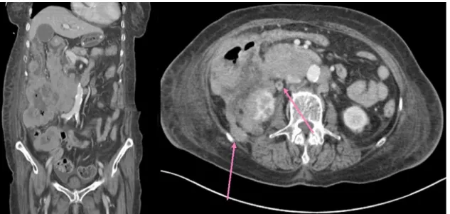
Fig. 1. Abdominal computed tomography scan shows a huge mass at the ascending colon with nearby multiple enlarged lymph nodes.
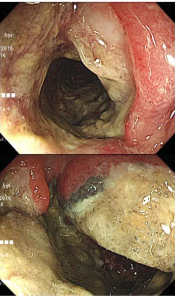
Fig. 2. Colonoscopy shows a huge ulcerative mass with luminal narrowing at hepatic flexure.
Fig. 3. (A) Histology examination showing inflammatory necrotic tissues (H&E, ×100). (B) Silver special staining revealing the filamentous bacterial colonies (arrows) (GMS, ×400).
room complaining of right lower quadrant pain with a mass.
She had been taking medication for hypertension and diabetes. Measurements of the vital signs were as follows:
blood pressure, 135/52 mmHg; pulse rate, 89 beats per mi- nute; respiratory rate, 20 breaths per minute; and body tem- perature, 36.3℃. A physical examination revealed a tender round mass in the right lower quadrant of the abdomen. No significantly enlarged lymph nodes were observed at the groin, neck, and axilla. A laboratory examination showed the follow- ing: leukocytes, 10,000/mm3; hemoglobin, 7.1 g/dL; platelets 525,000/mm3; BUN, 31.3 mg/dL; creatinine, 1.5 mg/dL;
LDH, 772 U/L; CRP, 4.058 mg/dL; total bilirubin, 0.3 mg/dL;
aspartate aminotransferase, 26 IU/L; alanine aminotransferase, 19 IU/L; sodium, 133 mEq/L; potassium, 5.1 mEq/L; and chloride, 103 mEq/L. Abdominal CT showed a huge mass with enlarged lymph nodes around the ascending colon; the mass
A
B
Fig. 4. In the follow-up abdominal computed tomography examination, size of the colonic mass increased significantly and multiple new nodules (arrows) were observed around the ascending colon.
Fig. 5. Pathological findings of the tumor revealed sheets of atypical large pleomorphic lymphoid cells with prominent nucleoli and apoptotic bodies (A; H&E, ×400) with pan-B cell marker expression (B; CD20, ×100).
was assumed to invade the right kidney (Fig. 1).
For a further evaluation of the colonic mass, a colonoscopy was performed, which revealed an ulcerative mass accom- panied by a luminal stricture at the hepatic flexure and as- cending colon (Fig. 2). Multiple biopsy was performed, and a histology examination revealed inflammatory necrotic tis- sues without neoplastic cells (Fig. 3A). Filamentous bacterial colonies, which were morphologically compatible with an Actinomyces species, were found (Fig. 3B).
Under the impression of abdominal actinomycosis, she re- ceived intravenous penicillin (penicillin G 15 million units/day in six divided doses, every 4 hours). On the other hand, two weeks of a penicillin injection did not improve the abdominal pain and mass, and the patient’s condition worsened. Follow up abdominal CT was performed, which showed an increase in the size of the mass and the additional enlargement of lymph nodes other than those previously found (Fig. 4). This led to the assumption of a misdiagnosis of this patient, and sono-guided percutaneous fine needle biopsy was performed for right retroperitoneal mass to obtain deep and sufficient tissue material. A histopathologic examination of the biopsy specimens showed sheets of large pleomorphic lymphocytes with prominent nucleoli and occasional apoptotic bodies (Fig. 5). The immunohistochemical study showed that the tu- mor cells were positive for CD20 (Fig. 5), CD79a, CD5, Bcl-6, Bcl-2, and MUM-1, but negative for pan-cytokeratin, CD3, and CD10. The tumor cells showed a high Ki-67 labeling index
A
B
(≥90%). These pathology features were compatible with high grade B-cell lymphoma, and the patient was diagnosed with colonic DLBCL, Lugano IIE with an international prognostic index score 3. She was scheduled for chemotherapy, but her general condition deteriorated rapidly due to the rapid pro- gression of DLBCL, and the patient expired a few weeks later.
DISCUSSION
Lymphoma is classified as primary if there is no invasion into the peripheral or mediastinal lymph nodes, no enlarge- ment of the liver or spleen, normal white blood cell counts, no spread to remote lymph nodes but restricted to the GI tract and regional lymph nodes.5 Primary malignant lympho- ma of the colon and rectum is rare, most of which occurs at the cecum (73%), followed by the ascending colon and rectum.6 The reason why more primary lymphoma cases are observed frequently at the cecum was assumed to be related to its larger amount of lymphoid structure.7 Recent trends show an increase in primary lymphoma in the GI tract;
this is assumed to be because of the increase in the number of patients suffering from acquired or iatrogenic immunodeficiency due to a range of conditions, such as inflammatory bowel disease, human immunodeficiency virus infections, and or- gan transplantation.8 Malignant lymphoma is classified as B cell and T cell lymphoma according to the revised European-American lymphoma classification. B-cell lymphoma generally forms a mass and can be misdiagnosed as ad- enocarcinoma of the colon. In particular, DLBCL tends to in- volve the long segment of the intestine and forms a large mass that accompanies an ulcer and stricture. On the other hand, T cell lymphoma generally leads to an ulcer but can be misdiagnosed as inflammatory diseases, such as inflammatory bowel disease, intestinal tuberculosis, or infectious colitis.9
Colonoscopy with a biopsy and abdominal CT scan are im- portant tools for diagnosing primary lymphoma of the colon.
Endoscopic morphology of colon lymphoma can be catego- rized into three types - fungating, ulcerative, and infiltrative - and two other mixed types - ulcerofungating and ulcer infiltrative.10 Fungating and ulcerofungating are the most com- mon types, but it is difficult to distinguish these types of lym- phoma from an adenocarcinoma. Some rare cases of multiple lymphomatous polyposis have also been encountered. A his- tology examination is critical for diagnosis. On the other hand,
lymphoma can frequently be missed from a biopsy if the possi- bility of the disease has not been taken into consideration.
In contrast to adenocarcinoma, GI lymphoma develops patho- logically from the lymphoid follicle at the submucosal layer, which leads to the spread of disease mainly within this layer, making it difficult to diagnosis by a mucosal biopsy.11 An ab- dominal CT examination may show a degree of infiltration by the tumor or enlargement of regional lymph node.12
Actinomycosis is a chronic suppurative granulomatous dis- ease caused by an anaerobic or microaerobic strain that nor- mally is found in the mouth, colon, and female genitals; the most common strain is Actinomyces israelii. Abdominal actino- mycosis is generally caused by abdominal surgery or external injury, but the disease can also develop in patients who have been treated with steroids for a long time, chemotherapy for leukemia, or renal transplant, all of which may result in dam- age to the mucosal layer.13 For this specific case, it was as- sumed that actinomycosis developed from a damaged colon mucosal layer caused by lymphoma. A diagnosis actino- mycosis may be difficult because a clinical and radiological manifestation of the disease may mimic that of a malignant tumor. Abdominal actinomycosis can be considered if a heter- ogeneous abdominal mass infiltrates through the serous membrane and shows fewer invasions into the lymph nodes compared to what is expected from the lesion size.14
Several cases similar to this one have been published. One is gastric DLBCL, which was first misdiagnosed as gastric acti- nomycosis by endoscopic biopsies.15 In this case, a persistent malignant appearing ulcer despite antimicrobial therapy was the clue of a misdiagnosis and the patient underwent a surgi- cal resection revealing DLBCL, followed by adjuvant chemotherapy. In addition, there are several cases of colon actinomycosis mimicking cancer, leading to an unnecessary surgical resection of a benign lesion.16 On the other hand, there is no case report of a colonic malignancy misdiagnosed as actinomycosis. This case emphasizes the importance of considering a possible hidden malignancy when abdominal actinomycosis is treated without surgery, particularly in cases of no response on effective antibiotics.
In conclusion, the final diagnosis in this case was colonic DLBCL, which was misdiagnosed as abdominal actinomycosis.
The diagnosis of GI lymphoma may be difficult by an endo- scopic biopsy alone, and can be obscured by actinomycosis, as in this case. If an actinomycotic mass that does not re-
spond to effective antibiotics is encountered, a hidden malig- nant tumor must be considered, which require a repeat biopsy by various modalities (e.g., fine needle aspiration or open bi- opsy) for a confirmatory diagnosis.
REFERENCES
1. Andrews CN, John Gill M, Urbanski SJ, Stewart D, Perini R, Beck P. Changing epidemiology and risk factors for gastrointestinal non-Hodgkin’s lymphoma in a North American population: pop- ulation-based study. Am J Gastroenterol 2008;103:1762-1769.
2. Katsumata R, Matsumoto H, Motoyasu O, et al. Primary colorectal lymphoma comprising both components of diffuse large B-cell lymphoma and mucosa-associated lymphoid tissue lymphoma combined with cytomegalovirus colitis. Clin J Gastroenterol 2016;9:59-62.
3. Choi MK, Kim GH. Diagnosis and treatment of gastric MALT lymphoma. Korean J Gastroenterol 2011;57:272-280.
4. Lim JA, Wong PS, Leong KN, Wong KL, Chow TS. Masking and mis- leading: concomitant actinomycosis and B-cell lymphoma - a case report and review of literature. Scott Med J 2018;63:
125-131.
5. Gill SS, Heuman DM, Mihas AA. Small intestinal neoplasms. J Clin Gastroenterol 2001;33:267-282.
6. Shepherd NA, Hall PA, Coates PJ, Levison DA. Primary malignant lymphoma of the colon and rectum. A histopathological and im- munohistochemical analysis of 45 cases with clinicopatho-
logical correlations. Histopathology 1998;12:235-252.
7. Wong MT, Eu KW. Primary colorectal lymphomas. Colorectal Dis 2006;8:586-591.
8. Kim YH, Lee JH, Yang SK, et al. Primary colon lymphoma in Korea:
a KASID (Korean Association for the Study of Intestinal Diseases) study. Dig Dis Sci 2005;50:2243-2247.
9. Ryu SH, Cheon JH, Kim JY, et al. The early diagnostic accuracy for gastrointestinal T-cell lymphoma from a perspective of gastroenterologists. Intest Res 2011;9:19-26.
10. Choe WH, Kim YH, Kim BJ, et al. The different colonoscopic mani- festations of primary colorectal lymphomas by their cellular origin. Intest Res 2013;1:22-30.
11. Jiang C, Gu L, Luo M, Xu Q, Zhou H. Primary rectal lymphoma: a case report and literature review. Oncol Lett 2015;10:43-44.
12. Lee HJ, Han JK, Kim TK, et al. Primary colorectal lymphoma: spec- trum of imaging findings with pathologic correlation. Eur Radiol 2002;12:2242-2249.
13. Valour F, Sénéchal A, Dupieux C, et al. Actinomycosis: etiology, clinical features, diagnosis, treatment, and management. Infect Drug Resist 2014;7:183-197.
14. Ercolak V, Paydas S, Ergin M, et al. Abdominal actinomycosis with multiple myeloma: a case report. Oncol Lett 2014;8:1876-1878.
15. Carneiro L, Ferreira R, Pinto J. The lymphoma hidden in actinomycosis. GE Port J Gastroenterol 2016;23:273-275.
16. Blanco-Vela CI, Luna-Ayala VM, Perez-Aguirre J. Colonic mass secondary to actinomycosis: a case report and literature review.
Rev Gastroenterol Mex 2014;79:206-208.