CASE REPORT
연조직염과 통풍 관절염으로 오인한 Pancreatitis Panniculitis Polyarthritis 증후군 1예
김이진, 박민수, 손형곤, 오원섭, 문기원, 박진명, 강창돈, 이승구1
강원대학교 의학전문대학원 내과학교실, 해부병리학교실1
Pancreatitis, Panniculitis, and Polyarthritis Syndrome Simulating Cellulitis and Gouty Arthritis
Ee Jin Kim, Min Soo Park, Hyung-Gon Son, Won Sup Oh, Ki Won Moon, Jin Myung Park, Chang Don Kang and Seungkoo Lee1 Departments of Internal Medicine and Anatomic Pathology1, Kangwon National University School of Medicine, Chuncheon, Korea
Pancreatitis, panniculitis, and polyarthritis (PPP) syndrome is a rare but critical disease with a high mortality rate. The diagnostic dilemma of PPP syndrome is the fact that symptoms occur unexpectedly. A 48-year-old man presented with fever and painful swelling of the left foot that was initially mistaken for cellulitis and gouty arthritis. The diagnosis of PPP syndrome was made based on the abdominal CT findings and elevated pancreatic enzyme levels, lobular panniculitis with ghost cells on a skin biopsy, and polyarthritis on a bone scan. The pancreatitis and panniculitis disappeared spontaneously over time, but the polyarthritis followed its own course despite the use of anti-inflammatory agents. In addition to this case, 30 cases of PPP syndrome in the English literature were reviewed.
Most of the patients had initial symptoms other than abdominal pain, leading to misdiagnosis. About one-third of them were finally diagnosed with a pancreatic tumor, of which pancreatic acinar cell carcinoma was the most dominant. They showed a mortality rate of 32.3%, associated mainly with the pancreatic malignancy. Therefore, PPP syndrome should be considered when cutaneous or osteoarticular manifestations occur in patients with pancreatitis. Active investigation and continued observations are needed for patients suspected of PPP syndrome. (Korean J Gastroenterol 2019;74:175-182)
Key Words: Pancreatitis; Panniculitis; Arthritis; Pancreatic neoplasms
Received February 15, 2019. Revised May 28, 2019. Accepted June 15, 2019.
CC This is an open access article distributed under the terms of the Creative Commons Attribution Non-Commercial License (http://creativecommons.org/licenses/
by-nc/4.0) which permits unrestricted non-commercial use, distribution, and reproduction in any medium, provided the original work is properly cited.
Copyright © 2019. Korean Society of Gastroenterology.
교신저자: 오원섭, 24341, 춘천시 강원대학길 1, 강원대학교 의학전문대학원 내과학교실
Correspondence to: Won Sup Oh, Department of Internal Medicine, Kangwon National University School of Medicine, 1 Kangwondaehak-gil, Chuncheon 24341, Korea.
Tel: +82-33-258-2013, Fax: +82-33-258-2455, E-mail: [email protected], ORCID: https://orcid.org/0000-0002-4992-4787 Financial support: None. Conflict of interest: None.
INTRODUCTION
Chiari first reported the association of pancreatitis with panniculitis in 1883.1 Pancreatic panniculitis is a rare compli- cation that occurs in approximately 2-3% of patients with pan- creatic diseases, mainly pancreatitis.2 Less frequently, these patients may also show polyarthritis, the combination of which is referred to as pancreatitis, panniculitis, and polyarthritis (PPP) syndrome.3
Pancreatitis, panniculitis, and polyarthritis, the triad of PPP
syndrome, can occur synchronously, but one may precede the others. For this reason, panniculitis can be mistaken for eryth- ema nodosum or subcutaneous abscess, whereas joint in- volvement can be mistaken for septic arthritis, crystal-induced arthropathy, or rheumatoid arthritis.
The prognosis of patients with PPP syndrome usually de- pends on the underlying pancreatic disorder. A delay in the diagnosis and treatment of the underlying pancreatic disease worsens the clinical outcomes, which can result in a mortality rate as high as 24%.3 This paper reports a case of PPP syn-
Fig. 1. Gross appearance of the left foot showing erythematous swelling and multiple erythematous nodules in both pretibial regions.
Fig. 2. Computed tomography of the abdomen demonstrating decreased volume of the pancreas, dilatation of the main pancreatic duct (white arrowhead), and low attenuated lesions with surrounding irregular infiltration in the left anterior pararenal space, suggesting chronic pancreatitis and superimposed peripancreatic necrosis, respectively.
Fig. 3. Erythematous swelling of the second proximal interphalangeal and fifth metacarpophalangeal joint of the right hand.
drome that was initially mistaken for cellulitis and gouty arthri- tis, along with a review of the English literature.
CASE REPORT
A 48-year-old man presented to the emergency room with a 7-day history of fever and painful swelling of the left foot.
The patient also complained of mild epigastric pain that oc- curred three weeks prior. He had continued to drink alcohol (mean 188 g/day) after suffering from pancreatitis at 18
months earlier. The patient was also a 30-pack-year smoker.
He had no history of diabetes or hypertension.
At the time of the initial presentation, the patient had a blood pressure of 110/70 mmHg, pulse rate of 114 beats/min, respiratory rate of 20 breaths/min, and temperature of 38.1℃.
Epigastric and periumbilical tenderness were evident upon palpation. The erythematous swelling on the left foot was com- bined with tenderness and a decreased range of motion.
Tender, erythematous nodules were observed in both pretibial regions (Fig. 1).
The laboratory tests showed a white blood cell count of 24,200/μL (reference range, 3,800-10,000), hemoglobin lev- el of 11.9 g/dL (13.3-16.5), platelet count of 205×103/μL (140-400×103), CRP level of 16.871 mg/dL (0.0-0.5), amylase level of >4,500 U/L (30-118), lipase level of >3,500 U/L (12-53), albumin level of 2.6 g/dL (3.2-4.8), AST level of 85 U/L (0-34), ALT level of 16 U/L (10-49), LDH level of 683 U/L (208-378), triglyceride level of 91 mg/dL (0-200), CA 19-9 level of 50.5 U/mL (0-37), and uric acid level of 4.3 mg/dL (3.7-9.2).
CT of the abdomen strongly suggested chronic pancreatitis based on the atrophic changes to the pancreas and dilatation of the main pancreatic duct, but there was no pancreatic calci- fication (Fig. 2). Thromboses in the portal veins were also visible.
Upon admission, the patient was treated with a combina-
Fig. 4. Plain radiographs of the right hand. (A) On hospital day 6, soft tissue swelling without a bone abnormality in the second proximal interphalangeal and fifth metacarpophalangeal joints (hollow arrowheads) is evident. (B) On hospitalization day 58, moth-eaten bone destruction is visible at the bases of the second middle and fifth proximal phalanges (white arrowheads).
Fig. 5. Histopathology examination of the lesion showed mostly lobular panniculitis without vasculitis, with adipocyte necrosis (“ghost cells,” black arrowheads) surrounded by neutrophils (H&E,
×100).
tion of ceftriaxone and clindamycin for presumed cellulitis, enoxaparin for portal vein thrombosis, and analgesics. On hos- pitalization day 6, he complained of painful swelling and ten- derness in the second proximal interphalangeal and the fifth metacarpophalangeal joints of the right hand (Fig. 3). The plain radiographs of the right hand revealed soft tissue swel- ling without bony abnormalities in the second proximal inter- phalangeal and fifth metacarpophalangeal joints (Fig. 4A).
The patient was then treated with colchicine (0.6 mg/day) and a non-steroidal anti-inflammatory drug (NSAID; celecoxib 400 mg/day) for presumed gouty arthritis. On hospitalization day 13, a histology examination of a skin biopsy of the left pretibial nodules revealed mostly lobular panniculitis and adi- pocyte necrosis (“ghost cells”, Fig. 5). Thereafter, such skin lesions in both pretibial areas disappeared spontaneously af- ter several days. On hospitalization day 15, the patient sud- denly developed a high fever and became drowsy with labored breathing. After blood culturing, the antibiotic agents were changed to meropenem. Owing to the growth of yeast on the
blood cultures, caspofungin was added, which then was changed to fluconazole after the identification of Candida
A B
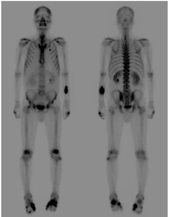
Fig. 6. Technetium-99m hydroxymethylene diphosphonate bone scan showing symmetrical uptake in the foot, hand, wrist, and elbow joints.
albicans.
After 21 days of hospitalization, his body temperature, white blood cell count, and pancreatic enzyme levels normalized. On the other hand, the painful swellings of the left foot and right hand were not resolved completely despite the long-term use of colchicine, NSAID, febuxostat (40 mg/day), or low-dose steroid (prednisolone 10 mg/day). On hospital- ization day 58, the plain radiographs of the hand showed moth-eaten bone destruction in the base of the second mid- dle and fifth proximal phalanges of the right hand (Fig. 4B).
A technetium-99m hydroxymethylene diphosphonate bone scan showed symmetrical uptake in the foot, hand, wrist, and elbow joints (Fig. 6). The blood tests for rheumatoid ar- thritis were negative. Ultrasound-guided aspiration of the left first metatarsophalangeal joint yielded a dry tap, i.e., failure to aspirate synovial fluid. As a result, samples could not be obtained for analysis. Finally, a diagnosis of PPP syn- drome was made based on the chronic pancreatitis on ab- dominal CT, mostly lobular panniculitis and adipocyte ne- crosis on a skin biopsy, and polyarthritis on a bone scan.
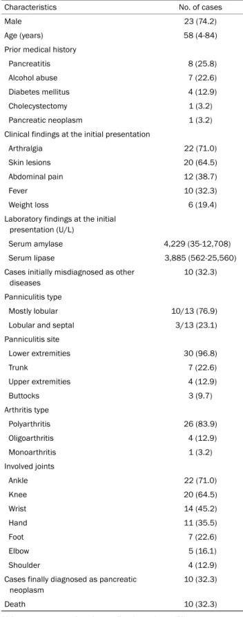
Table 1. Clinical Characteristics and Outcomes of 31 Cases of PPP Syndrome
Characteristics No. of cases
Male 23 (74.2)
Age (years) 58 (4-84)
Prior medical history
Pancreatitis 8 (25.8)
Alcohol abuse 7 (22.6)
Diabetes mellitus 4 (12.9)
Cholecystectomy 1 (3.2)
Pancreatic neoplasm 1 (3.2)
Clinical findings at the initial presentation
Arthralgia 22 (71.0)
Skin lesions 20 (64.5)
Abdominal pain 12 (38.7)
Fever 10 (32.3)
Weight loss 6 (19.4)
Laboratory findings at the initial presentation (U/L)
Serum amylase 4,229 (35-12,708)
Serum lipase 3,885 (562-25,560)
Cases initially misdiagnosed as other diseases
10 (32.3)
Panniculitis type
Mostly lobular 10/13 (76.9)
Lobular and septal 3/13 (23.1)
Panniculitis site
Lower extremities 30 (96.8)
Trunk 7 (22.6)
Upper extremities 4 (12.9)
Buttocks 3 (9.7)
Arthritis type
Polyarthritis 26 (83.9)
Oligoarthritis 4 (12.9)
Monoarthritis 1 (3.2)
Involved joints
Ankle 22 (71.0)
Knee 20 (64.5)
Wrist 14 (45.2)
Hand 11 (35.5)
Foot 7 (22.6)
Elbow 5 (16.1)
Shoulder 4 (12.9)
Cases finally diagnosed as pancreatic neoplasm
10 (32.3)
Death 10 (32.3)
Values are presented as the median (range) or n (%).
PPP, pancreatitis, panniculitis, and polyarthritis.
With respect to the treatment of chronic pancreatitis, opioid analgesics were used as needed to control the abdominal pain, and enoxaparin was administered to prevent the pro- gression of portal vein thrombosis. With these conservative treatments, abdominal pain and elevated levels of serum pancreatic enzyme improved gradually. The follow-up CT of the abdomen showed no interval change in pancreatic atro- phy, dilatation of the main pancreatic duct, and portal vein thrombosis except for the improvement in peripancreatic necrosis. Intravenous octreotide (1 mg/day) was administered to alleviate the osteoarticular symptoms but was dis- continued after three days because it was ineffective. This patient is currently under observation in the outpatient clin- ic with taking opioid analgesics in place of colchicine, NSAIDs, and steroids to control the joint pain.
LITERATURE REVIEW
English articles published between September 2009 and January 2019 were reviewed using the key words “pancreatitis”,
“panniculitis”, and “arthritis”. A total of 30 cases of PPP syn- drome were identified in the English literature.4-32 Table 1 lists the clinical characteristics and outcomes of 31 patients with PPP syndrome including the present case.
Of the total 31 patients with PPP syndrome, 23 (74.2%) were male and the median age was 58 years (range, 4-84).
At the time of the initial presentation, more than 70% of pa- tients had a chief complaint other than abdominal pain.
Arthralgia (71.0%) was the most common symptom, followed by skin lesions (64.5%), abdominal pain (38.7%), and fever (32.3%). Of the 31 patients with PPP syndrome, 10 (32.3%) were initially misdiagnosed as a subcutaneous abscess (four cases), gouty arthritis (two cases), septic arthritis (two cases), cellulitis (one case), compartment syndrome (one case), eryth- ema nodosum (one case), and sepsis (one case).
Of the 13 cases, in which panniculitis type was known, 10 (76.9%) had mostly lobular panniculitis and three (23.1%) had lobular and septal panniculitis. The lower extremities (96.8%) were the most common site of panniculitis, followed by the trunk (22.6%), upper extremities (12.9%), and buttocks (9.7%). Regarding the arthritis type, more than 80% of the patients showed polyarthritis (≥4 joints), which involved main- ly the ankle, knee, wrist, hand, and foot joints. No cases in- volved the simultaneous occurrence of PPP, i.e., the triad of
PPP syndrome. In addition, the time sequence of the three conditions varied from case to case, without certain rules.
Of the 31 patients with PPP syndrome, 10 (32.3%) were finally diagnosed with a pancreatic neoplasm, including acinar cell carcinoma (six cases), intraepithelial neoplasm (one case), neuroendocrine tumor (one case), pseudopapillary tu- mor (one case), and pancreatic tumor of unknown histology (one case). Of these, eight patients had a chief complaint of arthralgia or skin lesions other than abdominal pain at the time of the initial presentation. Overall, 10 (32.3%) out of 31 patients with PPP syndrome died, eight of whom had a pancreatic neoplasm.
DISCUSSION
This case highlights the challenge in the early diagnosis of PPP syndrome that presented initially with painful joint swelling and then was mistaken for cellulitis and gouty arthri- tis, resulting in the long-term use of broad-spectrum anti- biotics and anti-inflammatory agents. In a review of the liter- ature, the majority of cases of PPP syndrome also had chief complaints other than abdominal pain at the time of the initial presentation, often leading to a misdiagnosis. When a patient with pancreatic disease presents with cutaneous or osteo- articular lesions, a high clinical suspicion of PPP syndrome as well as a differential diagnosis of possible diseases with similar manifestations is needed. In addition, many patients with PPP syndrome are eventually diagnosed with a pancre- atic tumor and show a high mortality rate. This suggests that patients with PPP syndrome require careful observation and repeated investigations as needed, not only during treatment but also after recovery.
An early diagnosis of PPP syndrome is difficult when joint involvement or panniculitis dominate with a few symptoms of pancreatitis or vice versa. A delay in the diagnosis and treatment of the underlying pancreatitis can result in sig- nificant mortality.33,34 In addition, osteoarticular involvement in PPP syndrome can cause permanent destruction, leading to significant morbidity.
The pathogenesis of panniculitis and joint involvement in PPP syndrome is not well known. The most accepted hypoth- esis is fat necrosis caused by pancreatic lipolytic enzymes.35 In PPP syndrome, subcutaneous fat necrosis causes pancre- atic panniculitis, mainly in the lower extremities. The clinical
or histology findings of pancreatic panniculitis may vary ac- cording to the disease duration. Acute lesions usually appear as subcutaneous nodules (0.5-5 cm in diameter) and histolog- ically lobular panniculitis with necrosis of adipocytes (“ghost cells”), which is pathognomonic for pancreatic panniculitis and is subsequently replaced with granulomatous infiltration over time.2 In this case, a histology examination of the preti- bial skin lesions revealed mostly lobular panniculitis and adi- pocyte necrosis.
Osteoarticular involvement of PPP syndrome includes arthri- tis, serositis, or periarticular or intramedullary fat necrosis.15 In patients with PPP syndrome, arthritis can come from either periarticular fat necrosis or direct extension from the subcuta- neous fat necrosis to the adjacent joint space.36 Most joint involvement is polyarticular, involving mainly the foot, ankle, hand, or wrist joints. Aspiration of the involved joint fluid shows viscous and creamy to yellowish material on the gross appear- ance, positively birefringent lipid crystals or fat globules with a few cellular components on microscopy, and a high lipid content for cholesterol and triglyceride in a lipid analysis.35 In PPP syndrome, joint involvement usually follows its own course, without any response to treatment with an NSAID, steroid, or immunosuppressant. In this case, the foot, hand, wrist, and elbow joints were symmetrically involved. Aspiration of the involved joint yielded a dry tap with no crystals on microscopy. The pancreatitis and panniculitis improved sponta- neously with time, but the polyarthritis waxed and waned re- gardless of the long-term use of colchicine, an NSAID, a steroid, or octreotide.
In a review of the literature, most patients with PPP syn- drome presented with chief complaints other than symptoms of pancreatitis, such as abdominal pain. Therefore, the clini- cian focused more on the chief complaints rather than consid- ering their association with pancreatitis, making it easier to misdiagnose them as another disorder. In the present case, the patient presented with painful swelling of the left foot that was initially mistaken for cellulitis and then misdiagnosed as gouty arthritis. As a result, the patient was treated with colchicine, an NSAID, and a low-dose steroid for a long time.
The findings show that PPP syndrome should be considered when extrapancreatic manifestations, such as arthralgia or skin lesions occur in patients with pancreatitis.
In addition to acute or chronic pancreatitis, pancreatic can- cer or pseudocysts may be associated with PPP syndrome.3
In a previous review, the mortality rate of PPP syndrome was high at 24%.3 The review revealed a mortality rate of 30.0%.
Interestingly, more than 30% of the patients with PPP syn- drome were finally diagnosed with a pancreatic neoplasm, 80% of whom died. In these patients, the pancreatic cancer was more likely to be the underlying cause of PPP syndrome than pancreatitis. In the review, the mortality rate was highest in patients with PPP syndrome who were finally diagnosed with a pancreatic neoplasm, of which pancreatic acinar cell carcinoma was most common. Although pancreatic acinar cell carcinoma accounts for approximately 1% of all pancreatic tumors,37 it is the most common pancreatic neoplasm causing PPP syndrome. Therefore, PPP syndrome can be considered as a surrogate marker for pancreatic diseases with poor outcomes. In addition, patients with long-standing PPP syn- drome require careful monitoring for the possibility of pancre- atic cancers, such as acinar cell carcinoma.
In conclusion, clinicians should include PPP syndrome in a differential diagnosis when extrapancreatic manifestations, such as osteoarticular involvement or subcutaneous nodules occur in patients with pancreatic disorder(s). Considering the high mortality rate and possibility of a hidden pancreatic ma- lignancy in patients with PPP syndrome, continuous surveil- lance and active investigation are needed to improve the clin- ical outcomes.
ACKNOWLEDGEMENT
The authors thank professor Jin Cheon Kim, MD, Asan Medical Center, University of Ulsan College of Medicine, for proofreading this manuscript.
REFERENCES
1. Chiari H. Über die sogenannte fettnekrose. Prager Med Wochenschr 1883;8:284-286.
2. Requena L, Sánchez Yus E. Panniculitis. Part II. Mostly lobular panniculitis. J Am Acad Dermatol 2001;45:325-364.
3. Narváez J, Bianchi MM, Santo P, et al. Pancreatitis, panniculitis, and polyarthritis. Semin Arthritis Rheum 2010;39:417-423.
4. Agarwal S, Sasi A, Ray A, Jadon RS, Vikram N. Pancreatitis pan- niculitis polyarthritis syndrome with multiple bone infarcts. QJM 2019;112:43-44.
5. Chattopadhyay A, Mittal S, Sharma A, Jain S. Pancreatitis, pan- niculitis, and polyarthritis. J Clin Rheumatol 2018 Sep 27. [Epub ahead of print]
6. Fordham T, Sims HM, Farrant T. Unusual presentation of pan-
creatitis with extrapancreatic manifestations. BMJ Case Rep 2018;2018:bcr-2018-226440.
7. Sondhi AR, Wamsteker EJ, Piper MS. "Doc, I can't walk"-a classic presentation of a rare disease. Gastroenterology 2018;155:
1703-1705.
8. Fernández-Sartorio C, Combalia A, Ferrando J, et al. Pancreatic panniculitis: a case series from a tertiary university hospital in Spain. Australas J Dermatol 2018;59:e269-e272.
9. Graham PM, Altman DA, Gildenberg SR. Panniculitis, pan- creatitis, and polyarthritis: a rare clinical syndrome. Cutis 2018;
101:E34-E37.
10. Zundler S, Strobel D, Manger B, Neurath MF, Wildner D.
Pancreatic panniculitis and polyarthritis. Curr Rheumatol Rep 2017;19:62.
11. Dong E, Attam R, Wu BU. Board review vignette: PPP syndrome:
pancreatitis, panniculitis, polyarthritis. Am J Gastroenterol 2017;
112:1215-1216.
12. Rao P, Coffman N, Ferreira JP. A diagnostic dilemma in a patient with polyarthritis. Am J Med 2017;130:e497-e498.
13. Dieker W, Derer J, Henzler T, et al. Pancreatitis, panniculitis and polyarthritis (PPP-) syndrome caused by post-pancreatitis pseu- docyst with mesenteric fistula. Diagnosis and successful surgi- cal treatment. Case report and review of literature. Int J Surg Case Rep 2017;31:170-175.
14. Kang DJ, Lee SJ, Choo HJ, Her M, Yoon HK. Pancreatitis, pan- niculitis, and polyarthritis (PPP) syndrome: MRI features of intra- osseous fat necrosis involving the feet and knees. Skeletal Radiol 2017;46:279-285.
15. Ferri V, Ielpo B, Duran H, et al. Pancreatic disease, panniculitis, polyarthrtitis syndrome successfully treated with total pan- createctomy: case report and literature review. Int J Surg Case Rep 2016;28:223-226.
16. Loverdos I, Swan MC, Shekherdimian S, et al. A case of pan- creatitis, panniculitis and polyarthritis syndrome: elucidating the pathophysiologic mechanisms of a rare condition. J Pediatr Surg Case Rep 2015;3:223-226.
17. Langenhan R, Reimers N, Probst A. Osteomyelitis: a rare compli- cation of pancreatitis and PPP-syndrome. Joint Bone Spine 2016;83:221-224.
18. Naeyaert C, de Clerck F, De Wilde V. Pancreatic panniculitis as a paraneoplastic phenomenon of a pancreatic acinar cell carcinoma. Acta Clin Belg 2016;71:448-450.
19. Callata-Carhuapoma HR, Pato Cour E, Garcia-Paredes B, et al.
Pancreatic acinar cell carcinoma with bilateral ovarian meta- stases, panniculitis and polyarthritis treated with FOLFIRINOX chemotherapy regimen. A case report and review of the literature. Pancreatology 2015;15:440-444.
20. Arbeláez-Cortés A, Vanegas-García AL, Restrepo-Escobar M, Correa-Londoño LA, González-Naranjo LA. Polyarthritis and pan- creatic panniculitis associated with pancreatic carcinoma: re- view of the literature. J Clin Rheumatol 2014;20:433-436.
21. Laureano A, Mestre T, Ricardo L, Rodrigues AM, Cardoso J.
Pancreatic panniculitis - a cutaneous manifestation of acute pancreatitis. J Dermatol Case Rep 2014;8:35-37.
22. Azar L, Chatterjee S, Schils J. Pancreatitis, polyarthritis and pan- niculitis syndrome. Joint Bone Spine 2014;81:184.
23. Kashyap S, Shanker V, Kumari S, Rana L. Panniculitis-poly- arthritis-pancreatitis syndrome. Indian J Dermatol Venereol Leprol 2014;80:352-354.
24. Fraisse T, Boutet O, Tron AM, Prieur E. Pancreatitis, panniculitis, polyarthritis syndrome: an unusual cause of destructive polyarthritis. Joint Bone Spine 2010;77:617-618.
25. Vasdev V, Bhakuni D, Narayanan K, Jain R. Intramedullary fat ne- crosis, polyarthritis and panniculitis with pancreatic tumor: a case report. Int J Rheum Dis 2010;13:e74-e78.
26. Porcu A, Tilocca PL, Pilo L, Ruiu F, Dettori G. Pancreatic pseudo- cyst-inferior vena cava fistula causing caval stenosis, left renal vein thrombosis, subcutaneous fat necrosis, arthritis and dysfibrinogenemia. Ann Ital Chir 2010;81:215-220.
27. Borowicz J, Morrison M, Hogan D, Miller R. Subcutaneous fat ne- crosis/panniculitis and polyarthritis associated with acinar cell carcinoma of the pancreas: a rare presentation of pancreatitis, panniculitis and polyarthritis syndrome. J Drugs Dermatol 2010;9:1145-1150.
28. Harris MD, Bucobo JC, Buscaglia JM. Pancreatitis, panniculitis, polyarthritis syndrome successfully treated with EUS-guided cyst-gastrostomy. Gastrointest Endosc 2010;72:456-458.
29. Mustafa KN, Hadidy A, Shoumaf M, Razzuki SA. Polyarthritis with chondronecrosis associated with osteonecrosis, panniculitis and pancreatitis. Rheumatol Int 2010;30:1239-1242.
30. Jose T, Biju IK, Kumar A, et al. 'Pancreatitis, polyarthritis, pan- niculitis syndrome' (PPP syndrome) plus prolonged pyrexia--a rare presentation of chronic pancreatitis. Indian J Gastroenterol 2009;28:186-188.
31. Chee C. Panniculitis in a patient presenting with a pancreatic tu- mour and polyarthritis: a case report. J Med Case Rep 2009;
3:7331.
32. Kuwatani M, Kawakami H, Yamada Y. Osteonecrosis and pan- niculitis as life-threatening signs. Clin Gastroenterol Hepatol 2010;8:e52-e53.
33. Park SM. Recent advances in management of chronic pancreatitis. Korean J Gastroenterol 2015;66:144-149.
34. Park SM, Lee HS, Kim SY, et al. Clinical characteristics of chronic pancreatitis according to the history of pancreatitis. Korean J Gastroenterol 2009;53:239-245.
35. Dahl PR, Su WP, Cullimore KC, Dicken CH. Pancreatic panniculitis. J Am Acad Dermatol 1995;33:413-417.
36. Burns WA, Matthews MJ, Hamosh M, Weide GV, Blum R, Johnson FB. Lipase-secreting acinar cell carcinoma of the pancreas with polyarthropathy. A light and electron microscopic, histoche- mical, and biochemical study. Cancer 1974;33:1002-1009.
37. Klimstra DS, Heffess CS, Oertel JE, Rosai J. Acinar cell carcinoma of the pancreas. A clinicopathologic study of 28 cases. Am J Surg Pathol 1992;16:815-837.