Introduction
In typical scapular alignment, the vertebral border of the scapular and the vertebral spine are parallel, and the average distance between the vertebral border
of the scapular and the midline of the thoracic spine is about three inches (Sahrmann, 2002). Excessive downward rotation, elevated, depressed, abducted, ad- ducted, or winged scapulae are the frequently ob- served scapular alignment impairments (Kendall et al, Corresponding author: Hye-seon Jeon [email protected]
This study was in part supported by the “Brain Korea 21 PLUS Project (Grant No. 2016-51-0009),” the Korean Research Foundation for Department of Physical Therapy in the Graduate School of Yonsei University.
Comparative Effect of Modified Shrug Exercises With and Without Trunk Stabilization Exercise on Scapular Upward Rotator EMG and
Thickness in Subjects With Scapular Downward Rotation Syndrome
Ji-hyun Kim1, BPT, PT, Hyeo-bin Yoon1, BPT, PT, Joo-hee Park1, MSc, PT, Hye-seon Jeon2,3, PhD, PT
1Dept. of physical therapy, The Graduate School, Yonsei University
2Dept. of Physical Therapy, College of Health Science, Yonsei University
3Dept. of Ergonomic Therapy, The Graduate School of Health and Environment, Yonsei University
Abstract
1)Background: Scapular downward rotation syndrome (SDRS) is a common scapular alignment impairment that causes insufficient upward rotation and muscle imbalance, shortened levator scapulae (LS) and rhomboid, and lengthened serratus anterior (SA) and trapezius. A modified shrug exercise (MSE), performing a shrug exercise with the shoulders at 150° abduction, is known as an effective exercise to increase scapular stabilizer muscle activation. Previous studies revealed that scapular exercise are more effective when combined with trunk stabilization exercises in decreasing scapular winging and increasing scapular stabilizer muscle activation.
Objects: The purpose of our study was to clarify the effect of MSE with or without trunk stabilization exercises in subjects with SDRS.
Methods: Eighteen volunteer subjects (male=10, female=8) with SDRS were recruited for this experiment. All subjects performed MSE under 3 different conditions: (1) MSE, (2) MSE with an abdominal draw-in maneuver (ADIM), and (3) MSE with an abdominal expansion maneuver (AEM). The muscle thickness of the lower trapezius (LT) and the SA were measured using an ultrasonography in each condition. Electromyography (EMG) data were collected from the LT, LS, SA, and upper trapezius (UT) muscle activities. Data were statistically analysed using one-way repeated analysis of variance at a significance level of .05.
Results: The muscle thickness of the LT and the SA were the significant different in the MSE, MSE with ADIM (MSE+ADIM) and MSE with AEM (MSE+AEM) conditions (p<.05) In both LT and SA, the order of thick muscle thickness was MSE+AEM, MSE+ADIM, and MSE alone. No significant differences were found in the EMG activities of the SA, UT, LS, and LT in all condition.
Conclusion: In conclusion, MSE is more beneficial to people with SDRS when combined with trunk stabilization exercises by increased thickness of scapular stabilizer muscles.
Key Words: Abdominal draw-in maneuver; Abdominal expansion maneuver; Scapular downward rotation syndrome; Shoulder rehabilitation; Shrug exercise.
1983). Scapular downward rotation syndrome (SDRS) describes an atypical alignment of the scapula in which the inferior border of the scapular is positioned more centrally than the superior border of the scap- ula, thus positioning the shoulder lower with a down- ward slope at the acromial end (Caldwell et al, 2006).
Persons with SDRS tend to demonstrate insufficient upward rotation, shortened levator scapulae (LS) and rhomboids, weak and lengthened serratus anterior (SA) and trapezius, and a muscle imbalance between the scapular upward and downward rotators (Sahrmann, 2002).
The shrug exercise, the most common therapeutic exercise used for SDRS, is targeted to strengthen the trapezius muscles (Burkhead and Rockwood , 1992; Ekstrom et al, 2003; Hintermeister et al, 1998;
Pizzari et al, 2014). However, it has been reported that performing the shrug exercise in the traditional way (with the arms at the side of the trunk) strengthens the LS rather than the trapezius mus- cles, especially the upper trapezius (UT) (Moseley et al, 1992; Smith et al, 2004). Choi et al (2015) exam- ined the effects of the shrug exercise while subjects with SDRS performed shrug exercises at three differ- ent shoulder abduction positions. They measured the scapular downward rotation index (SDRI) and electro- myography (EMG) activation of the UT, LS, SA, and lower trapezius (LT) muscles and found that the shrug exercise at 150° of shoulder abduction was ef- fective for eliciting greater SA and LT muscle activ- ity, decreased SDRI, and improved muscle balance among the UT, LS, SA, and LT compared with 30°
and 90° shoulder abduction conditions. Additionally, Choi et al (2015) recommended performing shrug ex- ercises at 150° of shoulder abduction for people with relatively weak scapular upward rotators, especially SA and LT.
The abdominal draw-in maneuver (ADIM) has been widely used in clinics to increase lumbo-pelvic stability through selective activation of the transverse abdominis and internal oblique muscles (Macedo et al, 2009). The ADIM is also commonly performed during
arm and leg exercises to increase the stability of the lumbopelvic area. A prior study reported that the ef- fects of scapular stabilizer (UT, SA, and LT) activ- ities were significantly greater when the scapular exercise was superimposed onto the ADIM than when performing the scapular exercise alone (Kim et al, 2017). As activation of the abdominal muscles might affect the scapular muscle activity through the myofascial connections in the trunk, the abdominal expansion maneuver (AEM) was used to increase the stability of the deep trunk muscle and the lumbo- pelvic posture (Myers, 2009; Kim et al, 2012). Subjects were requested to hold their navel anteriorly and downwardly while expanding the lower abdomen but not the thoracic cage (Yoon et al, 2015). More re- cently, many studies have revealed a positive effect of AEM, indicating that AEM facilitates the co-con- traction of the diaphragm and the deep spinal stabil- izer muscles and increases the intra-abdominal pres- sure (Lee and Kim, 2015; Yoon et al, 2015).
However, no previous studies have compared ADIM and AEM with the shrug exercise in subjects with SDRS. Based on the findings of Choi et al (2015)’s experiment, we perform the shrug exercise at 150° of shoulder abduction, which we name the modified shrug exercise (MSE). The purpose of our study is to clarify the effect of MSE with and with- out trunk stabilization exercises (ADIM or AEM) in subjects with SDRS. More specifically, we aim to determine which trunk stabilization exercise is more effective at enhancing and normalizing scapular mus- cle activation during the MSE.
Methods
Subjects
Eighteen (10 males and 8 females) subjects with SDRS were recruited from community and university populations. The mean age of the subjects was 22.8 years (20-26 years). Prior to this study, we exam- ined 70 people to select subjects with SDRS. Based
on previous studies, we use the following inclusion criteria: (1) medial border of scapula is not parallel to thoracic spine, (2) a scapula abduction angle less than 60° during full shoulder abduction, (3) distance from the thoracic spine to medial boarder of the scapula is less than three inches, and (4) SDRI is greater than 10 (Figure 1.) (Lee et al, 2016). The exclusion criteria were (1) subjects with downward rotation syndrome due to a neurological problem; (2) a history of injury or surgery of the shoulder, trunk, and neck; (3) a positive result for the apprehension test; and (4) a positive result for the upper limb tension test. All subjects read an explanation about the experimental procedures and signed an informed consent form ap- proved by the Yonsei University Wonju Institutional Review Board (approval number: 1041849-201705- BM-053-02).
Instrumentation
Surface electromyography (EMG)
Noraxon TeleMyo 2400T (Noraxon Inc., Scottlsdale, AZ, USA) was used to collect EMG signals from the
UT, LS, SA, and LT muscles. The skin area was shaved and cleaned using rubbing alcohol. Bipolar surface electrodes (Ag/AgCl) were adhered at a 2 ㎝ inter-electrode distance. Electrodes for the UT were placed slightly laterally to and one-half the distance between the cervical spine at C-7 and the acromion.
Electrodes for the SA were determined by palpation while the patient flexed the arm against resistance;
they were placed just anterior to the border of the latissimus dorsi muscle at the level of the inferior tip of the scapula. The LT EMG electrode site was de- termined by palpating the inter-scapular region while the subjects flexed the arm to at least 90° of scap- ular retraction and depression. Electrodes were placed at an oblique angle, approximately 5㎝ down from the scapular spine. Electrodes of the LS were placed between the anterior margin of the UT and the pos- terior margin of the sternocleidomastoid (Criswell, 2010; Ludewig et al, 1996). EMG data were analyzed using Noraxon MyoResearch 1.06 software. The sampling rate was 1000 ㎐. A bandpass filter between 20 and 450 ㎐ was used. EMG data were processed into the root- mean-square with a window of 50 ㎳.
Subjects were asked to perform maximum volun- tary isometric contraction (MVIC) for 5 s for each muscle to normalize the EMG data of each muscle in individual. MVIC was repeated three times with in- tervals of 5 s between trials. The average value of three attempts was used for each subject’s final MVIC. A specific test position for the MVIC was selected based on Kendall et al (1983).
Ultrasonography measurement
Ultrasonography (Mysono U6, Medison, Seoul, Korea) was used to capture the linear depth of the LT and SA in each MSE condition. Before conducting the measurement, we identified the target muscles and placed the ultrasound (US) transducer at the scapular inferior angle at the same level as the vertebrae spine (O’sullivan et al, 2007). The thickness of the SA was measured between the pectoralis major and latissimus dorsi on a rib angle at the same level as Figure 1. Scapular downward rotation
index (a): distance between root of the scapular spine and spinous process in same level of the scapular spine, (b):
distance between inferior angle of scapula and spinous process in same level of the inferior angle.
the scapular inferior angle (Basmajian, 1983; Cuadros et al, 1995; Day and Uh, 2013). A 5-12㎒ linear transducer was placed transversely to measure the LT and vertically along the SA area. Screen calipers, which come with mysono U6, were used to measure the muscle thickness. The LT muscle thickness measurement was determined by the thickness value of a part 2 ㎝ away from the spinous process on the same level (O’sullivan et al, 2007). The SA thickness was measured from the superior border of the rib to the inside portion of the muscle border. The distance of the five parts, spanning the width of the rib, was measured and the average value was obtained and analyzed (Day and Uh, 2013).
Experimental procedure
In this study, the subjects performed the MSE un- der three different conditions in a random order. After receiving a verbal explanation of the experimental procedures, the subjects familiarized themselves with the MSE and the MSE combined with trunk stabili- zation exercises (ADIM and AEM).
First at all, the subjects were asked to perform the MSE by elevating their shoulders to an abduc- tion of 150° in sitting position on a backless chair and then shrug their shoulders as hard as they could. In the way of doing MSE with ADIM (MSE+ADIM), prior to initiating the MSE, the sub- jects were asked to pull their navel toward the spine and shrug their shoulders as hard as they could. The subjects maintained the shoulder muscle contraction
for 5 seconds while maintaining the ADIM. In MSE with AEM (MSE+AEM) condition, they expanded the lower abdomen and pushed their navel in an ante- rior-inferior direction (toward the symphysis pubis) without lateral expansion of the rib cage. This meth- od allowed the subject to achieve co-contraction of the diaphragm and deep spinal stabilizer muscles and increase the intra-abdominal pressure (Yoon et al, 2015).
No chest movements were allowed during the AEM.
They performed the MSE while maintaining the AEM.
These process were repeated three times in each conditions with a rest of 5 seconds between the trials. To confirm the contraction of the TrA muscle in all conditions, the examiner palpated the subject’s abdominal wall and utilized a visual bio-feedback using the US. While subjects conducted the ex- ercises, the examiner measured the SA and LT muscle thicknesses and the UT, LS, SA, and LT ac- tivities to record any changes in muscle activation and thickness. EMG data were collected during the middle 3 seconds period while the subjects main- tained the MSE for 5 seconds.
Statistical analysis
The data were analyzed using Windows SPSS version 24.0 (SPSS Inc., Chicago, IL, USA). A one-way repeated analysis of variance (ANOVA) was used to compare the muscle thickness and muscle activities in the three conditions. The Bonferroni test was used for the post-hoc analysis, and the sig- nificance level was set at .05.
Results
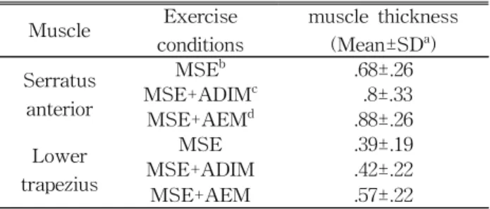
First, the muscle thickness of the LT and SA were found to be significantly different across the conditions (p<.05) (Figure 2)(Table 1). The thickness of the SA was greatest in the MSE+AEM condition and decreased toward the MSE+ADIM and MSE conditions (p<.05). The post-hoc analysis revealed that the SA muscle thicknesses were significantly Muscle Exercise
conditions
muscle thickness (Mean±SDa) Serratus
anterior
MSEb .68±.26
MSE+ADIMc .8±.33
MSE+AEMd .88±.26
Lower trapezius
MSE .39±.19
MSE+ADIM .42±.22
MSE+AEM .57±.22
amodified shrug exercise, bmodified shrug exercise with abdominal draw-in maneuver, cmodified shrug exercise with abdominal expansion maneuver, dstandard deviation.
Table 1. Muscle thickness
different in all paired comparisons (p<.05). The Thickness of the LT was also greatest in the MSE+AEM condition and decreased toward the MSE+ADIM and MSE conditions. However, the post-hoc analysis revealed that the LT muscle thick- ness in the MSE+ADIM was not statistically greater than that in the MSE condition (p<.05).
EMG amplitudes of the scapular upward rotators (UT, SA, and LT) were greater when the MSE was combined with the trunk muscle stabilization exercise than when the MSE was performed alone. However, for the LS, the downward rotator, the muscle activ- ity was greatest when the MSE was performed without the trunk muscle exercise. Nevertheless, am- plitudes of the EMG activities of all measured mus-
cles were not statistically different among the three conditions (p<.05) (Figure 3)(Table 2).
Discussion
The purpose of this study was to clarify the effect of the MSE with and without trunk stabilization ex- ercises (ADIM and AEM) in subjects with SDRS.
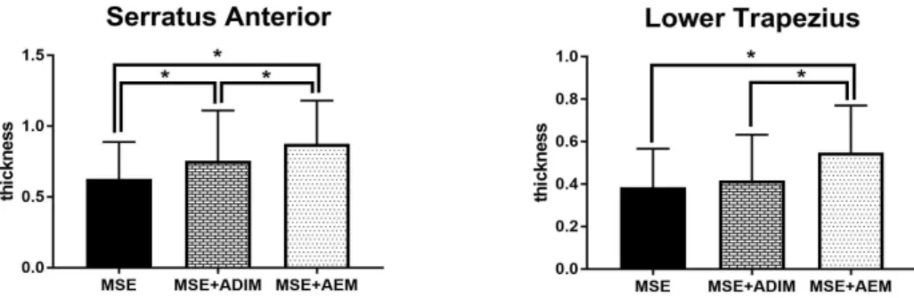
To our knowledge, this study is the first to evaluate the muscle thickness and activity of the UT, LS, SA, and LT muscles during the MSE with and without trunk stabilization exercises for subjects with SDRS. The main findings of this study indicate that, combining the MSE with trunk stabilization exercises Figure 2. muscle thickness of serratus anterior and lower trapezius (MSE: modified
shrug exercise, MSE+ADIM: modified shrug exercise with abdominal draw-in maneuver, MSE+AEM: modified shrug exercise with abdominal expansion maneuver).
Muscle Exercise conditions %MVICa (Mean±SDb)
Upper trapezius
MSEc 49.84±52.47
MSE+ADIMd 54.57±47.63
MSE+AEMe 62.92±37.29
Levator scapulae
MSE 45.02±57.14
MSE+ADIM 38.42±29.09
MSE+AEM 39.9±37.16
Serratus antreior
MSE 45.03±25.5
MSE+ADIM 53.33±59.3
MSE+AEM 54.59±36.12
Lower trapezius
MSE 19.68±24.21
MSE+ADIM 39.88±24.83
MSE+AEM 46.31±42.15
amaximal voluntary isometric contraction, bmean±standard deviation, cmodified shrug exercise, dmodified shrug exercise with abdominal draw-in maneuver, emodified shrug exercise with abdominal expansion maneuver.
Table 2. Surface EMG amplitude (Unit: %MVIC)
tends to increase the muscle activities and thick- nesses of the scapular upward rotators (UT, SA, and LT) and decrease the muscle activity of the scapular downward rotator (LS). In terms of changes in mus- cle thicknesses of the SA and LT during the MSE, the MSE+AEM condition was most effective, fol- lowed by the MSE+ADIM condition and MSE alone.
The tendency to increase the scapular upward ro- tator EMG activities and the statistical increase of muscle thickness in SA and LT during the MSE combined with trunk stabilization exercises were the main findings of this study. We are unable to reveal any significant differences in the EMG activities among the MSE conditions, which was likely be- cause of the small number of subjects and relatively large variability within the EMG data. However, the results of this study are consistent with previous findings by Kim et al (2017), ADIM greatly in- creased the activation of SA during push-ups plus exercise. It can be partially explained by the syner- gistic relationship between core stabilizers and scap-
ular stabilizer muscles via thoracolumbar fascia.
Thoracolumbar fascia, which consists of anterior, middle, and posterior layers, has an important bio- mechanical function in transferring the energy and load between the upper and lower extremities, be- tween the right and left sides of the body, and be- tween the abdominal wall and lumbo-pelvic area (Vleeming et al, 2014). The posterior layer of the thoracolumbar fascia consists of deep and superficial lamina. The superficial lamina is comprised of a fas- cia cover that includes muscles such as the rhom- boids, pectoralis major and minor, trapezius, SA, lat- issimus dorsi, and gluteus maximus (Willard et al, 2012). Therefore, a synergetic relationship exists be- tween the core stabilizers, originating from the deep lamina, and the scapular stabilizers, originating from the superficial lamina (Kanik et al, 2017). For these reasons, the SA and LT thicknesses might increase more when performing the MSE with trunk stabili- zation exercises than when performing MSE alone.
A previous study by Choi et al (2015) stated that Figure 3. muscle activation of upper trapezius, levator scapulae, serratus anterior and lower
trapezius. (MSE: modified shrug exercise, MSE+ADIM: modified shrug exercise with abdominal draw-in maneuver, MSE+AEM: modified shrug exercise with abdominal expansion maneuver, MVIC: maximal voluntary isometric contraction)
the SA and LT of subjects with SDRS were most facilitated during the shrug exercise at 150° of shoulder abduction. Based on their recommendation, we conducted this experiment to increase the in- volvement of SA and LT during the shrug exercise.
We named the shrug exercise at 150° of shoulder abduction the MSE. Because the UT is shortened in length at 150° of shoulder abduction, the MSE is mechanically less advantageous for the UT in gen- erating power compared to at 90° of shoulder abduction. The maximal muscle activity of the UT is achieved at 90° of shoulder abduction because both the lever arm of the UT and the external moment arm were longest at 90° of shoulder abduction (Moseley JR et al, 1992). However, the level of the UT increased by performing the MSE and the trunk muscle activation simultaneously. Therefore, adding trunk muscle stabilization exercises to the MSE con- siderably enhances the use of all measured upward rotators.
Conclusion
MSE for people with SDRS at 150° of shoulder abduction more effectively facilitates the use of the upward rotator thickness when combined with trunk stabilization exercises. Therefore, training the trunk stabilizer muscles and performing the shrug exercise is recommended in shoulder rehabilitation. Future studies should investigate why the AEM is more ef- fective than the ADIM.
References
Basmajian JV, Blumen R. Electrode Placement in Electromyographic Biofeedback. Biofeedback Principles and Practice for Clinicians. 1983.
Burkhead Wz Jr, Rockwood CA Jr. Treatment of instability of the shoulder with an exercise program. J Bone Joint Surg Am. 1992;74(6):
890-896.
Caldwell C, Sahrmann S, Van Dillen L, et al. use of a movement system impairment diagnosis for physical therapy in the examination and treatment of a patient with shoulder pain. J Ortho Sports PhysTher. 2007;37(9):551-563.
Choi WJ, Cynn HS, Lee CH, et al. Shrug exercises combined with shoulder abduction improve scapular upward rotator activity and scapular alignment in subjects with scapular downward rotation impairment. J Electromyogr Kinesiol.
2015;25(2):363-370. https://doi.org/10.1016/j.jelekin.
2014.12.001
Criswell E. Cramʼs Introduction to Surface Electromyo- graphy 2nd ed. Sudbury, Jones and Bartlett Publishers, 2010:268-298
Cuadros CL, Driscoll CL, Rothkopf DM. The anat- omy of the lower serratus anterior muscle: A fresh cadaver study. Plas Reconstr Surg. 1995;
95(1):93-97.
Day JM, Uhl T. Thickness of the lower trapezius and serratus anterior using ultrasound imaging during a repeated arm lifting task. Man Ther.
2013;18(6):588-593.
Ekstrom RA, Donatelli RA, Soderberg GL. Surface electromyographic analysis of exercises for the trapezius and serratus anterior muscles. J Orthop Sports Phys Ther. 2003;33(5):247-258.
Hintermeister RA, Lange GW, Schultheis JM, et al.
Electromyographic activity and applied load dur- ing shoulder rehabilitation exercises using elastic resistance. Am J Sports Med. 1998;26(2):
210-220.
Kanik ZH, Pala OO, Gunaydin G, et al. Relationship between scapular muscle and core endurance in healthy subjects. J Back Musculoskelet Rehabil.
2017;30(4):811-817. https://doi.org/10.3233/BMR- 150497
Kendall FP. Muscles Testing and Function. 3rd ed.
Lippincott Williams and Wilkins, 1983:222-228.
Kim DE, Shin AR, Lee JH, et al. Effect of the ab- dominal drawing-in maneuver on the scapular
stabilizer muscle activities and scapular wing- ing during push-up plus exercise in subjects with scapular winging. Physi Ther Korea. 2017;
24(1):61-70. https://doi.org/10.12674/ptk.2012.19.4.038 Kim KS, Lim OB, Yi CH, et al. Comparisons of
trunk muscle activity during arm lift in prone and standing positions with and without ab- dominal drawing-in maneuver. Phys Ther Korea.
2012;19(4):38-45. https://doi.org/10.12674/ptk.2012.
19.4.038
LEE HJ, KIM SY. Comparison of the effects of abdominal draw-in and expansion maneuvers on trunk stabilization in patients with low back pain and lumbar spine instability. Phys Ther Korea. 2015;22(1):37-48. https://doi.org/10.12674/
ptk.2015.22.1.037
Lee JH, Cynn HS, Choi WJ, et al. Various shrug exercises can change scapular kinematics and scapular rotator muscle activities in subjects with scapular downward rotation syndrome.
Hum Mov Sci. 2016;45:119-129. https://doi.org/
10.1016/j.humov.2015.11.016
Ludewig PM, Cook TM, Nawoczenski DA. Three-di- mensional scapular orientation and muscle activ- ity at selected positions of humeral elevation. J Orthop Sports Phys Ther. 1996;24(2):57-65.
https://doi.org10.2519/jospt.1996.24.2.57
Macedo LG, Maher CG, Latimer J, et al. Motor con- trol exercise for persistent, nonspecific low back pain: A systematic review. Phys Ther. 2009;89(1):
9-25.https://doi.org/10.2522/ptj.20080103
Moseley JB Jr, Jobe FW, Pink M, et al. EMG analysis of the scapular muscles during a shoulder rehabilitation program. Am J Sports Med. 1992;20(2):128-134.
Myers TW. Anatomy Trains: Myofascial meridians for manual and movement therapist, 3rd ed.
Edinburg, Elsevier, 2009:159-175.
O’sullivan C, Bentman S, Bennett K, et al.
Rehabilitative ultrasound imaging of the lower trapezius muscle: Technical description and reliability. J Orthop Sports Phys Ther.
2007;37(10):620-626.
Pizzari T, Wickham J, Balster S, et al. Modifying a shrug exercise can facilitate the upward rotator muscles of the scapula. Clin Biomech. 2014;29(2):
201-205. https://doi.org/10.1016/j.clinbiomech.2013.
11.011
Sahrmann S. Diagnosis and Treatment of Movement Impairment Syndromes. 1st ed. St. Louis, Mosby, 2002:199-252
Smith J, Padgett DJ, Kaufman KR, et al. Rhomboid muscle electromyography activity during 3 different manual muscle tests. Arch Phys Med Rehabil. 2004;85(6):987-992.
Vleeming A, Schuenke MD, Danneels L, et al. The functional coupling of the deep abdominal and paraspinal muscles: The effects of simulated paraspinal muscle contraction on force transfer to the middle and posterior layer of the thor- acolumbar fascia. J Anat. 2014;225(4):447-462.
Willard FH, Vleeming A, Schuenke MD, et al. The thoracolumbar fascia: Anatomy, function and clin- ical considerations. J Anat. 2012;221(6):507-536.
https://doi.org/10.1111/j.1469-7580.2012.01511.x Yoon MR, Choi HS, Shin WS. Effects of the ab-
dominal drawing-in maneuver and the abdominal expansion maneuver on grip strength, balance and pulmonary function in stroke patients. J Korean Phys Ther. 2015;27(3):147-153.
This article was received October 2, 2017, was reviewed October 2, 2017, and was accepted November 5, 2017.